This website uses cookies to ensure you get the best experience on our website.
- Table of Contents
Facts about Leucine-rich repeats and immunoglobulin-like domains protein 1.
Mouse | |
---|---|
Gene Name: | Lrig1 |
Uniprot: | P70193 |
Entrez: | 16206 |
Belongs to: |
---|
No superfamily |
D6Bwg0781e; Img; LIG1; LIG-1; LRIG1
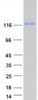
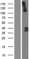
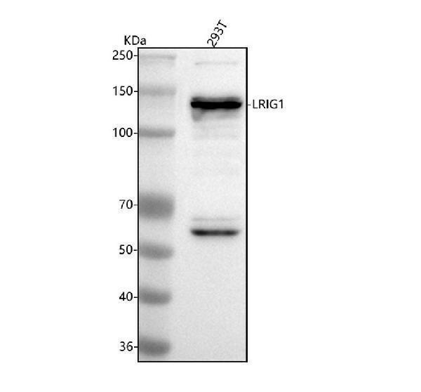
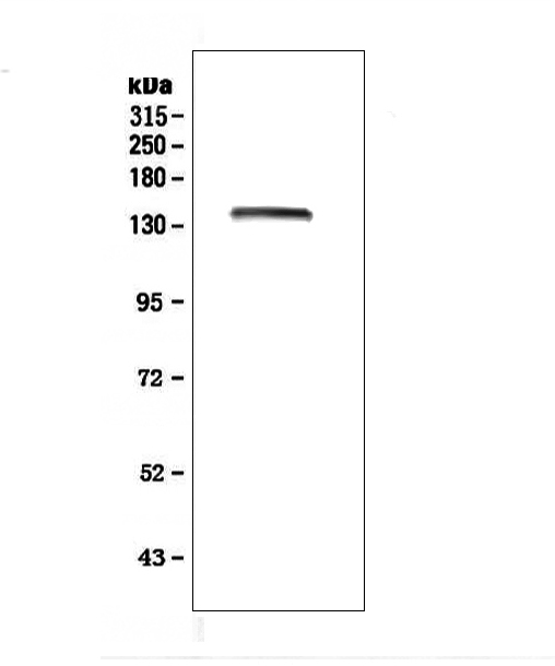
Mass (kDA):
119.156 kDA
Mouse | |
---|---|
Location: | 6 D2|6 43.15 cM |
Sequence: | 6; |
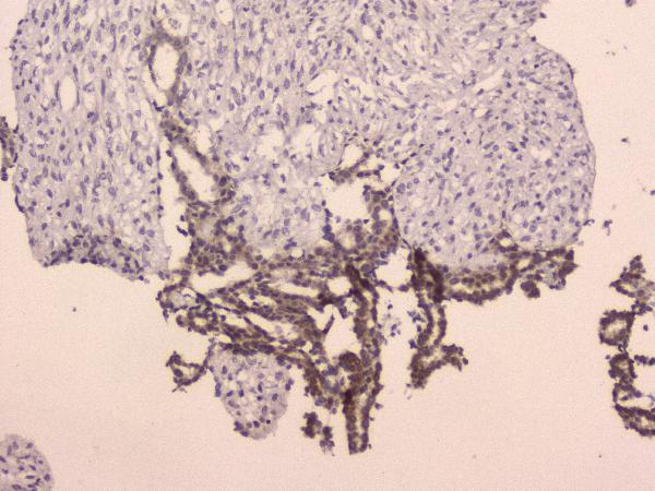
Detected in brain (at protein level) (PubMed:12067728). Predominantly expressed in the brain, restricted to a small subset of glial cells, such as Bergmann glial cells of the cerebellum and glial cells in the nerve fiber layer of the olfactory bulb. Expressed also in the skin. Low expression is detected in the thymus and heart. No expression in the kidney, liver, lung or small intestine.
This article is for you if you are looking for a breast-cancer marker that is specific for a particular disease. This article will talk about the LRIG1 mark and other proteins associated with breast cancer. The information in this article will help to determine the best LRIG1 marker to store for long-term storage. However, you will also learn about EGFR, PI3K/AKT MMP2, and EGFR.
Lrig1 acts as an inhibitor of panErbB, which is used to identify long-lived and non-cycling stem cell populations. Lrig1+ are different than Lgr5+ cells due to their upregulation of genes that suppress the cell's cycle. Lrig1+ cells are different from Lgr5+ cells because they have upregulated genes that suppress the cell cycle.
Lrig1-CreERT2/+ mice received 2mg of Tamoxifen. Then, their colon and small intestines had to be stained with a reporter for b-galactosidase (b-gal reporter). This indicates Lrig1 mRNA. Cells labeled with b-gal were mostly located in the lower one-third of the crypts. Some cells were labeled at top of the crypt.
These proteins were phosphorylation markers by the LRIG1 & EGFR marker in boster bio. These markers can be used to detect phosphorylated ERBB-receptors by Western blot analysis or densitometrical analyses. Both methods use GAPDH to control proteins. Data were analysed with Student's Test.
Janvier, Le Genest St Isle France bought transgenic mice in the C57BL/6N ancestry. The mice were kept in pathogen-free conditions at 23 degrees Celsius and 12-hour light/dark cycles. Mice were fed a standard rodent diet and water. LRIG1 was originally expressed on the skin of mice using a tetracycline-controlled transcriptional activation system.
During chemical carcinomagenesis, melanocytic tumors can develop from the overexpression of LRIG1. LRIG1 expression is also found in keratinocytes. DMBA treatment increases melanoma risk and burden. LRIG1 also is expressed in most cancer cells. Interestingly, LRIG1 is expressed in a small number of breast cancer patients.
LRIG1 is a regulator of cell proliferation. Overexpression of this gene causes a delay and altered expression of the cyclin-CDK protein proteins, which control cell cycle. Overexpression of LRIG1 causes U-251 MG cells to become less proliferative and migrate less. This gene may be a potential target in brain cancer treatments.
LRIG1 negatively regulates glioma cells' proliferation, migration, invasion, and migration. It also inhibits PI3K/AKT. This gene is abundant in many types, including gliomas. LRIG1 has been shown to have promising clinical applications by inhibiting the proliferation of glioma cells. This protein can also block the growth other types and cancer cells.
In this study, LRIG1 was shown to inhibit MMP2 (and MMP9) activity. This inhibition was confirmed by quantitative real-time PCR (qRT-PCR) on U-251 MG cells. It also suppressed MMP2 production and 9. This gene also regulates migration and invasion of U-251MG cells. It is not clear how LRIG1 may be useful for other purposes.
LRIG1 can be found in certain cells as a soluble peptide. Different types of cancer may be linked to the presence of this protein in different tissues. The best uses of the LRIG1 marker in liver cancer have yet to be determined. However, there have been several studies that suggest that the LRIG1 peptide could be used in prognostic purposes for various types of cancer, such as HCC.
Using the LRIG1 genome as a reference, a RNAi database was created from the three studied cell lines. Using the TRIZOL reagent to isolate the RNA, it was reverse transcribed to a final volume (30 uL). The LRIG1 PCR reactions were performed using a SYBR Masters Mix kit, which Takara in Kusatsu Japan provided. The primer sequences were 5'-ACTCTCTTAGGGAGA-3' and 5'-ACTCTCTTAGATTCACA-3'.
Lrig1 in the incisor marks two distinct pools stem cells: epithelial stem and mesenchymal. The LRIG1 -positive stem cells are responsible retaining periodontal msenchyme as well as forming dental pulp. It also distinguishes between incisors, molars. Lrig1 is responsible for signaling in the pulp.
LRIG1 is a transcription factor that is expressed by all spatial subtypes of the neurogenic stem cell population. This marker is expressed in particular by the a/b cell morphotype cells within the lateral walls. It is important that you understand that Lrig1 expression in SC populations differs from human tissues. However, evidence suggests that it could be useful in the study and treatment of neurogenic stem cells.
Interestingly, Lrig1 was previously used to identify brain quiescent neurogenic cells. Lrig1 expression was also associated to higher proliferating activity in neural stem cells in mouse models. It is important to determine Lrig1+ SC population live. However, the LRIG1 marker is only useful if the SC population has been studied in vitro or in vivo.
Next-generation sequencing is possible with the help of Molecular Inversion Probe array technology. These probes are capable of multiplexing several signatures simultaneously and are especially effective in identifying resistance and virulence genes. Researchers can now analyze thousands of samples with these innovative tools. These systems can be used to analyze thousands of samples in a single tube.
Molecular-inversion probes can be used to precisely target and adjust specific regions of your genome. These single-strand DNA molecules contain complementary sequences. To minimize off-target probe binding, they are joined by a linker. Target DNA is copied through PCR to close the loop. You can also create custom panels for specific genes, breakpoints, or other requirements. MIPs can be supplemented by amplicons for pseudogenes and fuzzy breakpoints. These amplicons may also be used to perform short repeat expansions.
Also, Molecular inversion probes are useful for characterizing alternative splicing. MIPs, single-stranded DNA molecules oligonucleotides, range in length between 130 and 150 bases. They are synthesized at home using standard methods. The cost per unit is about $0.08. Bulk amplification of single-stranded MIPs is necessary. Molecular inversion probes contain a sequence tag that distinguishes them from the original SNP MIPs.
The LRIG1 marker is a fluorescent label for enteroendocrine cells. It is expressed in a low frequency subset intestinal SCs (scCs). The labeled cells were often positive for the marker Ki67. Transcriptome profiling Lgr5+ as well as -negativeSCs revealed distinct gene expression profiles. The Lgr5+ cells expressed genes associated with cell cycle promotion, immune regulation, and oxidative stress.
The activity of MMP2 & MMP9 has been inhibited using the Lrig1 marker. This has been confirmed using quantitative real-timePCR. LRIG1 also suppressed mRNA levels in both MMPs. The invasion of and migration by U-251 MG MG cells is also affected through the LRIG1 mark. Although its use is currently limited, it is a promising biomarker for detecting multiple cell types.
The LRIG1 marker has many potential uses in research. It has been shown by experiments to inhibit glioma growth by preventing the proliferation of cells. It can also prevent cell division and migrate. By inhibiting cell growth, LRIG1 is particularly useful in tumorigenesis and drug development. Although LRIG1 can be found in cancer cells and its role is unclear in preventing tumorigenesis, it is expressed in some cancer cells.
In vitro studies showed that LRIG1 could suppress EGFR activitiy. A soluble form is made of the LRIG1 genes that expresses GFP fused to its COOH terminal side. In vitro experiments using mouse glioblastoma cellular cells have demonstrated a significant tumor-suppressive function. However, more research is needed on whether LRIG1 can inhibit EGFR activity in humans.
LRIG1 inhibits EGFR expression and phosphorylation, thereby preventing the growth of tumor cells. The signaling pathway between RTKs is affected by the protein, which blocks the signals and inhibits the growth tumor cells. Specifically, LRIG1 suppresses MET. HER2, HER3, IGF-1R, and HER3. These cancer-suppressing properties are crucial in diagnosing many types of tumors.
In vitro studies showed that LRIG1 suppresses lung carcinoma cell proliferation, invasiveness, migration, and migration. This gene has been examined using several cell lines including the HCC827, a mutant EGFR exon 19, deletion, and h2975 (a mutation L858R/T790M), and A549. Western blot analysis used a Boyden chamber as well as ImageJ software.
PMID: 8798419 by Suzuki Y., et al. cDNA cloning of a novel membrane glycoprotein that is expressed specifically in glial cells in the mouse brain. LIG-1, a protein with leucine-rich repeats and immunoglobulin-like domains.
PMID: 12067728 by Suzuki Y., et al. Targeted disruption of LIG-1 gene results in psoriasiform epidermal hyperplasia.