LRP1B / Low-density lipoprotein receptor-related protein 1B · IHC design guide

Design Immunohistochemistry for LRP1B

Plan chromogenic LRP1B IHC in paraffin sections with the catalog antibody at 2–5 μg/ml (datasheet A07425-1). Assess cytoplasmic staining in thyroid glandular and CNS neuronal cells, bearing in mind its low consistency with RNA expression (HPA tissue IHC).

Evidence assembled Oct 2026 · For research use; verify linked source records and product datasheet before use
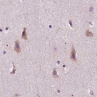
Immunohistochemistry protocol sheet for LRP1B (IHC for LRP1B): expected localisation Cytoplasmic staining in thyroid gland and CNS (HPA tissue IHC), antibody A07425-1, validated IHC image, and IHC protocol steps
Printable LRP1B IHC protocol sheet — expected localisation Cytoplasmic staining in thyroid gland and CNS (HPA tissue IHC), antibody A07425-1, controls and protocol steps. Open the full LRP1B IHC guide →

LRP1B Immunohistochemistry Experimental Design Guide

Expected localisation, validated protocols, controls and antibodies — the at-a-glance facts below, then the full design guide.

Must know before staining
Expected localisation Cytoplasmic staining in thyroid gland and CNS (HPA tissue IHC)
Staining pattern Thyroid glandular and CNS neuronal cytoplasm (HPA tissue IHC)
Antigen retrieval EDTA pH 8.0 HIER, heat-mediated (datasheet A07425-1)
Positive control ⓘ Caudate+2 more · see all
Negative control ⓘ Appendix+4 more · see all
Important caveats
Reasons your staining may differ from the expected pattern.
Fixation Keep paraffin-section fixation consistent (standard IHC practice; not target-specific); Matched tissue-IHC evidence does not establish the fixation claim. Validate the specimen-specific method before use. (selected-SKU IHC image A07425-1)
Caveat Staining has low consistency with RNA expression (HPA tissue IHC)
Regulation No staining regulator reported (UniProt)
Isoform / epitope No isoforms listed; extracellular vs cytoplasmic epitope matters (UniProt)
Section 1

Recommended LRP1B IHC & IF Protocols

The catalog antibody’s IHC-P protocol (datasheet A07425-1) is accompanied by four published LRP1B chromogenic IHC protocols (PMC12016830; PMC10741692; PMC9950470; PMC8335809).

Recommended immunohistochemistry (IHC-P) protocol parameters
SampleParaffin-embedded human breast cancer tissue; fixative not specified (datasheet A07425-1)
FixationImage fixative and duration unreported (datasheet A07425-1); verify before use.
Sectioning4–5 µm sections on charged slides (standard)
DeparaffinisationXylene, graded ethanol series to water (standard)
Antigen retrievalHeat retrieval: EDTA pH 8.0 (datasheet A07425-1); 20 min, 95–100 °C (standard)
Peroxidase block3% H2O2, 10 min, room temperature (standard)
Blocking10% goat serum (datasheet A07425-1)
Primary antibodyRabbit anti-LRP1B, 2-5μg/ml (datasheet A07425-1)
Primary incubationOvernight at 4 °C (datasheet A07425-1)
DetectionStreptavidin-biotin complex (SABC), DAB chromogen (datasheet A07425-1)
CounterstainHematoxylin, blue, dehydrate and mount (standard)
Expected resultLRP1B-positive staining in neuronal cells of caudate (HPA tissue IHC: Medium). HPA tissue profile: Cytoplasmic expression in thyroid gland and CNS. No signal in the no-primary control.
💡Decision noteStart with heat-mediated EDTA at pH 8.0 for the catalog antibody (datasheet A07425-1); use each article’s stated retrieval when reproducing its protocol.
Section 2

What Is the Expected LRP1B Staining Pattern?

In paraffin-section IHC, expect medium cytoplasmic staining in thyroid glandular cells and neuronal cells of the caudate and cerebral cortex (HPA tissue IHC). LRP1B is a single-pass membrane protein with a large extracellular region and a cytoplasmic tail (UniProt Q9NZR2 topology). HPA rates its tissue IHC pattern Approved but reports low consistency between antibody staining and RNA expression (HPA tissue IHC reliability).

What am I looking at on my slide?
Medium cytoplasmic staining in thyroid glandular cells or neuronal cells of the caudate or cerebral cortex (HPA tissue IHC).This matches the reported positive tissue pattern (HPA tissue IHC). Record the stained cell type and compartment alongside intensity; intensity alone cannot establish a match to the reference pattern (general IHC interpretation).
Predominantly nuclear staining, with little cytoplasmic signal in the expected cells.A nuclear-only pattern does not match HPA tissue IHC or UniProt membrane topology (HPA tissue IHC; UniProt Q9NZR2 topology). Treat it as suspect and compare it with the negative detection control before scoring (general IHC practice).
Prominent staining in cells reported as undetected, such as appendix glandular cells or bone-marrow hematopoietic cells (HPA tissue IHC).This departs from the reported cell-type pattern (HPA tissue IHC). Consider antibody cross-reactivity or endogenous detection activity, particularly if the negative detection control also stains (general IHC practice).
Diffuse color across tissue and spaces between cells, without a clear cell boundary or cytoplasmic pattern.That distribution is difficult to assign to LRP1B-positive cells (general IHC interpretation). Check background and detection controls before assigning a score; HPA's reported positive pattern is cellular and cytoplasmic (HPA tissue IHC).
No staining in thyroid glandular cells or caudate or cerebral-cortex neurons (HPA tissue IHC).An absent signal in a reported positive tissue makes the run inconclusive unless tissue preservation and the detection system pass controls (general IHC practice). HPA reports medium staining, but its antibody–RNA consistency is low (HPA tissue IHC).
💡Expected LRP1B appearanceCall positive a medium cytoplasmic signal in thyroid glandular cells or caudate or cerebral-cortex neurons (HPA tissue IHC); isolated nuclear staining or broad cell-type-independent color is suspect (HPA tissue IHC; general IHC interpretation).
How each factor affects the staining
Topology and observed compartmentLRP1B has an extracellular region at residues 25–4444, one transmembrane segment at 4445–4467, and a cytoplasmic tail at 4468–4599 (UniProt Q9NZR2 topology). HPA nevertheless describes tissue IHC staining as cytoplasmic; score the observed tissue pattern without assuming a sharp surface outline (HPA tissue IHC).
InternalizationUniProt describes ligand binding followed by internalization and degradation (UniProt Q9NZR2 subunit/function). Intracellular signal can therefore be biologically plausible, but this function alone does not verify that a particular IHC punctum contains LRP1B (general IHC interpretation).
Tissue pattern and confidenceHPA reports medium staining in thyroid glandular cells and caudate and cerebral-cortex neurons, while several other listed cell types are undetected (HPA tissue IHC). Its Approved rating includes a low antibody-staining versus RNA-consistency caveat; use tissue controls to interpret a new specimen (HPA tissue IHC; general IHC practice).
IHC versus ICC/IF: where should the signal appear?For tissue IHC, HPA reports cytoplasmic staining (HPA tissue IHC). For ICC/IF, HPA reports an approved vesicular location, with images in HEK293 and U2OS cells (HPA subcellular ICC/IF). These are observations from different applications; the ICC/IF result is not an IHC protocol or a required punctate tissue pattern.
Application-specific antibody validationHPA069094 is Approved for IHC, while HPA074788 is Approved for ICC/IF with no IHC status listed (HPA antibodies). Keep validation tied to the application used; the ICC/IF designation does not establish performance in paraffin-section IHC (HPA antibodies).
Why is my staining missing, weak or wrong?
SituationLikely causeNext action
Reported positive tissue is blank.The stain or detection run may have failed; absence alone cannot establish lack of LRP1B (general IHC practice).Inspect tissue quality and run-level detection controls, then repeat with the IHC-validated antibody and its validated IHC-P conditions; compare thyroid or listed CNS cells (HPA tissue IHC; HPA antibodies; general IHC practice).
Signal is mainly nuclear.The compartment conflicts with the reported cytoplasmic tissue pattern and membrane topology (HPA tissue IHC; UniProt Q9NZR2 topology).Check the negative detection control and review staining at cell level before scoring; seek an independent specificity check if the pattern persists (general IHC practice).
Color appears in many unexpected cell types.Cross-reactivity or endogenous detection activity is possible; HPA lists several cell types as undetected (HPA tissue IHC; general IHC practice).Compare the same tissue with a negative detection control and verify that the signal follows the reported cell-type distribution before calling it LRP1B (HPA tissue IHC; general IHC practice).
Diffuse background obscures cytoplasm.Nonspecific antibody binding or detection background can obscure cellular staining (general IHC practice).Review blocking, primary-antibody concentration, washes, and the detection control under the validated IHC-P workflow; judge whether cell-specific cytoplasmic signal emerges (general IHC practice; HPA tissue IHC).
An ICC/IF image looks vesicular, while tissue IHC looks broadly cytoplasmic.HPA reports vesicles in ICC/IF and cytoplasmic staining in tissue IHC; the observations come from different applications (HPA subcellular ICC/IF; HPA tissue IHC).Interpret each image against its application-specific reference pattern and antibody validation; do not require vesicle-like puncta to call tissue IHC positive (HPA tissue IHC; HPA subcellular ICC/IF; HPA antibodies).
A tissue stains despite weak or discordant RNA evidence.HPA explicitly reports low consistency between antibody staining and RNA expression data for this tissue IHC profile (HPA tissue IHC reliability).Report the observed compartment, cell type, and intensity with the reliability caveat; use appropriate tissue and detection controls before interpreting the stain as specific (HPA tissue IHC; general IHC practice).

Sample controls for LRP1B IHC & IF

🧪Run caudate first and score neuronal cells for staining (HPA: Medium in caudate neuronal cells). Use appendix glandular cells as a negative tissue (HPA: Not detected in appendix glandular cells); on the caudate slide, cells outside the neuronal compartment should show only background staining when assessed against the negative controls.
Positive control tissue: Caudate (Neuronal cells, HPA Medium)
Negative control tissue: Appendix (HPA Not detected)
ICC-IF cell lines (HPA subcellular resource): HPA ICC-IF images show LRP1B in HEK293, U2OS, with annotated localisation: Vesicles (approved) (HPA subcellular).
Technical controls: Include no-primary/secondary-only and host- and clonality-matched rabbit IgG isotype controls (selected-SKU caption: rabbit primary antibody); use LRP1B knockout tissue or cells as a biological specificity control. For chromogenic detection, quench endogenous peroxidase and check for endogenous biotin signal because the selected protocol uses a biotin-based detection system with DAB (selected-SKU caption: biotinylated secondary, SABC and DAB).
⚠️Feasibility: A target-specific fixation window or fixation effect is unreported, and the exact selected-SKU tissue-IHC caption does not state a fixative (selected-SKU caption: fixative not stated). Heat retrieval in EDTA at pH 8.0 is documented for a paraffin breast cancer section, but its necessity or performance in caudate is unreported (selected-SKU caption: heat-mediated EDTA retrieval); the available evidence does not establish whether frozen sections or IF would be easier. In caudate, assess background from endogenous pigment before scoring neuronal staining (standard IHC practice for brain tissue).

HPA tissue IHC evidence for LRP1B

Comprehensive Human Protein Atlas IHC scoring per tissue (reliability: Approved — Low consistency between antibody staining and RNA expression data.). Rows are taken directly from the HPA tissue chart — click any row's HPA link to view the source.

Positive expression · recommended positive controls

TissueCell typeLevelEvidenceSource
Caudate Neuronal cells Medium Protein (IHC) HPA →
Cerebral cortex Neuronal cells Medium Protein (IHC) HPA →
Thyroid gland Glandular cells Medium Protein (IHC) HPA →

Undetected expression · recommended negative controls

TissueCell typeLevelEvidenceSource
Appendix Glandular cells Not detected Protein (IHC) HPA →
Bone marrow Hematopoietic cells Not detected Protein (IHC) HPA →
Bronchus Respiratory epithelial cells Not detected Protein (IHC) HPA →
Cervix Glandular cells Not detected Protein (IHC) HPA →
Colon Endothelial cells Not detected Protein (IHC) HPA →
Section 3

Advanced LRP1B IHC Tips

Troubleshoot LRP1B staining in paraffin sections by checking retrieval, compartment, controls and scoring before interpreting chromogenic signal.

How should I optimize retrieval when LRP1B staining is weak in paraffin sections?
Begin with heat-mediated antigen retrieval in EDTA at pH 8.0 for sections stained with A07425-1 (datasheet A07425-1). Use the documented 2 μg/ml primary incubation overnight at 4°C as a starting condition, with thyroid gland or CNS tissue as a positive comparison (datasheet A07425-1; HPA: medium staining in thyroid gland and neurons). If staining remains weak, compare heating and cooling durations across adjacent sections while holding antibody concentration and detection constant; excessive retrieval can impair morphology (standard IHC practice). Evaluate cellular staining with the receptor’s membrane topology and reported vesicular location in mind (UniProt Q9NZR2 topology; HPA: vesicles).
Can fixation explain weak or uneven LRP1B staining?
The selected paraffin-section caption does not state a fixative, so target-specific fixation sensitivity is unknown (datasheet A07425-1). Record each specimen’s fixative, fixation duration and processing history before comparing stain intensity, because these variables can affect antigen access in IHC (standard IHC practice). Run a known-positive section through the same processing and EDTA pH 8.0 retrieval used for the test sections, then inspect both staining and tissue preservation (HPA: medium thyroid gland and neuronal staining; datasheet A07425-1; standard IHC practice). Treat a difference between batches as a processing clue requiring matched-section testing, rather than evidence of an LRP1B-specific fixation effect (standard IHC practice).
Should I expect membrane staining, cytoplasmic staining, or both?
LRP1B has an extracellular region at residues 25–4444, one transmembrane segment at 4445–4467 and a cytoplasmic tail at 4468–4599 (UniProt Q9NZR2 topology). HPA reports cytoplasmic staining in thyroid gland and CNS tissue and an approved vesicular location in cell imaging (HPA: tissue IHC and subcellular). Assess membrane-associated and punctate cytoplasmic patterns separately, retaining morphology and cell identity in the review (UniProt Q9NZR2 topology; HPA: vesicles; standard IHC practice). Diffuse nuclear staining alone conflicts with the supplied localization evidence; compare it with a no-primary control and a positive tissue section before scoring (UniProt Q9NZR2 topology; HPA: tissue IHC; standard IHC practice).
How do I troubleshoot discordant staining from antibodies against different LRP1B epitopes?
First map each antibody’s stated epitope against the extracellular residues 25–4444 and cytoplasmic residues 4468–4599; the intervening segment spans the membrane (UniProt Q9NZR2 topology). The supplied record lists 0 isoforms, which gives no basis here for assigning discordant staining to a particular splice product (UniProt Q9NZR2 isoform record). Its extracellular region contains repeated receptor domains and 46 listed glycosylation sites, so record epitope position when comparing retrieval responses without assuming a specific masking mechanism (UniProt Q9NZR2 domains and glycosylation). Compare antibodies on adjacent sections using matched retrieval, detection and controls, then report patterns by compartment rather than merging scores (standard IHC practice).
How can IF help assess an ambiguous LRP1B IHC pattern?
Use IF as a separate localization check, pairing LRP1B with a marker for the expected neuronal or thyroid glandular cell population and appropriate single-label controls (HPA: medium neuronal and thyroid glandular staining; standard IF practice). Choose fluorophores after examining unstained tissue autofluorescence, keeping the LRP1B channel in a relatively clear spectral range (standard IF practice). For an extracellular epitope, compare staining before and after permeabilisation; for a cytoplasmic-tail epitope, include permeabilisation to permit access (UniProt Q9NZR2 topology; standard IF practice). Interpret puncta against the reported vesicular location, but establish IF antibody performance independently of the paraffin-section IHC caption (HPA: vesicles; datasheet A07425-1; standard IF practice).
What should I check when DAB obscures the LRP1B signal?
Check a no-primary section for detection-system background and apply a peroxidase block before DAB development as part of the chromogenic workflow (standard IHC practice). The selected caption used 10% goat serum, biotinylated secondary antibody, a streptavidin–biotin complex and DAB; its reported primary concentration was 2 μg/ml (datasheet A07425-1). If background persists, compare primary dilution and incubation conditions on adjacent sections, and include controls for the biotin-based detection system (standard IHC practice; datasheet A07425-1). Preserve morphology when judging signal: broad staining across unrelated compartments is less persuasive than reproducible cellular staining in a positive comparison tissue (UniProt Q9NZR2 topology; HPA: tissue IHC; standard IHC practice).
How should I quantify heterogeneous LRP1B staining across paraffin sections? ⚠ ANSWER MARKED FOR VERIFICATION
Define the cell population and compartment before scoring, then report the percentage of positive cells and an H-score where intensity grades are reproducible (standard IHC practice). If counting puncta or stained cells, report density per mm² of viable tissue and normalize to the same eligible tissue area across sections (standard IHC practice). Score thyroid glandular or neuronal comparison tissue using the same thresholds, because HPA reports medium staining in those populations (HPA: tissue IHC; standard IHC practice). Keep membrane-associated and cytoplasmic or vesicular measurements separate, document excluded necrotic or damaged regions, and use identical acquisition and DAB development settings for comparisons (UniProt Q9NZR2 topology; HPA: vesicles; standard IHC practice).
When is an apparent LRP1B-positive cell likely to be artefactual?
Give greatest weight to reproducible staining in correctly identified cells, alongside a positive thyroid or neuronal comparison section and a clean no-primary control (HPA: medium thyroid gland and neuronal staining; standard IHC practice). Treat isolated nuclear signal cautiously because the supplied protein topology is membrane-associated and HPA reports vesicular and cytoplasmic patterns (UniProt Q9NZR2 topology; HPA: subcellular and tissue IHC). Exclude section-edge concentration, necrotic areas and staining attributable to endogenous enzyme activity before calling a cell positive (standard IHC practice). HPA labels its tissue IHC approved but reports low consistency with RNA expression, so present discordant cases with their controls and compartment-specific scores (HPA: reliability description; standard IHC practice).
Boster reagents

Best LRP1B / Low-density lipoprotein receptor-related protein 1B IHC Antibodies

A07425-1 has real IHC images from human paraffin sections and listed IHC reactivity in human, mouse, and rat (catalog IHC image captions; applications/reactivity).

Real IHC data IHC analysis of LRP1B using anti-LRP1B antibody (A07425-1). LRP1B was detected in a paraffin-embedded section of human breast cancer tissue. Heat mediated antigen retrieval was performed in EDTA buffer (pH 8.0, epitope retrieval solution). The tissue section was blocked with 10% goat serum. The tissue section was then incubated with 2 μg/ml rabbit anti-LRP1B Antibody (A07425-1) overnight at 4°C. Biotinylated goat anti-rabbit IgG was used as secondary antibody and incubated for 30 minutes at 37°C. The tissue section was developed using Strepavidin-Biotin-Complex (SABC) (Catalog # SA1022) with DAB as the chromogen.
Anti-LRP1B Antibody
Cat # A07425-1

A07425-1 is a rabbit polyclonal antibody listed for IHC in human, mouse, and rat (catalog host; dilution_raw; applications/reactivity). Its IHC images show human breast, liver, lung, and ovarian cancer paraffin sections stained at 2 μg/ml (catalog IHC image captions).

Which to pick: For paraffin-section tissue IHC, choose A07425-1; its own captions document EDTA pH 8.0 retrieval and DAB detection, but do not report the fixative (catalog IHC image captions). For mouse or rat tissue IHC, A07425-1 is the listed option, though its supplied images show human sections only (catalog applications/reactivity; IHC image captions). No SKU in this payload is validated for IF/ICC (catalog applications; IF image alts).

Each figure is that product's own IHC / IF validation image from its datasheet.

References

  1. UniProt Consortium. UniProt entry Q9NZR2 (LRP1B_HUMAN, Low-density lipoprotein receptor-related protein 1B).
  2. Human Protein Atlas. LRP1B tissue IHC expression (reliability: Approved).
  3. Human Protein Atlas. LRP1B subcellular location (ICC-IF): Localized to vesicles..
  4. Human Protein Atlas. LRP1B antibody validation summary (2 antibodies).
  5. Presence of RB1 or Absence of LRP1B Mutation Predicts Poor Overall Survival in Patients with Gastric Neuroendocrine Carcinoma and Mixed Adenoneuroendocrine Carcinoma. Cancer research and treatment 2025 — PMC12016830.
  6. Prognostic Implications of LRP1B and Its Relationship with the Tumor-Infiltrating Immune Cells in Gastric Cancer. Cancers 2023 — PMC10741692.
  7. The tumor biological significance of RNF43 and LRP1B in gastric cancer is complex and context-dependent. Scientific reports 2023 — PMC9950470.
  8. Screening and clinical significance of lymph node metastasis-related genes within esophagogastric junction adenocarcinoma. Cancer medicine 2021 — PMC8335809.
  9. PubMed PMID:10766186 — UniProt-cited evidence.
  10. PubMed PMID:11031110 — UniProt-cited evidence.
  11. PubMed PMID:11384978 — UniProt-cited evidence.