This website uses cookies to ensure you get the best experience on our website.


- Table of Contents
Plan LRRN1 chromogenic IHC in paraffin sections using high-staining fallopian tube glandular cells or skeletal muscle myocytes as positive tissue references (HPA tissue IHC). Compare cytoplasmic staining with cells reported as undetected, such as adipocytes, while accounting for medium consistency between antibody staining and RNA expression (HPA tissue IHC).

Expected localisation, validated protocols, controls and antibodies — the at-a-glance facts below, then the full design guide.
| Expected localisation | General cytoplasmic staining in tissue (HPA tissue IHC) | |
| Staining pattern | Cytoplasmic staining in positive cells (HPA tissue IHC) | |
| Antigen retrieval | EDTA pH 8.0 HIER, heat-mediated (datasheet A12074-1) | |
| Positive control | Fallopian tube+4 more · see all | |
| Negative control | Adipose tissue+4 more · see all |
| Fixation | Keep fixation consistent across sections. (standard IHC practice; not target-specific) | |
| Caveat | Antibody staining and RNA have medium consistency (HPA tissue IHC) | |
| Regulation | Expression regulation is not annotated (UniProt) | |
| Isoform / epitope | 0 isoforms annotated; epitope may be extracellular or cytoplasmic (UniProt) |
The catalog antibody has an IHC-P protocol (A12074-1 datasheet). Two published LRRN1 IHC studies provide sample preparation or detection details (PMC8479295; PMC6609808).
| Sample | Paraffin-embedded rat brain tissue; fixative not specified (datasheet A12074-1) |
| Fixation | Image fixative and duration unreported (datasheet A12074-1); verify before use. |
| Sectioning | 4–5 µm sections on charged slides (standard) |
| Deparaffinisation | Xylene, graded ethanol series to water (standard) |
| Antigen retrieval | Heat retrieval: EDTA pH 8.0 (datasheet A12074-1); 20 min, 95–100 °C (standard) |
| Peroxidase block | 3% H2O2, 10 min, room temperature (standard) |
| Blocking | 10% goat serum (datasheet A12074-1) |
| Primary antibody | Rabbit anti-LRRN1, 2-5 μg/ml (datasheet A12074-1) |
| Primary incubation | Overnight at 4 °C (datasheet A12074-1) |
| Detection | HRP-conjugated secondary, DAB chromogen (datasheet A12074-1) |
| Counterstain | Hematoxylin, blue, dehydrate and mount (standard) |
| Expected result | LRRN1-positive staining in glandular cells of fallopian tube (HPA tissue IHC: High). HPA tissue profile: General cytoplasmic expression. No signal in the no-primary control. |
In paraffin-section IHC, expect general cytoplasmic LRRN1 staining, with high staining reported in fallopian tube glandular cells, skeletal-muscle myocytes, testis spermatogonia and tonsil squamous epithelial cells (HPA: tissue IHC). LRRN1 is a membrane protein with an extracellular region at residues 26–631 and a transmembrane segment at 632–652 (UniProt Q6UXK5 topology). HPA rates the tissue IHC evidence Enhanced, with medium consistency between staining and RNA data (HPA: tissue IHC).
| Cytoplasmic staining is strongest in the expected cells, while nearby cells vary in intensity. | This fits HPA’s general cytoplasmic IHC profile. High staining is reported in fallopian tube glandular cells, myocytes, spermatogonia and tonsil squamous epithelial cells; medium staining is reported in caudate glial and cerebellar Purkinje cells (HPA: tissue IHC). Compare the named cell population, not just the tissue label, when judging a section. |
| Predominantly nuclear staining appears in cells expected to stain. | A dominant nuclear pattern conflicts with HPA’s general cytoplasmic IHC profile and UniProt’s membrane annotation (HPA: tissue IHC; UniProt Q6UXK5 subcellular location). Treat it as suspect until a positive-control section and appropriate detection controls support specificity. The available sources do not establish a nuclear IHC pattern for LRRN1. |
| Strong staining appears in cells reported as not detected, such as adipocytes or lymph-node germinal-center cells. | That distribution conflicts with the named HPA observations (HPA: not detected in adipocytes and lymph-node germinal-center cells). Cross-reactivity or endogenous detection activity are possible explanations, not diagnoses from appearance alone (general IHC practice). Check a matched positive section and a primary-antibody-omission control before assigning the signal to LRRN1. |
| Diffuse color covers tissue, cell-free spaces or much of the section without a clear cell pattern. | This does not reproduce the cell-specific HPA observations or its general cytoplasmic profile (HPA: tissue IHC). Background from detection chemistry or insufficient blocking or washing can obscure interpretation (general IHC practice). Judge the signal only after a control section shows that the diffuse color is acceptably low. |
| No staining appears in fallopian tube glandular cells or testis spermatogonia. | Both are reported as high-staining populations, so an entirely blank result warrants a technical check (HPA: High in fallopian tube glandular cells and testis spermatogonia). HPA’s Enhanced rating does not guarantee signal in every preparation (HPA: medium staining–RNA consistency). Verify tissue identity, detection reagents and the validated IHC conditions before calling the specimen negative. |
| Cell population and comparator tissue | HPA scores specific cells: tonsil squamous epithelial cells are High, whereas oral-mucosa squamous epithelial cells are Not detected (HPA: tissue IHC). Do not transfer an intensity expectation between tissues solely because their cell labels match. Use the annotated cell population as the comparison point. |
| Protein topology and epitope location | The mature chain begins at residue 26; UniProt places residues 26–631 extracellularly and 653–716 in the cytoplasmic region (UniProt Q6UXK5 topology and processing). Antibody epitope location is not supplied, so topology cannot predict this antibody’s staining intensity or prescribe retrieval conditions. |
| Antibody validation and staining confidence | HPA lists rabbit polyclonal HPA011071 as IHC Enhanced; its tissue page describes medium consistency between antibody staining and RNA expression (HPA: antibodies; HPA: tissue IHC reliability). This supports using its observed pattern as a reference while retaining section-level controls for discordant results. |
| IF/ICC question: where is LRRN1 seen? | HPA reports mainly endoplasmic-reticulum localization, with additional Golgi, vesicle, centriolar-satellite and basal-body localization in ICC-IF (HPA: subcellular ICC-IF). That is a separate cell-imaging observation; interpret paraffin-section chromogenic staining against HPA’s general cytoplasmic tissue IHC profile (HPA: tissue IHC). |
| Situation | Likely cause | Next action |
|---|---|---|
| A reported High population is blank in the test section. | A failed staining run or a section lacking the annotated cell population can explain the discrepancy; HPA reports High staining in fallopian tube glandular cells and testis spermatogonia (HPA: tissue IHC). | Confirm the cell population on the counterstained section, then check an HPA-reported positive tissue stained in the same run. Review the antibody’s IHC-validated conditions and detection steps before interpreting absence as biology (general IHC practice). |
| All sections, including the primary-antibody-omission control, show chromogenic color. | Signal present without primary antibody points to background from the detection workflow, including possible endogenous enzyme activity; it cannot establish LRRN1 staining (general IHC practice). | Check the detection system’s blocking and control steps, then repeat the matched control. Score LRRN1 only when the primary-antibody section has a distinct cell-associated pattern above control background (general IHC practice). |
| Color is widespread, but expected cells cannot be distinguished. | Diffuse background can mask the cell-specific comparison needed for HPA’s tissue IHC pattern (HPA: tissue IHC; general IHC practice). | Inspect the omission control and review blocking, washing and chromogen development. Compare expected positive cells with adjacent cells only after background permits a clear distinction (general IHC practice). |
| A section shows mainly nuclear staining. | That compartment differs from HPA’s general cytoplasmic tissue IHC profile and UniProt’s membrane annotation (HPA: tissue IHC; UniProt Q6UXK5 subcellular location). | Review morphology and the omission control, then compare with a same-run positive section. If the nuclear pattern persists without the expected cytoplasmic distribution, report it as discordant rather than confirmed LRRN1 localization (general IHC practice). |
| Strong signal appears in an HPA not-detected cell population. | Cross-reactivity or endogenous detection activity may account for unexpected color; HPA reports adipocytes and lymph-node germinal-center cells as Not detected (HPA: tissue IHC; general IHC practice). | Identify the stained cells on the counterstain, run a primary-antibody-omission control and compare an HPA-reported positive population in the same staining run. Avoid treating the unexpected signal alone as evidence of LRRN1 (general IHC practice). |
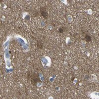
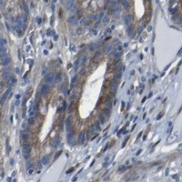
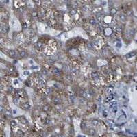
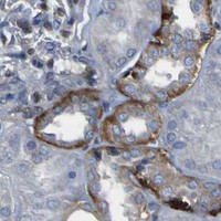


Comprehensive Human Protein Atlas IHC scoring per tissue (reliability: Enhanced — Medium consistency between antibody staining and RNA expression data.). Rows are taken directly from the HPA tissue chart — click any row's HPA link to view the source.
| Tissue | Cell type | Level | Evidence | Source |
|---|---|---|---|---|
| Fallopian tube | Glandular cells | High | Protein (IHC) | HPA → |
| Skeletal muscle | Myocytes | High | Protein (IHC) | HPA → |
| Testis | Spermatogonia cells | High | Protein (IHC) | HPA → |
| Tonsil | Squamous epithelial cells | High | Protein (IHC) | HPA → |
| Adrenal gland | Glandular cells | Medium | Protein (IHC) | HPA → |
| Tissue | Cell type | Level | Evidence | Source |
|---|---|---|---|---|
| Adipose tissue | Adipocytes | Not detected | Protein (IHC) | HPA → |
| Lymph node | Germinal center cells | Not detected | Protein (IHC) | HPA → |
| Oral mucosa | Squamous epithelial cells | Not detected | Protein (IHC) | HPA → |
| Prostate | Glandular cells | Not detected | Protein (IHC) | HPA → |
| Seminal vesicle | Glandular cells | Not detected | Protein (IHC) | HPA → |
Troubleshoot LRRN1 staining in paraffin sections by checking retrieval, tissue morphology, controls, and the cell type and compartment carrying the signal.
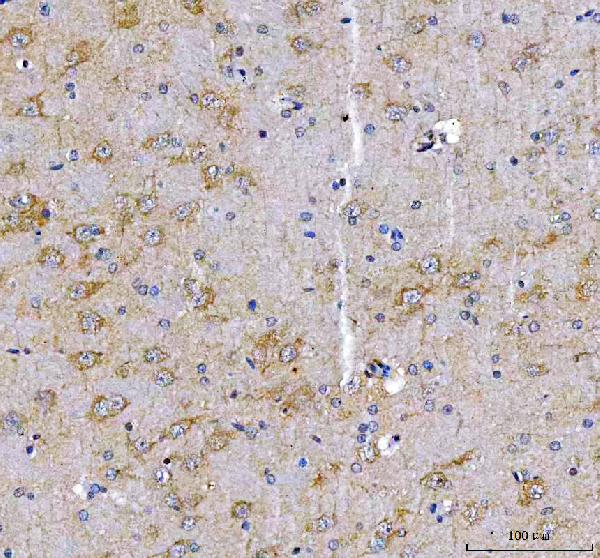
A12074-1 has real IHC data from paraffin-embedded rat brain (A12074-1 IHC image caption). Human, mouse, and rat are listed as reactive species (catalog reactivity); no IF image is supplied (catalog images).
A12074-1 is listed for IHC and for human, mouse, and rat reactivity (catalog applications and reactivity). Its figure shows chromogenic staining of paraffin-embedded rat brain after EDTA retrieval at pH 8.0 (A12074-1 IHC image caption).
Which to pick: For tissue IHC, choose A12074-1: its own paraffin-section image used 2 μg/ml primary antibody and DAB detection; the fixative is unreported (A12074-1 IHC image caption). No IF/ICC validation is supplied for A12074-1, so IF/ICC requires independent validation (catalog applications and images). For cross-species work, A12074-1 lists human, mouse, and rat reactivity, while its pictured IHC result covers rat only (catalog reactivity; A12074-1 IHC image caption).