LSM2 / U6 snRNA-associated Sm-like protein LSm2 · IHC design guide

Design Immunohistochemistry for LSM2

Plan chromogenic paraffin-section IHC for LSM2 using the catalog antibody's documented tissue protocol (datasheet A07586-2). Expect ubiquitous nuclear staining, with high signal in colon glandular cells and low signal in cardiomyocytes and adipocytes (HPA tissue IHC).

Evidence assembled Oct 2026 · For research use; verify linked source records and product datasheet before use
Immunohistochemistry protocol sheet for LSM2 (IHC for LSM2): expected localisation Ubiquitous nuclear tissue staining (HPA tissue IHC), antibody A07586-2, validated IHC image, and IHC protocol steps
Printable LSM2 IHC protocol sheet — expected localisation Ubiquitous nuclear tissue staining (HPA tissue IHC), antibody A07586-2, controls and protocol steps. Open the full LSM2 IHC guide →

LSM2 Immunohistochemistry Experimental Design Guide

Expected localisation, validated protocols, controls and antibodies — the at-a-glance facts below, then the full design guide.

Must know before staining
Expected localisation Ubiquitous nuclear tissue staining (HPA tissue IHC)
Staining pattern Nuclear staining across tissue cell types (HPA tissue IHC)
Antigen retrieval EDTA pH 8.0 HIER, heat-mediated (datasheet A07586-2)
Positive control ⓘ Adrenal gland+4 more · see all
Negative control ⓘ None in HPA (detected in all 45 tissues); use no-primary + isotype controls
Important caveats
Reasons your staining may differ from the expected pattern.
Fixation Keep fixation consistent across sections (standard IHC practice; not target-specific)
Caveat Cardiomyocytes and adipocytes show low staining (HPA tissue IHC)
Regulation Low tissue specificity (HPA tissue RNA)
Isoform / epitope No annotated isoforms; one chain spans residues 1–95 (UniProt)
Section 1

Recommended LSM2 IHC & IF Protocols

The catalog antibody protocol (datasheet A07586-2) is accompanied by published LSM2 chromogenic IHC methods for lung sections (PMC11871738) and melanoma tissue microarrays (PMC10262536).

Recommended immunohistochemistry (IHC-P) protocol parameters
SampleParaffin-embedded human breast cancer tissue; fixative not specified (datasheet A07586-2)
FixationImage fixative and duration unreported (datasheet A07586-2); verify before use.
Sectioning4–5 µm sections on charged slides (standard)
DeparaffinisationXylene, graded ethanol series to water (standard)
Antigen retrievalHeat retrieval: EDTA pH 8.0 (datasheet A07586-2); 20 min, 95–100 °C (standard)
Peroxidase block3% H2O2, 10 min, room temperature (standard)
Blocking10% goat serum (datasheet A07586-2)
Primary antibodyRabbit anti-LSM2, 2μg/ml (datasheet A07586-2)
Primary incubationOvernight at 4 °C (datasheet A07586-2)
DetectionStreptavidin-biotin complex (SABC), DAB chromogen (datasheet A07586-2)
CounterstainHematoxylin, blue, dehydrate and mount (standard)
Expected resultLSM2-positive staining in glandular cells of adrenal gland (HPA tissue IHC: High). HPA tissue profile: Ubiquitous nuclear expression. No signal in the no-primary control.
💡Decision noteStart with heat-mediated EDTA pH 8.0 retrieval for the catalog antibody (datasheet A07586-2); the published chromogenic methods do not specify a heat-retrieval condition (PMC11871738; PMC10262536).
Section 2

What Is the Expected LSM2 Staining Pattern?

LSM2 should stain nuclei across many tissues (UniProt Q9Y333: nucleus; HPA tissue IHC: ubiquitous nuclear expression). Expect strong staining in glandular cells of colon, breast and several other organs, and in bronchial respiratory epithelial cells (HPA tissue IHC: High). The tissue IHC profile is Supported, with high consistency between staining and RNA expression (HPA tissue IHC: Supported). LSM2 has no transmembrane segment (UniProt Q9Y333: topology).

What am I looking at on my slide?
Clear nuclear staining in colon glandular cells, with tissue structure still readable.This fits a known high-staining cell population (HPA tissue IHC: colon glandular cells, High). Score the nuclear signal within identified cells; broad nuclear staining elsewhere can also fit the reported ubiquitous profile (HPA tissue IHC: ubiquitous nuclear expression).
Strong cytoplasmic staining dominates while nuclei remain faint.This conflicts with the reported nuclear location (UniProt Q9Y333: nucleus; HPA subcellular: nucleoplasm). Review background and detection controls before treating cytoplasmic color as LSM2; compartment mismatch alone does not identify its cause (general IHC practice).
Colon stromal cells stain strongly, but glandular nuclei show little or no signal.The missing glandular signal conflicts with the reported High level in colon glandular cells (HPA tissue IHC: colon). Stromal color may reflect cross-reactivity or endogenous detection activity; check compartment and controls before assigning it to LSM2 (general IHC practice).
Color spreads diffusely across cells or tissue spaces, obscuring nuclear boundaries.Diffuse staining is difficult to score against a nuclear target (UniProt Q9Y333: nucleus). In chromogenic IHC, insufficient blocking, residual detection activity or overly strong development can produce background; assess matched controls and staining conditions (general IHC practice).
No nuclear signal appears in an otherwise intact colon glandular region.Colon glandular cells are reported High, making this a useful positive reference (HPA tissue IHC: colon). A blank result calls for review of the IHC-validated antibody, retrieval and detection workflow; it does not by itself establish absent LSM2 (general IHC practice).
💡Expected LSM2 appearanceCall a section positive when identifiable glandular or respiratory epithelial cells show predominantly nuclear staining that can be strong (HPA tissue IHC: High in listed cells); diffuse color or cytoplasmic-only staining is suspect against the nuclear reference (UniProt Q9Y333: nucleus).
How each factor affects the staining
Tissue and cell selectionHigh staining is reported in glandular cells of adrenal gland, appendix, breast, cervix, colon and duodenum, respiratory epithelial cells of bronchus, and cerebellar granular-layer cells (HPA tissue IHC: High). Cardiomyocytes and adipocytes are reported Low, not negative (HPA tissue IHC: Low).
Antibody evidence for IHCHPA066718 has Supported IHC validation; HPA052805 has no IHC status in the supplied record (HPA antibodies: IHC). An Enhanced ICC result for either antibody does not establish its performance in paraffin-section chromogenic IHC (HPA antibodies: ICC Enhanced; general IHC practice).
Location and molecular formThe expected compartment is nuclear, with nucleoplasmic enrichment observed by ICC-IF (UniProt Q9Y333: nucleus; HPA subcellular: nucleoplasm). The supplied sequence record has one 1–95 chain, no signal peptide and no transmembrane segment; it gives no basis for an expected membrane or secreted pattern (UniProt Q9Y333: processing and topology).
IF/ICC Q: what pattern is reported?A: Nucleoplasmic localization is reported in ICC-IF; image-bearing cell lines include HeLa, MCF-7, SK-MEL-30 and U2OS (HPA subcellular: nucleoplasm; ICC-IF images). Both listed antibodies have Enhanced ICC validation (HPA antibodies: ICC). This location reference supplies no IF/ICC protocol here.
Why is my staining missing, weak or wrong?
SituationLikely causeNext action
The positive reference has no nuclear stain.A missed workflow step, ineffective detection or an unsuitable antibody condition may suppress signal (general IHC practice).Check colon glandular cells against the reported High reference (HPA tissue IHC: colon); verify the catalog antibody's IHC-P instructions, retrieval, detection reagents and positive control together (general IHC practice).
Only weak nuclei are visible in heart muscle or adipose tissue.These cell populations are reported Low, so weak staining alone is compatible with the tissue profile (HPA tissue IHC: cardiomyocytes and adipocytes, Low).Compare with a High reference such as colon glandular cells before changing staining conditions; do not use a Low population as a required negative control (HPA tissue IHC: colon High; heart muscle and adipose tissue Low).
Cytoplasmic color is stronger than nuclear color.The pattern conflicts with nuclear and nucleoplasmic localization; the stain's identity remains unresolved (UniProt Q9Y333: nucleus; HPA subcellular: nucleoplasm).Inspect morphology and a detection control, then review blocking and chromogen development for background (general IHC practice). Score only a defensible cellular pattern, rather than assigning all visible color to LSM2 (general IHC practice).
Glandular cells look negative while adjacent structures are intensely colored.For colon, this reverses the reported High glandular pattern; cross-reactivity or endogenous detection activity is possible (HPA tissue IHC: colon; general IHC practice).Confirm cell identity with morphology and counterstain, inspect the no-primary control, and check whether the color is nuclear before interpreting the adjacent structures (general IHC practice).
Diffuse color obscures nuclei throughout the section.Background from blocking, washing or chromogen development can prevent nuclear scoring (general IHC practice).Review no-primary and reagent controls, washing, blocking and development against the same run; repeat only after the source of background is narrowed down (general IHC practice).
An antibody gives convincing ICC-IF nuclei but ambiguous paraffin-section IHC.ICC and IHC validation are application-specific: both listed antibodies are Enhanced for ICC, while only HPA066718 is Supported for IHC in the supplied record (HPA antibodies: validation status).Use IHC evidence when selecting the IHC-validated antibody, then judge the section against a High cell population and a nuclear compartment; keep the ICC-IF result as a location reference (HPA antibodies: IHC; HPA tissue IHC: High; HPA subcellular: nucleoplasm).

Sample controls for LSM2 IHC & IF

🧪Run breast first and expect staining in glandular cells (HPA: Breast glandular cells, High; selected-SKU IHC caption: breast cancer section). HPA detects LSM2 in all 45 scored tissues, so there is no documented negative tissue; use no-primary and isotype controls, and treat unstained cells within the breast section as candidate background only, not validated LSM2-negative cells (HPA: no negative rows; UniProt Q9Y333: nucleus).
Positive control tissue: Adrenal gland (Glandular cells, HPA High)
Negative control tissue: None in HPA: LSM2 is detected in all 45 scored tissues. Use a no-primary (secondary-only) and an isotype control instead.
ICC-IF cell lines (HPA subcellular resource): HPA ICC-IF images show LSM2 in HeLa, MCF-7, SK-MEL-30, U2OS, with annotated localisation: Nucleoplasm (enhanced) (HPA subcellular).
Technical controls: Include a secondary-only slide, a concentration-matched rabbit IgG isotype slide, and LSM2-knockout material as a biological negative (selected-SKU IHC caption: rabbit primary; standard IHC controls). For breast sections developed with the caption’s biotin-based DAB system, check endogenous peroxidase and biotin background (selected-SKU IHC caption: SABC with DAB; standard IHC practice).
⚠️Feasibility: A target-specific fixation window and fixation effect are unreported; the selected-SKU paraffin-section caption also does not report the fixative (selected-SKU IHC caption: fixative not stated). Heat retrieval in EDTA at pH 8.0 was used for the pictured breast cancer section, but the evidence does not establish that retrieval is required (selected-SKU IHC caption: EDTA pH 8.0). Frozen-section performance is unreported; ICC-IF images support evaluating nucleoplasmic localisation in the listed cell lines, while breast IHC with biotin-based DAB warrants the background checks above (HPA subcellular: nucleoplasm, ICC-IF images; selected-SKU IHC caption: SABC with DAB).

HPA tissue IHC evidence for LSM2

Comprehensive Human Protein Atlas IHC scoring per tissue (reliability: Supported — High consistency between antibody staining and RNA expression data.). Rows are taken directly from the HPA tissue chart — click any row's HPA link to view the source.

Positive expression · recommended positive controls

TissueCell typeLevelEvidenceSource
Adrenal gland Glandular cells High Protein (IHC) HPA →
Appendix Glandular cells High Protein (IHC) HPA →
Breast Glandular cells High Protein (IHC) HPA →
Bronchus Respiratory epithelial cells High Protein (IHC) HPA →
Cerebellum Cells in granular layer High Protein (IHC) HPA →

Undetected expression · recommended negative controls

TissueCell typeLevelEvidenceSource
None in HPA: LSM2 is detected in all 45 scored tissues. Use a no-primary (secondary-only) and an isotype control instead.
Section 3

Advanced LSM2 IHC Tips

Troubleshoot LSM2 staining in paraffin sections by checking retrieval, nuclear localisation, controls, and cell-level scoring (datasheet A07586-2; HPA tissue IHC).

What should I change if nuclear LSM2 staining is weak after antigen retrieval?
Use heat-mediated retrieval in EDTA at pH 8.0 for paraffin-section LSM2 IHC (datasheet A07586-2). Run a known positive section alongside the test section, keeping heating and cooling conditions consistent so retrieval can be assessed separately from detection (standard IHC practice). The selected antibody stained a paraffin section after this retrieval at 2 μg/mL with overnight incubation at 4°C (datasheet A07586-2). If staining remains weak, check primary-antibody exposure and detection reagents before testing another retrieval condition on matched sections (standard IHC practice). Judge improvement by clearer nuclear staining without a comparable rise in diffuse background (HPA subcellular; standard IHC practice).
Could fixation explain variable LSM2 staining across my paraffin sections?
The selected paraffin-section caption does not state a fixative, so LSM2-specific fixation sensitivity is unknown (datasheet A07586-2). Record each specimen’s fixative, fixation duration, processing history, and section age before comparing staining intensity across cases (standard IHC practice). Process matched control sections together and begin with EDTA retrieval at pH 8.0 to limit variation from the staining workflow (datasheet A07586-2; standard IHC practice). If one processing batch loses nuclear signal, compare its control tissue with a previously successful batch before changing antibody concentration (HPA tissue IHC; standard IHC practice). Nuclear localisation identifies where to score LSM2, but does not establish how fixation affects its epitope (UniProt Q9Y333; standard IHC practice).
Should LSM2 staining be nuclear, cytoplasmic, or both?
Prioritise nuclear staining when evaluating LSM2 in paraffin sections: UniProt assigns it to the nucleus, and HPA reports ubiquitous nuclear expression (UniProt Q9Y333; HPA tissue IHC). HPA further places the protein in the nucleoplasm in cell images (HPA subcellular). Use a counterstain and inspect individual intact cells so cytoplasmic chromogen deposits are not counted as nuclear signal (standard IHC practice). Compare the same cell compartment across test and control sections, especially when overall staining intensity differs (standard IHC practice). Predominantly cytoplasmic or extracellular staining warrants checks of background and tissue integrity before it is interpreted as LSM2 localisation (UniProt Q9Y333; HPA subcellular; standard IHC practice).
How do LSM2 sequence features affect interpretation of an uncertain epitope?
The supplied record lists a single 95-residue LSM2 chain and 0 isoforms, so it provides no isoform-specific staining pattern to distinguish (UniProt Q9Y333). Its Sm domain spans residues 2–76, while a modified threonine is reported at residue 79 (UniProt Q9Y333). The antibody epitope is not specified in the selected staining caption; therefore, do not attribute a staining difference to that modification or to epitope masking without direct evidence (datasheet A07586-2; standard IHC practice). LSM2 has no annotated transmembrane segment or signal peptide, consistent with assessing intracellular nuclear staining (UniProt Q9Y333). Compare matched retrieval and antibody conditions before assigning biological meaning to a negative section (standard IHC practice).
How should I check LSM2 localisation in a separate multiplex IF experiment?
For IF/ICC, evaluate LSM2 in the nucleoplasm and include a marker for the expected cell population, such as glandular cells in breast tissue (HPA subcellular; HPA tissue IHC). Choose spectrally separated fluorophores, placing a weaker channel away from the tissue’s strongest autofluorescence after inspecting unstained controls (standard IF practice). Use an intracellular-antigen permeabilisation step and confirm that it preserves nuclear morphology; LSM2 has no annotated transmembrane segment (UniProt Q9Y333; standard IF practice). Include single-stain controls to assess bleed-through and a no-primary control for nonspecific fluorescence (standard IF practice). Optimise IF fixation and permeabilisation independently, because the selected paraffin IHC caption supplies no IF/ICC preparation conditions (datasheet A07586-2).
What causes diffuse or granular background in LSM2 chromogenic IHC?
First compare the test slide with a no-primary control and inspect whether DAB deposits follow tissue edges, damaged areas, or reagent pooling (standard IHC practice). The selected paraffin-section example used 10% goat-serum blocking, a biotinylated secondary antibody, and streptavidin-biotin detection with DAB (datasheet A07586-2). Include a peroxidase-blocking step and assess endogenous biotin when using that detection system; these are general chromogenic workflow checks, not evidence of LSM2-specific interference (standard IHC practice). If background persists, check washing, blocking, and primary concentration on matched sections while preserving EDTA retrieval at pH 8.0 initially (datasheet A07586-2; standard IHC practice). Score convincing nuclear signal separately from diffuse nonnuclear colour (HPA tissue IHC; standard IHC practice).
How should I quantify LSM2 staining across tissue regions? ⚠ ANSWER MARKED FOR VERIFICATION
Define viable tissue regions and the cell population of interest before scoring, then evaluate nuclear staining because HPA describes ubiquitous nuclear expression (HPA tissue IHC). Report the percentage of positive nuclei and intensity categories, or calculate an H-score by multiplying each intensity grade 0–3 by its cell percentage, yielding 0–300 (standard IHC practice). Normalise positive counts to all evaluable nuclei in the specified population, or report positive-cell density per mm² of viable tissue (standard IHC practice). Apply the same threshold, counterstain assessment, and region-selection rules to every section (standard IHC practice). Record cell type and region because HPA reports differing staining levels among cell populations (HPA tissue IHC).
How can I distinguish true LSM2 positivity from staining artefacts?
Seek reproducible nuclear staining in intact cells: LSM2 is nuclear in UniProt, and HPA describes ubiquitous nuclear tissue staining (UniProt Q9Y333; HPA tissue IHC). HPA reports high staining in breast glandular cells but low staining in cardiomyocytes and adipocytes, so identify the cell population before comparing signal across tissues (HPA tissue IHC). Treat edge-restricted colour, necrotic regions, and signal outside intact cells cautiously, and compare them with a no-primary control (standard IHC practice). Check endogenous peroxidase and, for the selected biotin-based detection method, endogenous biotin when chromogen appears without a credible nuclear pattern (datasheet A07586-2; standard IHC practice). A clean negative control supports interpretation, but cannot alone establish antibody specificity (standard IHC practice).
Boster reagents

Best LSM2 / U6 snRNA-associated Sm-like protein LSm2 IHC Antibodies

Two anti-LSM2 antibodies have real IHC images from human paraffin sections (catalog IHC captions); A07586-2 also has IF data from MCF-7 cells (catalog IF caption). Listed reactivity spans human, monkey, mouse, and rat (catalog).

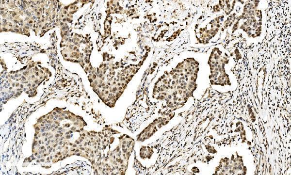
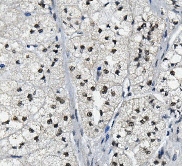
Real IHC data IHC analysis of LSM2 using anti-LSM2 antibody (A07586-2). LSM2 was detected in paraffin-embedded section of human breast cancer tissue. Heat mediated antigen retrieval was performed in EDTA buffer (pH8.0, epitope retrieval solution). The tissue section was blocked with 10% goat serum. The tissue section was then incubated with 2μg/ml rabbit anti-LSM2 Antibody (A07586-2) overnight at 4°C. Biotinylated goat anti-rabbit IgG was used as secondary antibody and incubated for 30 minutes at 37°C. The tissue section was developed using Strepavidin-Biotin-Complex (SABC) (Catalog # SA1022) with DAB as the chromogen.
Anti-LSM2 Antibody ®
Cat # A07586-2
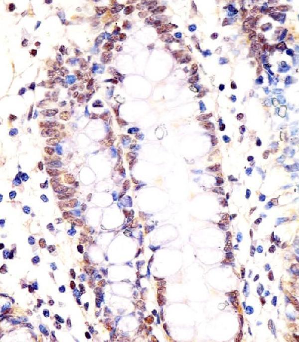
Real IHC data M07586 staining LSM2 in human colon tissue sections by Immunohistochemistry (IHC-P -paraformaldehyde-fixed, paraffin-embedded sections). Tissue was fixed with formaldehyde and blocked with 3% BSA for 0. 5 hour at room temperature; antigen retrieval was by heat mediation with a citrate buffer (pH6). Samples were incubated with primary antibody (1/25) for 1 hours at 37°C. A undiluted biotinylated goat polyvalent antibody was used as the secondary antibody.
Anti-LSM2 Antibody (C-Term)
Cat # M07586

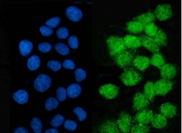
A07586-2 has IHC images from human breast, adrenal, lung, and rectal cancer paraffin sections, plus IF data from MCF-7 cells (A07586-2 image captions). M07586 has an IHC-P image from formaldehyde-fixed, paraffin-embedded human colon tissue and lists human and mouse reactivity (M07586 IHC caption; catalog reactivity).

Which to pick: For tissue IHC with documented fixation, choose the polyclonal M07586 at 1:25 for paraffin-section, paraffin-embedded human colon sections (catalog: polyclonal; M07586 IHC caption; datasheet: 1:25). For IF/ICC, choose A07586-2: its IF image shows MCF-7 cells stained at 5 µg/mL (A07586-2 IF caption). A07586-2 has the broader listed species reactivity—human, monkey, mouse, and rat—and IHC images from human paraffin sections; the IHC captions do not report a fixative (catalog reactivity; A07586-2 IHC captions). The selected A07586-2 tissue-IHC caption documents paraffin sections, but does not specify the fixative (selected-SKU IHC image A07586-2).

Each figure is that product's own IHC / IF validation image from its datasheet.

References

  1. UniProt Consortium. UniProt entry Q9Y333 (LSM2_HUMAN, U6 snRNA-associated Sm-like protein LSm2).
  2. Human Protein Atlas. LSM2 tissue IHC expression (reliability: Supported).
  3. Human Protein Atlas. LSM2 subcellular location (ICC-IF): Localized to the nucleoplasm..
  4. Human Protein Atlas. LSM2 antibody validation summary (2 antibodies).
  5. Lsm2 is critical to club cell proliferation and its inhibition aggravates COPD progression. Respiratory research 2025 — PMC11871738.
  6. LSM2 is associated with a poor prognosis and promotes cell proliferation, migration, and invasion in skin cutaneous melanoma. BMC medical genomics 2023 — PMC10262536.
  7. PubMed PMID:10523320 — UniProt-cited evidence.
  8. PubMed PMID:14656967 — UniProt-cited evidence.
  9. PubMed PMID:10931946 — UniProt-cited evidence.