This website uses cookies to ensure you get the best experience on our website.


- Table of Contents
Plan LSM7 IHC in paraffin sections using the catalog antibody’s 1–2 μg/mL range (datasheet A09236-1). Compare cytoplasmic staining with the HPA tissue profile, while accounting for its uncertain reliability and UniProt’s nuclear annotation (HPA tissue IHC; UniProt).

Expected localisation, validated protocols, controls and antibodies — the at-a-glance facts below, then the full design guide.
| Expected localisation | Cytoplasmic tissue staining (HPA tissue IHC); nuclear location (UniProt) | |
| Staining pattern | Cytoplasmic staining in most tissues; high in alveolar cells (HPA tissue IHC) | |
| Antigen retrieval | EDTA pH 8.0 HIER, heat-mediated (datasheet A09236-1) | |
| Positive control | Adrenal gland+4 more · see all | |
| Negative control | Cervix+4 more · see all |
| Fixation | Keep fixation consistent across sections. (standard IHC practice; not target-specific) | |
| Caveat | Tissue staining has uncertain reliability (HPA tissue IHC) | |
| Regulation | No specific regulator annotated (UniProt) | |
| Isoform / epitope | No isoforms annotated; mature chain spans residues 2–103 (UniProt) |
The catalog antibody’s IHC-P protocol (datasheet: A09236-1) appears alongside one published paraffin-section LSM7 IHC protocol (PMC12816532: methods).
| Sample | Paraffin-embedded human bladder cancer tissue; fixative not specified (datasheet A09236-1) |
| Fixation | Image fixative and duration unreported (datasheet A09236-1); verify before use. |
| Sectioning | 4–5 µm sections on charged slides (standard) |
| Deparaffinisation | Xylene, graded ethanol series to water (standard) |
| Antigen retrieval | Heat retrieval: EDTA pH 8.0 (datasheet A09236-1); 20 min, 95–100 °C (standard) |
| Peroxidase block | 3% H2O2, 10 min, room temperature (standard) |
| Blocking | 10% goat serum (datasheet A09236-1) |
| Primary antibody | Rabbit anti-LSM7, 1-2μg/ml (datasheet A09236-1) |
| Primary incubation | Overnight at 4 °C (datasheet A09236-1) |
| Detection | Streptavidin-biotin complex (SABC), DAB chromogen (datasheet A09236-1) |
| Counterstain | Hematoxylin, blue, dehydrate and mount (standard) |
| Expected result | LSM7-positive staining in glandular cells of adrenal gland (HPA tissue IHC: High). HPA tissue profile: Cytoplasmic expression in most tissues. No signal in the no-primary control. |
For paraffin IHC, expect predominantly cytoplasmic LSM7 staining in several cell types, including adrenal glandular cells, hippocampal neurons, lung alveolar cells, skin fibroblasts and cells in testis seminiferous ducts (HPA tissue IHC: High; cytoplasmic expression in most tissues). Treat this as a provisional pattern: the tissue IHC assessment is Uncertain, while UniProt places LSM7 in the nucleus and reports no transmembrane segment (HPA tissue IHC: Uncertain; UniProt Q9UK45: subcellular location and topology).
| Cytoplasmic staining in the listed high-staining cell populations, with identifiable cell boundaries and limited background (HPA tissue IHC: High; cytoplasmic profile). | This matches the reported paraffin tissue pattern. Compare signal within the relevant cells and against a no-primary control; a strong signal alone does not establish antibody specificity because the tissue IHC assessment remains Uncertain (HPA tissue IHC: Uncertain; general IHC practice). |
| Predominantly nuclear or nucleolar staining in paraffin sections, without the reported cytoplasmic pattern (HPA tissue IHC: cytoplasmic profile). | Check for a compartment artefact or detection problem before calling it the expected IHC result (general IHC practice). The discrepancy needs care: UniProt assigns LSM7 to the nucleus, and HPA ICC-IF reports an approved nucleoli fibrillar center location (UniProt Q9UK45: nucleus; HPA ICC-IF: approved location). |
| Strong staining in cell types listed as not detected, such as pancreatic exocrine glandular cells or esophageal squamous epithelial cells (HPA tissue IHC: Not detected). | Consider cross-reactivity or endogenous detection activity, particularly if the same signal appears in controls (general IHC practice). Do not label the tissue positive from this slide alone: HPA's negative observations come from an IHC assessment rated Uncertain (HPA tissue IHC: Uncertain). |
| Diffuse color covers cells, extracellular areas or section edges and obscures where individual cells stain (general IHC practice). | This pattern cannot be scored reliably as LSM7 localisation. Review blocking, washing, detection and counterstain, then compare a no-primary control; HPA reports cellular cytoplasmic staining rather than a diffuse tissue-wide deposit (general IHC practice; HPA tissue IHC: cytoplasmic profile). |
| Little or no signal in adrenal glandular cells or lung alveolar cells despite intact tissue morphology (HPA tissue IHC: High). | First check the IHC-validated antibody and staining run using an appropriate positive control, then review retrieval, dilution and detection as general workflow variables (general IHC practice). A negative result in one section cannot settle LSM7 absence because HPA rates its tissue IHC assessment Uncertain (HPA tissue IHC: Uncertain). |
| Assay-dependent localisation | HPA describes cytoplasmic staining in tissue IHC, whereas its ICC-IF result places LSM7 in the nucleoli fibrillar center and UniProt lists the nucleus. Record the assay when comparing compartments; this evidence does not resolve the difference (HPA tissue IHC: cytoplasmic profile; HPA ICC-IF: approved location; UniProt Q9UK45: nucleus). |
| IF/ICC: what pattern should be expected? | For IF/ICC interpretation, HPA reports localisation to the nucleoli fibrillar center, with images from A-431, U-251MG and U2OS. This is an IF observation, not a paraffin IHC protocol or a reason to require nucleolar chromogenic staining (HPA ICC-IF: approved location and image cell lines; HPA tissue IHC: cytoplasmic profile). |
| Strength of tissue evidence | The tissue profile reports low RNA tissue specificity and staining across most tissues, but its antibody staining versus RNA consistency is medium and pending external verification. Use the named high-staining cells as provisional comparators, not as independently validated specificity controls (HPA tissue IHC: low RNA tissue specificity; Uncertain reliability). |
| Topology and processing | UniProt reports no transmembrane segment or signal peptide and identifies a chain spanning residues 2–103. Those annotations give no basis to expect membrane or secreted-tissue staining, and they do not establish how fixation affects epitope access (UniProt Q9UK45: topology and processing). |
| Situation | Likely cause | Next action |
|---|---|---|
| No stain appears in an HPA high-staining cell population (HPA tissue IHC: High). | A failed staining run, unsuitable retrieval, antibody dilution or detection can suppress signal (general IHC practice). | Check run controls and tissue integrity, then optimise retrieval and dilution under the IHC-validated antibody's instructions; treat the HPA comparator as provisional (general IHC practice; HPA tissue IHC: Uncertain). |
| The slide shows predominantly nuclear stain instead of the reported cytoplasmic IHC pattern (HPA tissue IHC: cytoplasmic profile). | Compartment misassignment or staining artefact is possible, while UniProt and ICC-IF support nuclear or nucleolar localisation (general IHC practice; UniProt Q9UK45: nucleus; HPA ICC-IF: approved location). | Review cell morphology, counterstain and controls; document the assay-specific discrepancy before interpreting the nuclear signal as LSM7 (general IHC practice; HPA tissue IHC: Uncertain). |
| Cells listed as not detected stain strongly (HPA tissue IHC: Not detected). | Cross-reactivity or endogenous detection activity may contribute; the HPA negative observation is provisional (general IHC practice; HPA tissue IHC: Uncertain). | Compare no-primary and detection controls, then assess whether the pattern persists with an independently validated approach (general IHC practice). |
| Diffuse deposit makes cell-level scoring difficult (general IHC practice). | Insufficient blocking or washing, excessive reagent concentration, or detection background can obscure localisation (general IHC practice). | Inspect the no-primary control and adjust blocking, washing and reagent concentration according to the detection system; score only interpretable cellular stain (general IHC practice). |
| Staining is uneven across the paraffin section (general IHC practice). | Uneven reagent coverage, section adhesion or retrieval conditions can create regional variation unrelated to cell type (general IHC practice). | Check section quality and reagent coverage, and repeat with consistent retrieval conditions before comparing tissue regions (general IHC practice). |
| Color develops in the no-primary control (general IHC practice). | Endogenous detection activity or background from detection reagents is possible without primary antibody (general IHC practice). | Resolve the control signal using the detection system's blocking and control steps before assigning positive LSM7 staining (general IHC practice). |
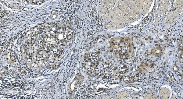
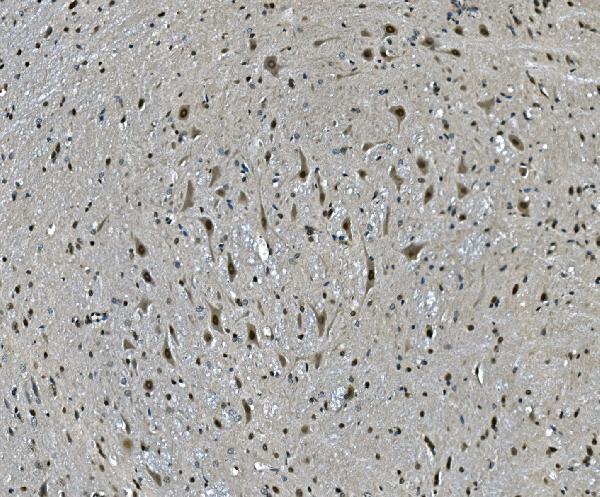

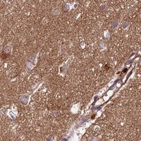


Comprehensive Human Protein Atlas IHC scoring per tissue (reliability: Uncertain — Medium consistency between antibody staining and RNA expression data. Pending external verification.). Rows are taken directly from the HPA tissue chart — click any row's HPA link to view the source.
| Tissue | Cell type | Level | Evidence | Source |
|---|---|---|---|---|
| Adrenal gland | Glandular cells | High | Protein (IHC) | HPA → |
| Hippocampus | Neuronal cells | High | Protein (IHC) | HPA → |
| Lung | Alveolar cells | High | Protein (IHC) | HPA → |
| Skin | Fibroblasts | High | Protein (IHC) | HPA → |
| Testis | Cells in seminiferous ducts | High | Protein (IHC) | HPA → |
| Tissue | Cell type | Level | Evidence | Source |
|---|---|---|---|---|
| Cervix | Glandular cells | Not detected | Protein (IHC) | HPA → |
| Duodenum | Glandular cells | Not detected | Protein (IHC) | HPA → |
| Epididymis | Glandular cells | Not detected | Protein (IHC) | HPA → |
| Esophagus | Squamous epithelial cells | Not detected | Protein (IHC) | HPA → |
| Oral mucosa | Squamous epithelial cells | Not detected | Protein (IHC) | HPA → |
Troubleshoot LSM7 staining in paraffin sections by checking retrieval, controls, compartment patterns, and scoring against the reported evidence.
A09236-1 has IHC images from human bladder cancer and mouse and rat brain paraffin sections (catalog IHC captions), plus IF data from human MCF-7 cells (catalog IF caption).
A09236-1 is listed for IHC in human, mouse and rat (catalog applications and reactivity), with paraffin-section images from human bladder cancer and mouse and rat brain (catalog IHC captions). The same SKU is listed for IF/ICC (catalog applications), with an IF image from human MCF-7 cells (catalog IF caption).
Which to pick: Choose A09236-1 for tissue IHC in paraffin sections: its captions document EDTA retrieval at pH 8.0 and rabbit primary antibody at 2 μg/ml (catalog IHC captions); the fixative is unreported (catalog IHC captions). Choose A09236-1 for IF/ICC in human cells: IF/ICC is listed, and its MCF-7 IF caption documents 5 μg/ml primary antibody (catalog applications; catalog IF caption). For cross-species IHC, A09236-1 lists human, mouse and rat reactivity and has paraffin-section images for all three (catalog reactivity; catalog IHC captions); its host is rabbit and clonality is unreported (catalog host and clone fields).