This website uses cookies to ensure you get the best experience on our website.


- Table of Contents
This guide helps plan chromogenic IHC for LSM8, which shows general nuclear staining in tissue (HPA tissue IHC). Adrenal gland glandular cells show high staining, while ovarian stromal cells have no detected staining (HPA tissue IHC); interpret this comparison in light of the uncertain staining reliability (HPA tissue IHC).

Expected localisation, validated protocols, controls and antibodies — the at-a-glance facts below, then the full design guide.
| Expected localisation | General nuclear staining observed (HPA tissue IHC) | |
| Staining pattern | Nuclear staining across diverse tissue cell types (HPA tissue IHC) | |
| Antigen retrieval | EDTA pH 8.0 HIER, heat-mediated (datasheet A10947) | |
| Positive control | Adrenal gland+4 more · see all | |
| Negative control | Caudate+2 more · see all |
| Fixation | Keep fixation conditions consistent across sections (standard IHC practice; not target-specific) | |
| Caveat | Staining and RNA expression show medium consistency (HPA tissue IHC) | |
| Regulation | No specific regulation annotated (UniProt) | |
| Isoform / epitope | No annotated isoforms; epitope map unknown (UniProt) |
The catalog antibody has an IHC-P protocol (datasheet A10947). The published gastric tissue IHC workflow below selected samples by LSM8 expression but stained immune cell markers (PMC10064066).
| Sample | Paraffin-embedded human skin cancer tissue; fixative not specified (datasheet A10947) |
| Fixation | Image fixative and duration unreported (datasheet A10947); verify before use. |
| Sectioning | 4–5 µm sections on charged slides (standard) |
| Deparaffinisation | Xylene, graded ethanol series to water (standard) |
| Antigen retrieval | Heat retrieval: EDTA pH 8.0 (datasheet A10947); 20 min, 95–100 °C (standard) |
| Peroxidase block | 3% H2O2, 10 min, room temperature (standard) |
| Blocking | 10% goat serum (datasheet A10947) |
| Primary antibody | Rabbit anti-LSM8, 1-2μg/ml (datasheet A10947) |
| Primary incubation | Overnight at 4 °C (datasheet A10947) |
| Detection | Streptavidin-biotin complex (SABC), DAB chromogen (datasheet A10947) |
| Counterstain | Hematoxylin, blue, dehydrate and mount (standard) |
| Expected result | LSM8-positive staining in glandular cells of adrenal gland (HPA tissue IHC: High). HPA tissue profile: General nuclear expression. No signal in the no-primary control. |
LSM8 is a nuclear spliceosome component without a transmembrane segment (UniProt O95777). In paraffin sections, expect mainly nuclear staining across several cell types, with high staining reported in glandular cells of the adrenal, parathyroid and thyroid glands and in bronchial respiratory epithelium (HPA: tissue IHC). Treat these as reference patterns, since the tissue IHC reliability is Uncertain, with medium consistency between antibody staining and RNA expression (HPA: reliability).
| Clear nuclear staining in glandular cells of the thyroid or parathyroid gland. | This fits the reported high staining in those cells (HPA: tissue IHC) and the nuclear location of LSM8 (UniProt O95777). Compare cells within the same section before judging intensity. |
| Predominantly cytoplasmic or membranous chromogen, with little nuclear signal. | This conflicts with the general nuclear IHC profile (HPA: tissue IHC) and nuclear annotation (UniProt O95777). Review morphology and controls; nonspecific staining or detection artefact may explain the pattern (general IHC practice). |
| Strong staining in ovarian stromal cells or soft tissue fibroblasts. | Those cells were reported as not detected (HPA: tissue IHC). If staining persists after controls, consider antibody cross-reactivity or endogenous detection activity (general IHC practice); HPA's Uncertain reliability limits a definitive negative call. |
| Haze or chromogen deposits across nuclei and surrounding tissue. | A diffuse pattern is difficult to score against the expected nuclear distribution (HPA: tissue IHC). Check the no-primary control, blocking and detection reagents for background (general IHC practice). |
| No nuclear signal in thyroid glandular cells. | HPA reports high staining in these cells, so the result warrants a technical check (HPA: tissue IHC). Confirm tissue integrity, reagent performance and scoring against a concurrent control (general IHC practice); the HPA result is not a guaranteed positive. |
| Tissue and cell selection | High staining was reported in adrenal, cervical, gallbladder, parathyroid and thyroid glandular cells, bronchial respiratory epithelium and testicular Leydig cells (HPA: tissue IHC). Score the named cells, rather than an entire organ, against that reference. |
| Lower-signal comparisons | Cerebellar granular-layer cells were medium; several listed neuronal, glial and glandular populations were low (HPA: tissue IHC). A weak result in those populations alone gives less information about assay performance. |
| Antibody evidence | Tissue IHC reliability is Uncertain, and the listed antibody HPA020116 is rated Uncertain for IHC (HPA: reliability; HPA: antibody validation). Interpret agreement with the atlas pattern as supportive, not proof of specificity. |
| Molecular location | LSM8 is nuclear, lacks a transmembrane segment and participates in the LSM2–8 spliceosomal complex (UniProt O95777). These annotations support a nuclear reading; they do not establish antigen retrieval conditions or fixation sensitivity. |
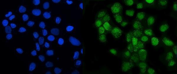
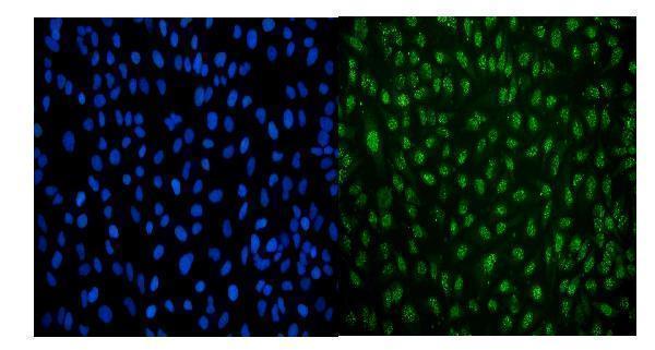
| IF/ICC Q: What location is reported? | A: Nucleoplasm and vesicles are approved locations in ICC-IF images, including A-431 cells (HPA: subcellular; HPA: antibody validation). That IF observation does not make vesicular chromogen a required tissue IHC result. |
| Situation | Likely cause | Next action |
|---|---|---|
| Nuclei are blank in a reported high-staining tissue. | Assay failure or an unrepresentative section is possible (general IHC practice); HPA tissue IHC is Uncertain (HPA: reliability). | Check tissue morphology and a concurrent control, then review the existing retrieval and detection workflow (general IHC practice). No LSM8-specific fixation sensitivity is reported here. |
| Cytoplasmic staining dominates the slide. | The distribution conflicts with nuclear LSM8 (UniProt O95777) and general nuclear tissue IHC (HPA: tissue IHC). Background or misidentified cell boundaries may contribute (general IHC practice). | Inspect counterstained nuclei and compare a no-primary control; reassess any signal that remains outside nuclei (general IHC practice). |
| Negative-comparison cells stain strongly. | Ovarian stromal cells and soft tissue fibroblasts were not detected in the atlas (HPA: tissue IHC). Cross-reactivity or endogenous detection activity is possible (general IHC practice). | Compare the same cell types with no-primary and detection controls, and confirm cell identity before calling the signal specific (general IHC practice). |
| Every region has diffuse brown background. | A broad deposit obscures the expected nuclear distribution (HPA: tissue IHC); inadequate blocking, washing or detection control may contribute (general IHC practice). | Inspect the no-primary control and review blocking, washes and chromogen development within the established assay (general IHC practice). |
| Only weak signal appears in a low-staining population. | Some neuronal, glial and glandular populations were reported low (HPA: tissue IHC), so their weak signal alone cannot establish assay failure. | Assess a concurrently processed tissue and cell population reported high by HPA, while keeping the Uncertain IHC reliability in mind (HPA: tissue IHC; HPA: reliability). |
| Vesicular signal appears in tissue IHC. | Vesicles are approved in ICC-IF (HPA: subcellular), while the tissue IHC profile is generally nuclear (HPA: tissue IHC). The methods provide different evidence. | Score the nuclear tissue pattern first; use tissue controls to assess vesicular chromogen before assigning it to LSM8 (HPA: tissue IHC; general IHC practice). |




Comprehensive Human Protein Atlas IHC scoring per tissue (reliability: Uncertain — Medium consistency between antibody staining and RNA expression data.). Rows are taken directly from the HPA tissue chart — click any row's HPA link to view the source.
| Tissue | Cell type | Level | Evidence | Source |
|---|---|---|---|---|
| Adrenal gland | Glandular cells | High | Protein (IHC) | HPA → |
| Bronchus | Respiratory epithelial cells | High | Protein (IHC) | HPA → |
| Cervix | Glandular cells | High | Protein (IHC) | HPA → |
| Gallbladder | Glandular cells | High | Protein (IHC) | HPA → |
| Parathyroid gland | Glandular cells | High | Protein (IHC) | HPA → |
Troubleshoot LSM8 staining by checking nuclear localisation, section quality and controls before interpreting differences in chromogenic signal (UniProt O95777; HPA tissue IHC).
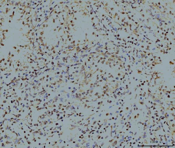
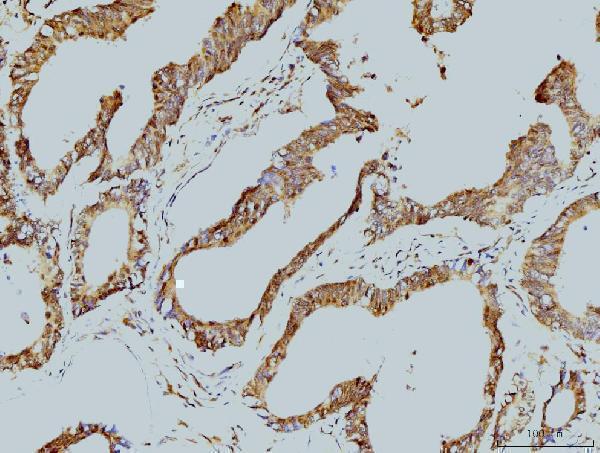
Anti-LSM8 antibodies have paraffin-section IHC images from human tissues and mouse and rat testis, plus ICC/IF images from A431 and HeLa cells (catalog image captions).
A10947 has IHC images from human skin and ovary cancers and mouse and rat testis, plus an ICC/IF image from A431 cells (A10947 image captions). M10947 has IHC images from human renal and colonic cancers, placenta, and spleen, plus an ICC/IF image from HeLa cells (M10947 image captions).
Which to pick: For paraffin-section tissue IHC across human, mouse, and rat samples, choose rabbit antibody A10947: its IHC captions show all three species, and its listed reactivity includes all three (A10947 IHC captions; catalog reactivity). For human tissue IHC where a monoclonal is preferred, choose mouse clone 6B11, M10947; its IHC captions show human paraffin sections, while A10947 has the stronger cross-species IHC image record (M10947 catalog clone and IHC captions; A10947 IHC captions). Both list IF/ICC and have cell images, so choose A10947 for A431 or M10947 for HeLa as the documented starting point; the fixative is unreported for both paraffin-section IHC examples (catalog applications; A10947 and M10947 image captions).