LSR / Lipolysis-stimulated lipoprotein receptor · IHC design guide

Design Immunohistochemistry for LSR

Plan LSR chromogenic IHC on paraffin sections with 2–5 μg/ml catalog antibody (datasheet A02742-1). Use colon or stomach glandular cells as positive references (HPA tissue IHC), and assess observed cytoplasmic and nuclear staining (HPA tissue IHC) alongside the expected tricellular junction location (UniProt).

Evidence assembled Oct 2026 · For research use; verify linked source records and product datasheet before use
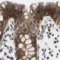
Immunohistochemistry protocol sheet for LSR (IHC for LSR): expected localisation Cytoplasmic and nuclear tissue staining (HPA tissue IHC), antibody A02742-1, validated IHC image, and IHC protocol steps
Printable LSR IHC protocol sheet — expected localisation Cytoplasmic and nuclear tissue staining (HPA tissue IHC), antibody A02742-1, controls and protocol steps. Open the full LSR IHC guide →

LSR Immunohistochemistry Experimental Design Guide

Expected localisation, validated protocols, controls and antibodies — the at-a-glance facts below, then the full design guide.

Must know before staining
Expected localisation Cytoplasmic and nuclear tissue staining (HPA tissue IHC)
Staining pattern Glandular cells show cytoplasmic and nuclear staining (HPA tissue IHC)
Antigen retrieval EDTA pH 8.0 HIER, heat-mediated (datasheet A02742-1)
Positive control ⓘ Colon+4 more · see all
Negative control ⓘ Adipose tissue+4 more · see all
Important caveats
Reasons your staining may differ from the expected pattern.
Fixation Keep fixation consistent across sections (standard IHC practice; not target-specific)
Caveat Antibody staining has medium consistency with RNA data (HPA tissue IHC)
Regulation Target-specific regulation not annotated (UniProt)
Isoform / epitope 6 isoforms; verify whether the epitope is extracellular or cytoplasmic (UniProt)
Section 1

Recommended LSR IHC & IF Protocols

Start with the catalog antibody’s IHC-P protocol (datasheet A02742-1), then compare the published endometrial tissue protocol (PMC5053684).

Recommended immunohistochemistry (IHC-P) protocol parameters
SampleParaffin-embedded mouse colon tissue; fixative not specified (datasheet A02742-1)
FixationImage fixative and duration unreported (datasheet A02742-1); verify before use.
Sectioning4–5 µm sections on charged slides (standard)
DeparaffinisationXylene, graded ethanol series to water (standard)
Antigen retrievalHeat retrieval: EDTA pH 8.0 (datasheet A02742-1); 20 min, 95–100 °C (standard)
Peroxidase block3% H2O2, 10 min, room temperature (standard)
Blocking10% goat serum (datasheet A02742-1)
Primary antibodyRabbit anti-LSR, 2-5 μg/ml (datasheet A02742-1)
Primary incubationOvernight at 4 °C (datasheet A02742-1)
DetectionStreptavidin-biotin complex (SABC), DAB chromogen (datasheet A02742-1)
CounterstainHematoxylin, blue, dehydrate and mount (standard)
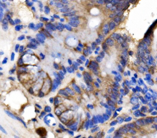
Expected resultLSR-positive staining in glandular cells of colon (HPA tissue IHC: High). HPA tissue profile: Cytoplasmic and nuclear expression in several tissues. No signal in the no-primary control.
💡Decision noteTry EDTA pH 8.0 heat retrieval first (datasheet A02742-1); the published protocol used ER2 solution with autoclave retrieval (PMC5053684).
Section 2

What Is the Expected LSR Staining Pattern?

LSR is a single-pass membrane protein expected at cell junctions, especially tricellular contacts (UniProt Q86X29 topology and localisation). Colon and stomach glandular cells show High IHC staining (HPA tissue IHC). HPA also describes cytoplasmic and nuclear staining in several tissues; its IHC reliability is Approved, with medium consistency between staining and RNA expression (HPA tissue IHC). Interpret compartment and cell type together.

What am I looking at on my slide?
Sharp membrane or junctional staining in colon or stomach glandular cells.This fits the expected LSR location at tricellular contacts (UniProt Q86X29 localisation) and a cell type with High observed staining (HPA tissue IHC). Junctional localisation is an expectation, not a claim that HPA resolved tricellular contacts in those sections.
Predominantly nuclear or diffuse cytoplasmic staining, with little discernible membrane signal.This is less consistent with LSR topology (UniProt Q86X29), but HPA reports cytoplasmic and nuclear IHC staining in several tissues (HPA tissue IHC). Treat an isolated compartment pattern as uncertain; compare it with controls and an independent antibody before calling it artefact.
Strong staining in adipocytes or esophageal squamous epithelial cells.Those cells are listed as Not detected (HPA tissue IHC). Check for cross-reactivity or endogenous chromogen-generating activity using appropriate controls (general IHC practice); a tissue-level negative reference does not prove that every stained cell is a false positive.
Broad colour deposition across cells and surrounding tissue without cell borders or junctions.This lacks the spatial definition expected for a membrane and tight-junction protein (UniProt Q86X29 localisation). Review background and detection controls, including reagent-only sections, before assigning the deposit to LSR (general IHC practice).
No detectable stain in colon or stomach glandular cells.Both are High reference cell populations (HPA tissue IHC), so a blank result raises concern about assay performance. Confirm tissue preservation, detection and counterstain, then review retrieval and antibody conditions as general IHC troubleshooting steps.
💡Expected LSR appearanceA persuasive positive is defined membrane or junctional staining in colon or stomach glandular cells, where HPA reports High IHC signal (HPA tissue IHC; UniProt Q86X29 localisation); widespread featureless deposit or strong signal confined to HPA Not detected cell types calls for control review.
How each factor affects the staining
Epitope side and topologyLSR has an extracellular region, one transmembrane segment and a cytoplasmic region (UniProt Q86X29 topology). Interpret membrane staining with the antibody's documented epitope in mind; an epitope location cannot be inferred from the tissue pattern alone.
Tissue reference and confidenceColon and stomach glandular cells are High, whereas adipocytes and esophageal squamous cells are Not detected (HPA tissue IHC). HPA rates IHC Approved with medium RNA–staining consistency, so use these as reference patterns rather than absolute truth.
Antibody validation scopeHPA007270 is IHC Approved and ICC Enhanced; HPA075309 has ICC Enhanced status with no IHC status supplied (HPA antibodies). Enhanced ICC evidence should not be read as enhanced validation of a paraffin-section IHC result.
Isoforms and modified residuesUniProt lists 6 isoforms and multiple phosphorylation sites (UniProt Q86X29). An antibody's epitope may affect which forms it detects, but the supplied record gives no epitope mapping or evidence that phosphorylation changes this IHC pattern.
Retrieval and detectionAntigen retrieval and endogenous enzyme blocking are general considerations in chromogenic paraffin IHC (general IHC practice). The supplied UniProt and HPA records do not establish LSR-specific retrieval conditions or fixation sensitivity; optimise against positive tissue and negative controls.
IF/ICC Q&A: should its pattern match IHC exactly?HPA ICC-IF localises LSR to plasma membrane and cell junctions in imaged cell lines (HPA subcellular). That supports a compartment check, but HPA tissue IHC also reports cytoplasmic and nuclear signal; compare each application within its own validated context.
Why is my staining missing, weak or wrong?
SituationLikely causeNext action
Positive reference section is blank.Assay failure is possible when High colon or stomach glandular staining is expected (HPA tissue IHC); the record does not identify a specific failed step.Check section integrity and detection controls, then optimise retrieval and antibody conditions using general IHC practice.
Only nuclear staining is visible.HPA reports nuclear IHC signal, while UniProt places LSR at membrane junctions (HPA tissue IHC; UniProt Q86X29 localisation). The sources do not resolve that discrepancy.Record the compartment separately, inspect a positive tissue control, and seek concordance with an independently validated antibody before attributing it to LSR.
Unexpected signal appears in a negative reference cell type.Adipocytes and esophageal squamous epithelial cells are Not detected in HPA tissue IHC; nonspecific binding or endogenous detection activity may contribute (general IHC practice).Compare with no-primary and detection controls, assess tissue morphology, and review antibody dilution and blocking (general IHC practice).
Colour deposit obscures cell borders throughout the section.Diffuse deposit prevents assessment of the junctional location expected from UniProt Q86X29; its cause cannot be assigned from appearance alone.Inspect negative controls and detection timing; adjust blocking, reagent concentration or development time as indicated by those controls (general IHC practice).
Signal varies between adjacent glandular areas.HPA reports tissue-level staining categories, which do not specify uniform staining of every glandular cell (HPA tissue IHC). Section quality or local assay variation may also contribute (general IHC practice).Score intensity and localisation by cell population, check morphology and controls, and avoid treating a patchy field as proof of an LSR-specific change.
An ICC-IF image looks junctional, but paraffin IHC looks cytoplasmic.HPA ICC-IF reports membrane and junction localisation, while HPA tissue IHC describes cytoplasmic and nuclear staining in several tissues (HPA subcellular; HPA tissue IHC).Interpret the paraffin section with its IHC controls and cell-type references; do not transfer an ICC-IF validation status or visual pattern directly to IHC.

Sample controls for LSR IHC & IF

🧪Run colon first: glandular cells should stain (HPA: High in colon glandular cells). Use adipose tissue as the negative comparator (HPA: Not detected in adipocytes); on the colon slide, assess morphologically non-glandular cells as internal background comparators rather than assuming they are LSR-negative.
Positive control tissue: Colon (Glandular cells, HPA High)
Negative control tissue: Adipose tissue (HPA Not detected)
ICC-IF cell lines (HPA subcellular resource): HPA ICC-IF images show LSR in A-431, CACO-2, OE19, RT-4, U2OS, with annotated localisation: Plasma membrane (enhanced), Cell Junctions (enhanced) (HPA subcellular).
Technical controls: Include no-primary (secondary-only) and host-matched rabbit control immunoglobulin controls (selected-SKU caption: rabbit primary), plus LSR-knockout tissue or a validated immunizing-peptide block as a biological specificity control. Check endogenous peroxidase and biotin background in colon when using the caption’s biotin-based, DAB detection (selected-SKU caption: SABC–DAB).
⚠️Feasibility: A target-specific fixation window or fixation effect is unreported, and the exact selected-SKU paraffin-section caption does not state a fixative (selected-SKU caption: fixative not stated). The illustrated IHC result used heat retrieval in EDTA at pH 8.0, so retrieval conditions should be checked for the chosen specimen preparation (selected-SKU caption: EDTA heat retrieval); the supplied evidence does not establish that frozen sections or IF are easier. In colon, distinguish junctional staining from luminal or detection-system background when scoring glandular cells (UniProt Q86X29: tricellular junction localization; selected-SKU caption: SABC–DAB).

HPA tissue IHC evidence for LSR

Comprehensive Human Protein Atlas IHC scoring per tissue (reliability: Approved — Medium consistency between antibody staining and RNA expression data.). Rows are taken directly from the HPA tissue chart — click any row's HPA link to view the source.

Positive expression · recommended positive controls

TissueCell typeLevelEvidenceSource
Colon Glandular cells High Protein (IHC) HPA →
Stomach Glandular cells High Protein (IHC) HPA →
Caudate Neuronal cells Medium Protein (IHC) HPA →
Cerebral cortex Endothelial cells Medium Protein (IHC) HPA →
Duodenum Glandular cells Medium Protein (IHC) HPA →

Undetected expression · recommended negative controls

TissueCell typeLevelEvidenceSource
Adipose tissue Adipocytes Not detected Protein (IHC) HPA →
Esophagus Squamous epithelial cells Not detected Protein (IHC) HPA →
Smooth muscle Smooth muscle cells Not detected Protein (IHC) HPA →
Soft tissue Fibroblasts Not detected Protein (IHC) HPA →
Vagina Squamous epithelial cells Not detected Protein (IHC) HPA →
Section 3

Advanced LSR IHC Tips

Use the catalog antibody’s paraffin-section result as the IHC starting point, then assess staining against LSR’s junctional localisation and tissue controls.

Which retrieval conditions should I start with for LSR in paraffin sections?
Start with heat-mediated retrieval in EDTA at pH 8.0 (datasheet A02742-1). This condition preceded LSR detection in a paraffin-embedded mouse colon section, followed by overnight primary incubation at 4°C (datasheet A02742-1). If staining is weak, first check heating consistency and section adhesion, then vary retrieval duration on matched sections while keeping detection conditions constant (standard IHC practice). Compare glandular staining with a no-primary control and inspect junctional detail, because LSR is reported at cell membranes and tricellular contacts (HPA subcellular; UniProt Q86X29 localisation). Record the retrieval time used for every section (standard IHC practice).
How should I assess whether fixation is masking LSR staining?
Target-specific fixation sensitivity is unknown: the catalog image identifies a paraffin-embedded mouse colon section but does not state its fixative (datasheet A02742-1). Record the fixative and fixation duration for each specimen, and compare sections processed together before attributing weak staining to fixation (standard IHC practice). Hold EDTA retrieval at pH 8.0 and the primary concentration at 2 μg/ml while assessing variation among those sections (datasheet A02742-1). If fixation histories differ, run matched positive controls and interpret intensity cautiously, since processing differences can change antigen accessibility in chromogenic IHC (standard IHC practice).
Should LSR staining be junctional, cytoplasmic, or nuclear?
Give particular attention to membrane and cell-junction staining, including tricellular contacts, because these are reported LSR locations (UniProt Q86X29 localisation; HPA subcellular). The receptor has an extracellular region at residues 1–259, a transmembrane segment at 260–280, and a cytoplasmic region at 281–649 (UniProt Q86X29 topology). HPA also describes cytoplasmic and nuclear tissue staining, so those patterns require comparison with tissue context and controls rather than automatic acceptance or rejection (HPA tissue IHC profile). Inspect adjacent cells at high magnification and document membrane, cytoplasmic, and nuclear staining separately (standard IHC practice).
How could an unknown antibody epitope affect LSR IHC interpretation?
LSR has 6 listed isoforms, but the supplied catalog caption does not identify this antibody’s epitope or establish which isoforms it detects (UniProt Q86X29 isoforms; datasheet A02742-1). Map any subsequently documented immunogen against the extracellular 1–259 and cytoplasmic 281–649 regions before drawing isoform-specific conclusions (UniProt Q86X29 topology; standard IHC practice). The cytoplasmic region includes annotated phosphorylation sites, including residues 336 and 365, but their effect on this antibody’s staining is unreported (UniProt Q86X29 modified residues; datasheet A02742-1). Use epitope information and independent controls to investigate discordant staining; do not assign an isoform from chromogenic appearance alone (standard IHC practice).
How can I use IF to investigate an ambiguous LSR IHC pattern?
Use IF as a separate localisation check and multiplex LSR with an epithelial marker when examining glandular cells, which show high colon and stomach tissue staining (HPA tissue IHC; standard IF practice). Include a junction marker when testing the predicted tricellular pattern, and choose fluorophores after checking tissue autofluorescence in unstained sections (UniProt Q86X29 localisation; standard IF practice). If a documented epitope lies within cytoplasmic residues 281–649, assess permeabilisation; an extracellular epitope within residues 1–259 may need different access conditions (UniProt Q86X29 topology; standard IF practice). The supplied IHC caption gives no IF fixation or permeabilisation validation for this antibody (datasheet A02742-1).
What should I change if LSR DAB staining is diffuse or widespread?
Start with a no-primary control and examine whether the DAB signal persists, then check endogenous peroxidase blocking and secondary-antibody background (standard chromogenic IHC practice). The catalog example used 10% goat serum, 2 μg/ml primary antibody overnight at 4°C, and a biotin-based detection system with DAB (datasheet A02742-1). When background is widespread, compare a primary-antibody dilution series and blocking conditions on adjacent sections, changing one variable at a time (standard IHC practice). Evaluate diffuse signal against cell boundaries: LSR is reported at plasma membranes and junctions, although HPA also reports cytoplasmic and nuclear tissue staining (HPA subcellular; HPA tissue IHC profile).
How should I score LSR staining when localisation and intensity both vary? ⚠ ANSWER MARKED FOR VERIFICATION
Define the compartment before scoring: record junctional or membranous staining separately from cytoplasmic and nuclear staining (UniProt Q86X29 localisation; HPA tissue IHC profile; standard IHC practice). For glandular cells, report the percentage positive and an H-score based on the percentages at intensity grades 0–3, using the same threshold across sections (HPA tissue IHC; standard IHC practice). Normalise counts to the number of evaluable cells in the defined compartment, or report positive-cell density per mm² of viable tissue when architecture varies (standard IHC practice). Exclude damaged areas consistently and record control performance alongside each batch (standard IHC practice).
How do I distinguish credible LSR staining from section or detection artefacts?
A credible pattern should match the cell population and compartment under study: colon and stomach glandular cells show high HPA tissue staining, while LSR is reported at membranes and junctions (HPA tissue IHC; HPA subcellular). Compare suspicious nuclear or diffuse staining with controls, because HPA also reports cytoplasmic and nuclear expression in several tissues (HPA tissue IHC profile; standard IHC practice). Review section edges, necrotic areas, and staining retained in a no-primary control for processing or endogenous-enzyme artefacts (standard chromogenic IHC practice). Do not infer specific LSR detection from DAB intensity alone; require reproducible localisation and appropriate control behaviour (standard IHC practice).
Boster reagents

Best LSR / Lipolysis-stimulated lipoprotein receptor IHC Antibodies

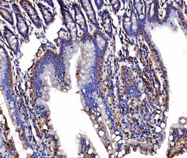
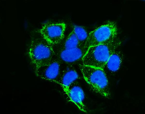
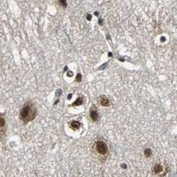
Anti-LSR antibody A02742-1 has IHC images from paraffin sections of mouse and rat colon and human breast and liver cancers, plus IF data from A431 cells (catalog image captions).

Real IHC data IHC analysis of LSR using anti-LSR antibody (A02742-1). LSR was detected in a paraffin-embedded section of mouse colon tissue. Heat mediated antigen retrieval was performed in EDTA buffer (pH 8.0, epitope retrieval solution). The tissue section was blocked with 10% goat serum. The tissue section was then incubated with 2 μg/ml rabbit anti-LSR Antibody (A02742-1) overnight at 4°C. Biotinylated goat anti-rabbit IgG was used as secondary antibody and incubated for 30 minutes at 37°C. The tissue section was developed using Strepavidin-Biotin-Complex (SABC) (Catalog # SA1022) with DAB as the chromogen.
Anti-LSR Antibody ®
Cat # A02742-1

A02742-1 is the sole SKU shown; its IHC captions document staining in paraffin sections of mouse and rat colon and human breast and liver cancers (catalog IHC image captions). Its IF caption documents staining in A431 cells (catalog IF image caption).

Which to pick: Choose A02742-1 for paraffin-section IHC; its IHC images document staining in human, mouse, and rat samples, but do not report the fixative (catalog IHC image captions). The same SKU is listed for IF and ICC, with an IF image from A431 cells (catalog applications; catalog IF image caption). For work across species, the catalog lists human, mouse, and rat reactivity; clonality is unreported (catalog reactivity; catalog clone field).

Each figure is that product's own IHC / IF validation image from its datasheet.

References

  1. UniProt Consortium. UniProt entry Q86X29 (LSR_HUMAN, Lipolysis-stimulated lipoprotein receptor).
  2. Human Protein Atlas. LSR tissue IHC expression (reliability: Approved).
  3. Human Protein Atlas. LSR subcellular location (ICC-IF): Localized to the plasma membrane and cell junctions..
  4. Human Protein Atlas. LSR antibody validation summary (2 antibodies).
  5. Anti-lipolysis-stimulated lipoprotein receptor monoclonal antibody as a novel therapeutic agent for endometrial cancer. BMC cancer 2022 — PMC9210735.
  6. Lipolysis-stimulated lipoprotein receptor overexpression is a novel predictor of poor clinical prognosis and a potential therapeutic target in gastric cancer. Oncotarget 2018 — PMC6152476.
  7. The roles of tricellular tight junction protein lipolysis-stimulated lipoprotein receptor in malignancy of human endometrial cancer cells. Oncotarget 2016 — PMC5053684.
  8. Lipolysis-stimulated lipoprotein receptor-targeted antibody-drug conjugate demonstrates potent antitumor activity against epithelial ovarian cancer. Neoplasia (New York, N.Y.) 2023 — PMC9679668.
  9. PubMed PMID:14702039 — UniProt-cited evidence.
  10. PubMed PMID:16303743 — UniProt-cited evidence.
  11. PubMed PMID:15057824 — UniProt-cited evidence.