LUC7L2 / Putative RNA-binding protein Luc7-like 2 · IHC design guide

Design Immunohistochemistry for LUC7L2

Plan chromogenic LUC7L2 IHC in paraffin sections around the reported nuclear tissue pattern (HPA tissue IHC). Start antibody titration at 1:100–1:300 (datasheet) and include controls for possible off-target staining (HPA tissue IHC).

Evidence assembled Oct 2026 · For research use; verify linked source records and product datasheet before use
Immunohistochemistry protocol sheet for LUC7L2 (IHC for LUC7L2): expected localisation Nuclear staining in tissue (HPA tissue IHC), antibody A11514, validated IHC image, and IHC protocol steps
Printable LUC7L2 IHC protocol sheet — expected localisation Nuclear staining in tissue (HPA tissue IHC), antibody A11514, controls and protocol steps. Open the full LUC7L2 IHC guide →

LUC7L2 Immunohistochemistry Experimental Design Guide

Expected localisation, validated protocols, controls and antibodies — the at-a-glance facts below, then the full design guide.

Must know before staining
Expected localisation Nuclear staining in tissue (HPA tissue IHC)
Staining pattern Widespread nuclear staining across cell types (HPA tissue IHC)
Antigen retrieval Tris-EDTA pH 9.0 HIER, 95–98 °C, 20 min (rule: nuclear antigen)
Positive control ⓘ Appendix+4 more · see all
Negative control ⓘ Adipose tissue
Important caveats
Reasons your staining may differ from the expected pattern.
Fixation Keep paraffin tissue fixation consistent. (standard IHC practice; not target-specific)
Caveat Presumed off-target binding may mimic staining (HPA tissue IHC)
Regulation No specific expression regulator annotated (UniProt)
Isoform / epitope 3 isoforms; confirm antibody epitope coverage (UniProt)
Section 1

Recommended LUC7L2 IHC & IF Protocols

Compare the catalog antibody’s IHC-P protocol (datasheet) with three published LUC7L2 IHC protocols (PMC8671510; PMC13178732; PMC11109612).

Recommended immunohistochemistry (IHC-P) protocol parameters
SampleParaffin-embedded human colon carcinoma tissue; fixative not specified (datasheet A11514)
FixationImage fixative and duration unreported (datasheet A11514); verify before use.
Sectioning4–5 µm sections on charged slides (standard)
DeparaffinisationXylene, graded ethanol series to water (standard)
Antigen retrievalHeat-induced epitope retrieval in Tris-EDTA buffer, pH 9.0, 20 min at 95–98 °C (standard rule: nuclear antigen)
Peroxidase block3% H2O2, 10 min, room temperature (standard)
Blocking10% normal serum of the secondary host, 30 min, room temperature (standard)
Primary antibodyRabbit anti-LUC7L2, 1:100-1:300 (datasheet A11514)
Primary incubationOvernight at 4 °C (standard)
DetectionHRP-polymer secondary, DAB chromogen 5–10 min (standard)
CounterstainHematoxylin, blue, dehydrate and mount (standard)
Expected resultLUC7L2-positive staining in glandular cells of appendix (HPA tissue IHC: Medium). HPA tissue profile: Ubiquitous nuclear expression. No signal in the no-primary control.
💡Decision noteStart with Tris-EDTA pH 9.0 at 95–98 °C for 20 min (page retrieval rule); a published alternative uses citrate at 95 °C for 30 min (PMC11109612).
Section 2

What Is the Expected LUC7L2 Staining Pattern?

LUC7L2 is expected in nuclei, including nucleoplasm and nuclear speckles (UniProt Q9Y383: subcellular location). HPA describes ubiquitous nuclear tissue staining, with medium staining in several listed cell populations and no detected staining in adipocytes (HPA: tissue IHC). Treat the pattern as provisional: HPA rates tissue IHC Approved with medium RNA–staining consistency and warns of presumed off-target binding (HPA: reliability). LUC7L2 has no transmembrane segment (UniProt Q9Y383: topology).

What am I looking at on my slide?
Medium nuclear staining in appendix glandular cells or bone marrow hematopoietic cells (HPA: tissue IHC).This matches reported IHC-positive examples and the expected nuclear compartment (HPA: tissue IHC; UniProt Q9Y383: subcellular location). Score nuclei within the named cell population, rather than treating every stained structure as confirmation (general IHC practice).
Predominantly cytoplasmic or membrane-like signal, with little nuclear signal.That distribution conflicts with the reported nuclear location and lack of a transmembrane segment (UniProt Q9Y383: subcellular location and topology). Check staining controls, morphology and detection background before assigning the signal to LUC7L2 (general IHC practice).
Strong staining in adipocytes, where HPA reports not detected (HPA: adipose tissue IHC).Consider cross-reactivity or endogenous chromogenic activity and inspect the matching control section (general IHC practice). This is a discrepancy with one reported cell population, not proof that every adipocyte must be negative (HPA: adipose tissue IHC; HPA: reliability caveat).
A diffuse haze extends across tissue, nuclei and cell-free areas.That appearance is poorly suited to judging the reported nuclear pattern (UniProt Q9Y383: subcellular location). Review blocking, washes, chromogen development and detection controls as general IHC checks; HPA supplies no LUC7L2-specific cause for diffuse background (general IHC practice).
No convincing nuclear signal in appendix glandular cells despite visible, intact nuclei (HPA: appendix IHC).The result differs from HPA's medium staining reference (HPA: appendix IHC). Check tissue identity, antibody and detection controls, and the section's overall staining quality before calling LUC7L2 absent (general IHC practice).
💡Expected LUC7L2 appearanceCall a result consistent when identifiable nuclei in a reported positive population show medium staining, as in appendix glandular cells; prominent cytoplasmic, membrane-like or adipocyte staining warrants review (HPA: appendix and adipose tissue IHC; UniProt Q9Y383: subcellular location and topology).
How each factor affects the staining
Subnuclear locationUniProt places LUC7L2 in nucleoplasm and nuclear speckles (UniProt Q9Y383: subcellular location). A chromogenic section may support a nuclear call without resolving individual speckles; score the compartment visible at the available resolution (general IHC practice).
Choice of comparison tissueHPA reports medium staining in the listed appendix, bone marrow, breast, bronchus, caudate, cerebellum, cerebral cortex and cervix cell populations; adipocytes are not detected (HPA: tissue IHC). Compare the same cell types, since whole-section averages can hide them (general IHC practice).
Strength of validationThe listed antibody HPA051631 is IHC Approved, while the tissue assessment notes medium RNA–staining consistency, pending external verification and presumed off-target binding (HPA: antibody validation and reliability). An Approved label therefore does not independently establish the identity of every stained cell.
Isoforms and antibody coverageUniProt lists 3 LUC7L2 isoforms (UniProt Q9Y383: isoforms). No epitope or isoform coverage is supplied here, so the observed IHC pattern cannot establish which isoform contributes; check antibody documentation before making an isoform-specific interpretation (general IHC practice).
IF/ICC: what should a separate assay show?HPA approves a nucleoplasmic IF location and lists images from HeLa, MCF-7 and U2OS, while warning that the antibodies target proteins from multiple genes (HPA: subcellular ICC-IF). The IF/ICC assay has its own guide; this comparison provides a location check, not an IHC protocol.
Why is my staining missing, weak or wrong?
SituationLikely causeNext action
A reported positive population has no nuclear stain.The result conflicts with HPA's medium reference for that population; the record does not identify a specific technical cause (HPA: tissue IHC).Verify the cell population, a known-positive section, antibody preparation and chromogenic detection controls before interpreting absence (general IHC practice).
Signal is mainly cytoplasmic or traces cell outlines.That pattern disagrees with nuclear localization and no annotated transmembrane segment (UniProt Q9Y383: subcellular location and topology).Recheck nuclear morphology and background on the same slide; use a detection control to assess nonspecific signal (general IHC practice).
Adipocytes stain strongly.HPA reports adipocytes as not detected and cautions about presumed off-target binding (HPA: adipose tissue IHC and reliability).Compare adipocytes with nearby nuclei and the appropriate detection control; avoid counting adipocyte signal as confirmed LUC7L2 (general IHC practice).
The entire section has diffuse brown background.Background obscures the expected nuclear distribution; no LUC7L2-specific mechanism is supplied (UniProt Q9Y383: subcellular location; general IHC practice).Review blocking, washing and chromogen development, then reassess whether nuclei remain distinguishable from surrounding tissue (general IHC practice).
A low-staining population looks negative.HPA reports low staining in adrenal glandular cells, skeletal myocytes, peripheral nerve and splenic white-pulp cells (HPA: tissue IHC).Identify the relevant cell population and compare it with a reported medium-staining reference before treating weak signal as assay failure (HPA: tissue IHC; general IHC practice).
Several cell types show nuclear staining, but specificity remains uncertain.Broad nuclear staining is reported, while HPA notes medium consistency and presumed off-target binding (HPA: tissue IHC and reliability).Record compartment and cell type separately, compare HPA's positive and adipocyte observations, and describe unresolved staining conservatively (HPA: tissue IHC; general IHC practice).

Sample controls for LUC7L2 IHC & IF

🧪Run appendix first: its glandular cells should show nuclear staining (HPA: Medium in appendix glandular cells; UniProt Q9Y383: nuclear localization). Use adipose tissue as the negative comparator because adipocytes are listed as not detected (HPA: Not detected in adipocytes); within the appendix slide, glandular cytoplasm should provide a low-signal compartment, while other cell types should not be presumed negative (UniProt Q9Y383: nuclear localization).
Positive control tissue: Appendix (Glandular cells, HPA Medium)
Negative control tissue: Adipose tissue (HPA Not detected)
ICC-IF cell lines (HPA subcellular resource): HPA ICC-IF images show LUC7L2 in HeLa, MCF-7, U2OS, with annotated localisation: Nucleoplasm (approved) (HPA subcellular).
Technical controls: Include a no-primary, secondary-only control and a host-species- and clonality-matched isotype control (standard IHC practice); use a knockout sample if available, or the peptide-block condition shown for the catalog antibody (A11514 tissue-IHC caption). Block endogenous peroxidase and check for residual chromogenic signal in appendix inflammatory cells (standard IHC practice).
⚠️Feasibility: A target-specific fixation window or fixation effect is unreported, and the fixative in the A11514 paraffin-section caption is unreported (A11514 tissue-IHC caption). Antigen retrieval dependence is unreported; optimize retrieval for the paraffin IHC assay (supplied evidence; standard IHC practice). Neither frozen sections nor IF can be judged easier from the supplied evidence; HPA reports nucleoplasmic ICC-IF localization but cautions that its summary is based on antibodies targeting proteins from multiple genes (HPA subcellular summary).

HPA tissue IHC evidence for LUC7L2

Comprehensive Human Protein Atlas IHC scoring per tissue (reliability: Approved — Medium consistency between antibody staining and RNA expression data. Pending external verification. Caution, targets protein from more than one gene. Presumed off target binding observed and disregarded.). Rows are taken directly from the HPA tissue chart — click any row's HPA link to view the source.

Positive expression · recommended positive controls

TissueCell typeLevelEvidenceSource
Appendix Glandular cells Medium Protein (IHC) HPA →
Bone marrow Hematopoietic cells Medium Protein (IHC) HPA →
Breast Glandular cells Medium Protein (IHC) HPA →
Bronchus Respiratory epithelial cells Medium Protein (IHC) HPA →
Caudate Neuronal cells Medium Protein (IHC) HPA →

Undetected expression · recommended negative controls

TissueCell typeLevelEvidenceSource
Adipose tissue Adipocytes Not detected Protein (IHC) HPA →
Section 3

Advanced LUC7L2 IHC Tips

Troubleshoot chromogenic LUC7L2 IHC in paraffin sections by checking nuclear localisation, retrieval, controls, and scoring before interpreting signal.

How should I retrieve LUC7L2 in paraffin sections when nuclear staining is weak?
Start with Tris-EDTA at pH 9.0 for heat-induced retrieval at 95–98 °C for 20 min (page retrieval rule: nuclear antigen). Keep section thickness, heating vessel, and cooling time consistent across the comparison, because these variables affect antigen exposure in paraffin sections (standard IHC practice). Compare the weak section with a concurrently processed positive control and a no-primary control, then adjust one retrieval variable at a time (standard IHC practice). Score nuclear signal separately from diffuse cytoplasmic colour, since LUC7L2 is reported in the nucleoplasm and nuclear speckles (HPA: subcellular; UniProt Q9Y383: subcellular).
Could fixation explain inconsistent LUC7L2 nuclear staining across blocks?
Target-specific fixation sensitivity for LUC7L2 is unknown from the supplied evidence; the catalog image identifies paraffin-embedded human colon carcinoma but does not state its fixative (A11514 tissue-IHC caption). Record each block’s fixative and fixation duration before comparing staining, and process control and test sections together where possible (standard IHC practice). When blocks differ in handling, compare intact nuclear morphology and staining within similarly preserved regions before changing antibody conditions (standard IHC practice). Treat an apparent fixation effect as a working hypothesis until matched material confirms it; nuclear localisation and reported modifications alone do not establish fixation sensitivity (UniProt Q9Y383: subcellular and modified residues).
Where should convincing LUC7L2 chromogenic signal appear?
Prioritise staining within morphologically intact nuclei: LUC7L2 is reported in the nucleoplasm, with a nuclear-speckle annotation in UniProt (HPA: subcellular; UniProt Q9Y383: subcellular). Inspect higher magnification before calling puncta speckles, since a chromogenic precipitate can obscure fine nuclear structure (standard IHC practice). HPA describes ubiquitous nuclear tissue expression, but its tissue-IHC assessment has medium staining-to-RNA consistency and cautions about presumed off-target binding (HPA: tissue IHC). Compare nuclear signal with the no-primary control and local tissue morphology; cytoplasmic-only or extracellular colour warrants investigation before assignment to LUC7L2 (standard IHC practice; HPA: subcellular).
Can this stain distinguish LUC7L2 isoforms or modified epitopes?
Do not assign a chromogenic pattern to a particular isoform without epitope mapping: the record lists 3 isoforms but supplies no isoform-specific antibody epitope (UniProt Q9Y383: isoforms; supplied antibody evidence). LUC7L2 includes phosphoserine at position 18 and JMJD6-associated hydroxylysines at positions 266 and 269 (UniProt Q9Y383: modified residues). Those annotations do not show whether the catalog antibody recognises or loses recognition of a modified form (supplied antibody evidence). If isoform or modification specificity matters, establish it with a mapped epitope and independently characterised material before interpreting differences between sections (standard IHC practice).
How can I assess LUC7L2 alongside a cell marker by IF?
For a separate IF experiment, pair LUC7L2 with a validated marker of the cell population being examined and include single-stain controls for each channel (standard IF practice). Choose a fluorophore channel with low background in the actual tissue, checking unstained sections for autofluorescence and spectral overlap before multiplexing (standard IF practice). LUC7L2 is nuclear and has no annotated transmembrane segment, so use nuclear access as the basis for optimising permeabilisation after fixation (UniProt Q9Y383: subcellular and topology; standard IF practice). Interpret overlap within segmented nuclei, while keeping in mind that the HPA nucleoplasmic IF assignment cautions about antibodies targeting proteins from multiple genes (HPA: subcellular).
What should I check when brown staining obscures LUC7L2 nuclei?
Run a no-primary control through the same detection steps to locate background arising from the tissue or detection system (standard IHC practice). For peroxidase-based chromogenic IHC, check the peroxidase block and compare background before and after DAB development; these are general workflow checks (standard IHC practice). Review blocking, washes, and primary-antibody concentration systematically, changing one condition at a time while retaining a concurrently stained positive section (standard IHC practice). Be especially cautious with widespread colour outside nuclei, because the expected compartment is nuclear and HPA reports presumed off-target binding in its tissue-IHC assessment (UniProt Q9Y383: subcellular; HPA: tissue IHC).
How should I quantify LUC7L2 staining across tissue sections? ⚠ ANSWER MARKED FOR VERIFICATION
Define the cell population and viable analysis area before scoring, then use nuclear percentage positive or a nuclear H-score with fixed intensity thresholds (standard IHC practice; HPA: nuclear tissue profile). Report the number of evaluable nuclei and normalise positive counts to all evaluable nuclei in that population, rather than to section area alone (standard IHC practice). If measuring positive-cell density per mm², also report tissue area and cell density so changes in cellularity remain visible (standard IHC practice). Keep retrieval, chromogen development, imaging, and thresholds matched across samples, and exclude folds, edges, necrosis, and no-primary background consistently (standard IHC practice).
When does apparent LUC7L2 positivity warrant further validation?
Give greatest weight to reproducible staining in intact nuclei, consistent with reported nucleoplasmic and nuclear-speckle localisation (HPA: subcellular; UniProt Q9Y383: subcellular). Investigate cytoplasmic-only staining, extracellular deposits, section-edge gradients, and signal confined to necrotic tissue before calling a region positive (standard IHC practice). A no-primary control helps identify detection background, including endogenous enzyme activity in peroxidase workflows, while the A11514 caption reports peptide blocking of its paraffin-section image (standard IHC practice; A11514 tissue-IHC caption). Peptide blocking alone cannot settle specificity when cross-reactivity is plausible; HPA explicitly cautions that its tissue-IHC assessment includes presumed off-target binding and awaits external verification (HPA: tissue IHC).
Boster reagents

Best LUC7L2 / Putative RNA-binding protein Luc7-like 2 IHC Antibodies

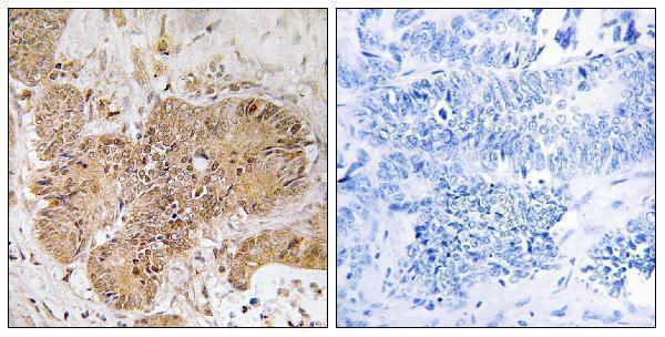
A11514 is listed for IHC and IF in human, mouse and rat; its IHC image shows a human paraffin-embedded colon carcinoma section (catalog: applications and reactivity; IHC image caption).

Real IHC data Immunohistochemistry analysis of paraffin-embedded human colon carcinoma tissue, using LUC7L2 Antibody. The picture on the right is blocked with the synthesized peptide.
Anti-LUC7L2 Antibody
Cat # A11514

A11514 will render with an IHC image of a human paraffin-embedded colon carcinoma section and a peptide-blocked comparison (IHC image caption). IF is listed as an application, but no IF image is supplied; human, mouse and rat are listed as reactive species (catalog: applications, IF image alts and reactivity).

Which to pick: For tissue IHC, choose A11514 because its own image caption documents staining in a paraffin-embedded human section; the fixative is unreported (IHC image caption). For IF, A11514 is the listed option, although ICC and an IF image are not documented (catalog: applications and IF image alts). It is also the listed cross-species option based on human, mouse and rat reactivity, but the supplied IHC image documents human tissue only (catalog: reactivity; IHC image caption).

Each figure is that product's own IHC / IF validation image from its datasheet.

References

  1. UniProt Consortium. UniProt entry Q9Y383 (LC7L2_HUMAN, Putative RNA-binding protein Luc7-like 2).
  2. Human Protein Atlas. LUC7L2 tissue IHC expression (reliability: Approved).
  3. Human Protein Atlas. LUC7L2 subcellular location (ICC-IF): Localized to the nucleoplasm. Caution: Based on antibodies targeting proteins from multiple genes..
  4. Human Protein Atlas. LUC7L2 antibody validation summary (1 antibodies).
  5. CRISPR/Cas9 genome-wide screening identifies LUC7L2 that promotes radioresistance via autophagy in nasopharyngeal carcinoma cells. Cell death discovery 2021 — PMC8671510.
  6. CircRAD18 promotes glioblastoma proliferation, migration and invasion via the miR‑1231/LUC7L2 axis. International journal of molecular medicine 2026 — PMC13178732.
  7. Histone H3K9 Lactylation Confers Temozolomide Resistance in Glioblastoma via LUC7L2-Mediated MLH1 Intron Retention. Advanced science (Weinheim, Baden-Wurttemberg, Germany) 2024 — PMC11109612.
  8. PubMed PMID:10810093 — UniProt-cited evidence.
  9. PubMed PMID:14702039 — UniProt-cited evidence.
  10. PubMed PMID:12853948 — UniProt-cited evidence.