This website uses cookies to ensure you get the best experience on our website.


- Table of Contents
Plan chromogenic LY6D IHC on paraffin sections using the catalog antibody at 2–5 μg/ml (datasheet A09680). Use esophageal squamous epithelium as a high-staining reference and assess membranous and cytoplasmic signal (HPA tissue IHC).

Expected localisation, validated protocols, controls and antibodies — the at-a-glance facts below, then the full design guide.
| Expected localisation | Membranous and cytoplasmic in squamous epithelium (HPA tissue IHC) | |
| Staining pattern | Squamous epithelial cells show membranous and cytoplasmic staining (HPA tissue IHC) | |
| Antigen retrieval | EDTA pH 8.0 HIER, heat-mediated (datasheet A09680) | |
| Positive control | Cervix+4 more · see all | |
| Negative control | Adipose tissue+4 more · see all |
| Fixation | Keep fixation consistent across paraffin sections (standard IHC practice; not target-specific) | |
| Caveat | Normal squamous epithelium can stain strongly (HPA tissue IHC) | |
| Regulation | Higher in squamous epithelia (HPA tissue IHC) | |
| Isoform / epitope | No isoforms annotated; mature chain spans aa 21–98 and is surface-facing, with no cytoplasmic tail (UniProt) |
The catalog antibody protocol (datasheet: A09680) is accompanied by published LY6D IHC protocols for bladder cancer tissue microarrays (PMC7539380) and pancreatic cancer tissue microarrays (PMC8715959).
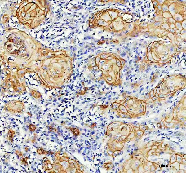
| Sample | Paraffin-embedded human skin cancer tissue; fixative not specified (datasheet A09680) |
| Fixation | Image fixative and duration unreported (datasheet A09680); verify before use. |
| Sectioning | 4–5 µm sections on charged slides (standard) |
| Deparaffinisation | Xylene, graded ethanol series to water (standard) |
| Antigen retrieval | Heat retrieval: EDTA pH 8.0 (datasheet A09680); 20 min, 95–100 °C (standard) |
| Peroxidase block | 3% H2O2, 10 min, room temperature (standard) |
| Blocking | 10% goat serum (datasheet A09680) |
| Primary antibody | Rabbit anti-LY6D, 2-5 μg/ml (datasheet A09680) |
| Primary incubation | Overnight at 4 °C (datasheet A09680) |
| Detection | HRP-conjugated secondary, DAB chromogen (datasheet A09680) |
| Counterstain | Hematoxylin, blue, dehydrate and mount (standard) |
| Expected result | LY6D-positive staining in squamous epithelial cells of cervix (HPA tissue IHC: High). HPA tissue profile: Membranous and cytoplasmic expression in squamous epithelium. No signal in the no-primary control. |
LY6D is a cell membrane protein with no transmembrane segment; UniProt describes it at the outer surface of transitional and stratified squamous epithelia (UniProt Q14210 topology; tissue specificity). In paraffin-section IHC, expect membranous and some cytoplasmic staining in squamous epithelial cells, with medium staining reported in skin basal-layer cells and urothelium (HPA tissue IHC). HPA rates the tissue pattern Enhanced for agreement between staining and RNA expression (HPA tissue IHC).
| Clear membrane outlines, with some cytoplasmic color, in squamous epithelial cells. | This fits the observed IHC profile (HPA tissue IHC: membranous and cytoplasmic expression). High staining is reported in squamous epithelial cells of cervix, esophagus, oral mucosa, tonsil and vagina (HPA tissue IHC). Judge the pattern within the relevant cells as well as its intensity. |
| Predominantly nuclear staining, with no convincing epithelial membrane pattern. | Nuclear localization conflicts with the cell membrane assignment (UniProt Q14210 subcellular location) and the observed tissue profile (HPA tissue IHC). Treat it as a possible staining artefact and check controls and detection conditions before interpreting the nuclei as LY6D. |
| Strong color in cell types reported as negative, such as adipocytes or respiratory epithelial cells. | Those cells are reported as not detected in the sampled tissues (HPA tissue IHC: adipose tissue and bronchus). Unexpected staining raises possible cross-reactivity or endogenous detection activity; the slide alone cannot distinguish them. Compare with a no-primary control and the expected epithelial distribution. |
| Diffuse color across epithelium, stroma and empty areas, without distinct cell borders. | A field-wide haze does not establish the reported membranous and cytoplasmic epithelial pattern (HPA tissue IHC). In routine IHC, excess background can arise from detection reagents, inadequate blocking or overly concentrated primary antibody. Evaluate control sections before assigning weak diffuse color to LY6D. |
| No staining in a well-preserved, known-positive squamous epithelial section. | An absent signal conflicts with reported high staining in several squamous epithelia (HPA tissue IHC: cervix, esophagus, oral mucosa, tonsil and vagina). First verify tissue identity, slide quality, antibody performance and detection controls. This record does not establish LY6D-specific fixation sensitivity or a retrieval requirement. |
| Cellular compartment | UniProt assigns LY6D to the cell membrane and reports no transmembrane segment (UniProt Q14210 topology). HPA observes both membranous and cytoplasmic tissue IHC staining (HPA tissue IHC). Assess membrane localization alongside cell identity; cytoplasmic color alone is less specific than the combined reported pattern. |
| Choice of reference tissue | Squamous epithelial cells score High in cervix, esophagus, oral mucosa, tonsil and vagina; skin basal-layer cells and urinary bladder urothelial cells score Medium (HPA tissue IHC). Use the reported cell type and level when choosing a comparison section, and avoid requiring identical intensity across tissues. |
| Strength and limits of validation | HPA calls tissue IHC reliability Enhanced because staining is highly consistent with RNA expression (HPA tissue IHC). Three listed antibodies carry Enhanced IHC status, while their ICC fields are empty (HPA antibodies). These ratings support the reported tissue pattern but do not validate every unexpected compartment or cell type. |
| Protein processing and epitope uncertainty | UniProt lists a signal peptide at residues 1–20, a mature chain at 21–98 and a propeptide at 99–128 (UniProt Q14210 processing). The supplied record gives no antibody epitope. It therefore cannot establish which processed form the IHC-validated antibody recognizes or predict a retrieval condition. |
| IF/ICC Q&A: is a membrane signal expected? | Yes as a localization expectation: HPA summarizes LY6D as membrane-associated (HPA subcellular), consistent with UniProt (UniProt Q14210 subcellular location). HPA supplies no ICC-IF images or listed antibody ICC validation (HPA subcellular; HPA antibodies). Consult the separate IF/ICC guide for experimental design. |
| Situation | Likely cause | Next action |
|---|---|---|
| Known-positive squamous epithelium is blank. | This disagrees with reported High staining in several squamous epithelia (HPA tissue IHC). The cause remains undetermined from a blank slide. | Confirm that the section contains the specified epithelial cells, then review the catalog antibody's IHC-P instructions, detection reagents and positive control. Do not infer a LY6D-specific fixation or retrieval defect from this result. |
| Signal is cytoplasmic, with no discernible cell-border staining. | Cytoplasmic staining is reported, but the reference pattern also includes membranes (HPA tissue IHC). The isolated pattern has weaker localization support. | Compare a reported-positive section and no-primary control. Review staining strength and morphology before scoring; record the observed compartment rather than calling a membrane-positive result. |
| Nuclei are the strongest stained structures. | A dominant nuclear pattern differs from the membrane assignment and tissue IHC profile (UniProt Q14210 subcellular location; HPA tissue IHC). | Check whether counterstain or detection color is being mistaken for target signal. Inspect controls, then reassess epithelial borders at higher magnification. |
| Reported-negative cells stain as strongly as the epithelial positive control. | Unexpected staining could reflect cross-reactivity or endogenous detection activity; HPA reports no detection in several listed negative cell types (HPA tissue IHC). | Run a no-primary control on the same tissue and review blocking appropriate to the detection system. If color persists, resolve the background before attributing it to LY6D. |
| The whole section has weak, diffuse color. | Diffuse background obscures the cell-specific membrane and cytoplasmic pattern (HPA tissue IHC); routine IHC reagent or blocking issues are possible. | Inspect no-primary and negative-tissue controls. Check detection reagent handling, blocking and antibody dilution against the chosen IHC-P instructions; score only localized staining above background. |
| Skin or urinary bladder looks weaker than a squamous epithelial positive control. | HPA reports Medium staining in skin basal-layer cells and urothelium, versus High staining in several squamous epithelia (HPA tissue IHC). | Compare the correct cell populations and section quality before changing conditions. Medium staining can agree with the reference; a completely blank result still warrants control review. |

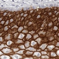
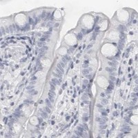

Comprehensive Human Protein Atlas IHC scoring per tissue (reliability: Enhanced — High consistency between antibody staining and RNA expression data.). Rows are taken directly from the HPA tissue chart — click any row's HPA link to view the source.
| Tissue | Cell type | Level | Evidence | Source |
|---|---|---|---|---|
| Cervix | Squamous epithelial cells | High | Protein (IHC) | HPA → |
| Esophagus | Squamous epithelial cells | High | Protein (IHC) | HPA → |
| Oral mucosa | Squamous epithelial cells | High | Protein (IHC) | HPA → |
| Skin | Cells in basal layer | Medium | Protein (IHC) | HPA → |
| Tonsil | Squamous epithelial cells | High | Protein (IHC) | HPA → |
| Tissue | Cell type | Level | Evidence | Source |
|---|---|---|---|---|
| Adipose tissue | Adipocytes | Not detected | Protein (IHC) | HPA → |
| Adrenal gland | Glandular cells | Not detected | Protein (IHC) | HPA → |
| Appendix | Glandular cells | Not detected | Protein (IHC) | HPA → |
| Bone marrow | Hematopoietic cells | Not detected | Protein (IHC) | HPA → |
| Breast | Adipocytes | Not detected | Protein (IHC) | HPA → |
These LY6D questions focus on interpreting chromogenic staining in paraffin sections, using the catalog antibody’s tissue image and the supplied protein expression evidence.
A09680 has real IHC data from a paraffin-embedded human skin cancer section (catalog IHC image caption); the catalog lists human reactivity (catalog: reactivity). No IF data are supplied (catalog: IF images).
A09680 will render with its IHC figure from a paraffin-embedded human skin cancer section (catalog IHC image caption). It is listed for human IHC, WB and ELISA, with no IF application or image listed (catalog: applications, reactivity and IF images).
Which to pick: Choose A09680 for human paraffin-section IHC: it is a rabbit polyclonal antibody listed for human IHC (catalog: host, clonality, applications and reactivity), and its own figure shows EDTA pH 8.0 retrieval and 2 μg/ml primary antibody (A09680 IHC image caption). The caption does not report the fixative (A09680 IHC image caption). No SKU in the supplied catalog is listed for IF/ICC or cross-species use (catalog: applications and reactivity).