This website uses cookies to ensure you get the best experience on our website.


- Table of Contents
Plan LYPD3 IHC in paraffin sections using squamous epithelium as a positive tissue reference (HPA tissue IHC). This guide covers membrane localization expected from UniProt and practical controls for interpreting tissue staining (UniProt; HPA tissue IHC).

Expected localisation, validated protocols, controls and antibodies — the at-a-glance facts below, then the full design guide.
| Expected localisation | Cell membrane expected (UniProt); squamous epithelia stain (HPA tissue IHC) | |
| Staining pattern | Selective squamous epithelial staining; compartment unspecified (HPA tissue IHC) | |
| Antigen retrieval | EDTA pH 8.0 HIER, heat-mediated (datasheet A08396) | |
| Positive control | Cervix+4 more · see all | |
| Negative control | Adipose tissue+4 more · see all |
| Fixation | Matched tissue-IHC evidence does not establish the fixation claim. Validate the specimen-specific method before use. (selected-SKU IHC image A08396) | |
| Caveat | Presumed off-target staining was observed and disregarded (HPA tissue IHC) | |
| Regulation | Present in chronic-wound keratinocytes (UniProt) | |
| Isoform / epitope | No isoforms listed; check epitope against mature chain 31–326 (UniProt) |
The catalog antibody’s IHC-P protocol (datasheet A08396) is accompanied by published LYPD3 protocols for HNSCC and breast cancer tissue microarrays (PMC11250566; PMC6874102).
| Sample | Paraffin-embedded human skin cancer tissue; fixative not specified (datasheet A08396) |
| Fixation | Image fixative and duration unreported (datasheet A08396); verify before use. |
| Sectioning | 4–5 µm sections on charged slides (standard) |
| Deparaffinisation | Xylene, graded ethanol series to water (standard) |
| Antigen retrieval | Heat retrieval: EDTA pH 8.0 (datasheet A08396); 20 min, 95–100 °C (standard) |
| Peroxidase block | 3% H2O2, 10 min, room temperature (standard) |
| Blocking | 10% goat serum (datasheet A08396) |
| Primary antibody | Rabbit anti-LYPD3, 1-2 μg/ml (datasheet A08396) |
| Primary incubation | Overnight at 4 °C (datasheet A08396) |
| Detection | HRP-conjugated secondary, DAB chromogen (datasheet A08396) |
| Counterstain | Hematoxylin, blue, dehydrate and mount (standard) |
| Expected result | LYPD3-positive staining in squamous epithelial cells of cervix (HPA tissue IHC: High). HPA tissue profile: Selective expression in squamous epithelia. No signal in the no-primary control. |
In paraffin-section IHC, expect LYPD3 chiefly along the cell membrane of squamous epithelial cells; the protein has no transmembrane segment and is GPI anchored (UniProt O95274 topology; UniProt O95274 keywords). HPA reports high staining in several squamous epithelia and rates its tissue IHC profile Enhanced, while noting presumed off-target binding that was disregarded (HPA tissue IHC). Interpret the staining by its cell type and location together.
| Strong staining outlines squamous epithelial cells in cervix, esophagus, oral mucosa, tonsil or vagina (HPA tissue IHC). | This fits the reported high epithelial staining and the expected membrane location (HPA tissue IHC; UniProt O95274 subcellular location). Compare stained cells with the section's morphology before scoring intensity (general IHC practice). |
| Predominantly nuclear staining appears in squamous epithelium. | A nuclear-only pattern conflicts with the reported cell-membrane location (UniProt O95274 subcellular location). Treat it as suspect IHC signal and review controls and antibody specificity before assigning it to LYPD3 (general IHC practice). |
| Cells outside the reported epithelial pattern stain, particularly in a comparison tissue. | Compare the exact cell type with HPA's tissue entries: adipocytes in adipose tissue and respiratory epithelial cells in bronchus were not detected (HPA tissue IHC). Unexpected staining can reflect antibody cross-reactivity or endogenous detection activity (general IHC practice). |
| Color spreads across epithelium, stroma and blank areas without clear cell borders. | This is diffuse background rather than an interpretable membrane pattern (UniProt O95274 subcellular location; general IHC practice). Review blocking, washes, detection chemistry and the no-primary control before scoring (general IHC practice). |
| No staining appears in an adequately sampled HPA high-staining squamous epithelium. | This conflicts with HPA's reported high staining in the specified epithelial cell types (HPA tissue IHC). First verify tissue identity and the positive control, then check primary-antibody use and detection steps (general IHC practice). |
| Cell location and processing | LYPD3 is a GPI-anchored membrane protein with no transmembrane segment; its annotated mature chain spans residues 31–326 (UniProt O95274 topology; UniProt O95274 processing). Use membrane association as the IHC interpretation anchor. |
| Epitope location | UniProt lists a signal peptide at residues 1–30 and a propeptide at 327–346 (UniProt O95274 processing). Antibody epitope information is absent here, so those annotations cannot predict this antibody's staining. |
| Glycosylation | Four glycosylation sites are annotated at residues 118, 163, 176 and 183 (UniProt O95274 glycosylation). Their effect on this IHC antibody or antigen retrieval is unreported in the supplied sources. |
| Tissue and cell-type controls | HPA reports high staining in squamous epithelial cells at several sites and no detection in adipocytes from adipose tissue or respiratory epithelial cells from bronchus (HPA tissue IHC). These are cell-specific comparisons, not whole-tissue guarantees. |
| Esophagus evidence | HPA reports high esophageal squamous-cell IHC staining, whereas UniProt describes esophageal expression as weak (HPA tissue IHC; UniProt O95274 tissue specificity). Keep the source and measurement context attached to each claim. |
| IF/ICC Q: Should intracellular fluorescence be scored like IHC membrane staining? | A: No. HPA's ICC-IF summary localizes LYPD3 to approved endoplasmic-reticulum and vesicle sites (HPA subcellular ICC-IF), while UniProt lists the cell membrane (UniProt O95274 subcellular location). Assess IF/ICC on its own guide page. |
| Situation | Likely cause | Next action |
|---|---|---|
| The expected squamous epithelium is blank. | Tissue selection or an IHC workflow step may have failed; the listed squamous epithelial cells stain high in HPA IHC (HPA tissue IHC; general IHC practice). | Confirm the epithelial cells are present and inspect a concurrently stained positive section, primary-antibody step and detection controls (general IHC practice). |
| Only nuclei stain in a nominally positive section. | That compartment conflicts with LYPD3's reported membrane location (UniProt O95274 subcellular location). | Compare with a positive squamous epithelial section and a no-primary control; withhold a positive call if the nuclear pattern persists (general IHC practice). |
| Color appears in HPA non-detected comparison cells. | Unexpected cell staining can arise from antibody cross-reactivity or endogenous detection activity (HPA tissue IHC; general IHC practice). | Check cell identity, a no-primary control and endogenous-enzyme blocking appropriate to the chromogenic system (general IHC practice). |
| Diffuse color obscures epithelial borders. | Background may reflect insufficient blocking, washing or overly strong detection conditions (general IHC practice). | Review blocking and washes, and compare the no-primary control before adjusting assay conditions (general IHC practice). |
| Staining is weak or uneven across a section. | Section handling or staining steps may vary across the slide (general IHC practice); the supplied sources give no LYPD3-specific fixation sensitivity. | Check section coverage and reagent delivery, then assess antigen retrieval with routine assay controls; do not infer an LYPD3-specific retrieval requirement (general IHC practice). |
| A tumor stains while adjacent tissue has little signal. | UniProt reports LYPD3 in transitional cell carcinomas and breast cancer tissue, with absent staining in adjacent normal tissue in that account (UniProt O95274 tissue specificity). | Score tumor and adjacent cells separately, record membrane-associated staining, and avoid treating one tissue compartment as the control for every other compartment (UniProt O95274 subcellular location; general IHC practice). |
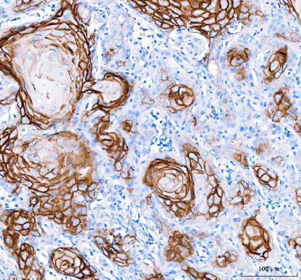
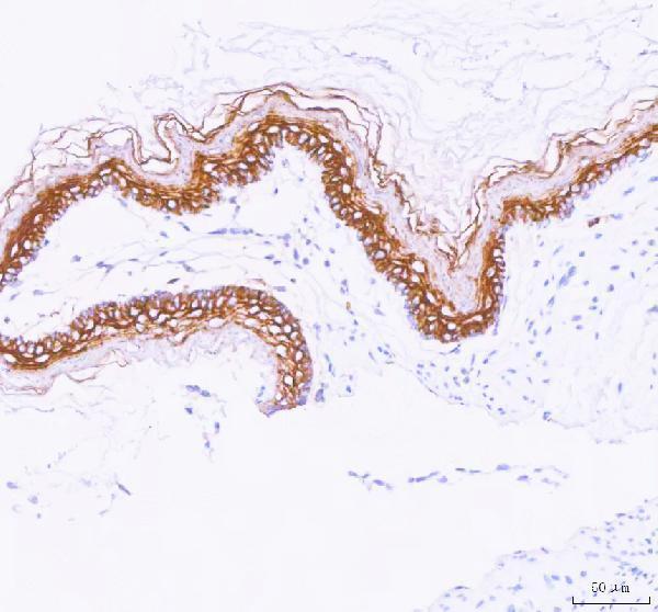
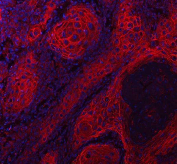


Comprehensive Human Protein Atlas IHC scoring per tissue (reliability: Enhanced — High consistency between antibody staining and RNA expression data. Presumed off target binding observed and disregarded.). Rows are taken directly from the HPA tissue chart — click any row's HPA link to view the source.
| Tissue | Cell type | Level | Evidence | Source |
|---|---|---|---|---|
| Cervix | Squamous epithelial cells | High | Protein (IHC) | HPA → |
| Esophagus | Squamous epithelial cells | High | Protein (IHC) | HPA → |
| Oral mucosa | Squamous epithelial cells | High | Protein (IHC) | HPA → |
| Skin | Cells in corneal layer | High | Protein (IHC) | HPA → |
| Tonsil | Squamous epithelial cells | High | Protein (IHC) | HPA → |
| Tissue | Cell type | Level | Evidence | Source |
|---|---|---|---|---|
| Adipose tissue | Adipocytes | Not detected | Protein (IHC) | HPA → |
| Adrenal gland | Glandular cells | Not detected | Protein (IHC) | HPA → |
| Appendix | Glandular cells | Not detected | Protein (IHC) | HPA → |
| Bone marrow | Hematopoietic cells | Not detected | Protein (IHC) | HPA → |
| Breast | Adipocytes | Not detected | Protein (IHC) | HPA → |
Troubleshoot LYPD3 staining in paraffin sections by checking retrieval, compartment, cell type, and controls before comparing signal intensity (UniProt O95274; HPA tissue IHC).
A08396 has IHC images from human skin cancer and rat stomach paraffin sections, plus an IF image from human skin cancer paraffin sections (A08396 image captions).
A08396 will render with its human skin cancer paraffin-section IHC figure; its captions also document rat stomach IHC and human skin cancer IF (A08396 image captions). M08396-1 will render as a human-reactive IHC antibody, although no IHC image is supplied for it (M08396-1 catalog applications, reactivity and image list).
Which to pick: For tissue IHC, choose A08396 when an illustrated paraffin-section workflow is useful; its IHC captions document human skin cancer and rat stomach sections, and do not report the fixative (A08396 IHC image captions). For IF, choose A08396 based on its human paraffin-section IF image; ICC validation is not documented (A08396 IF image caption and applications). For work across species, A08396 lists human, mouse and rat reactivity, while the rabbit monoclonal M08396-1 lists human reactivity and IHC without a supplied figure (catalog reactivity, host, clonality, applications and image lists).