This website uses cookies to ensure you get the best experience on our website.


- Table of Contents
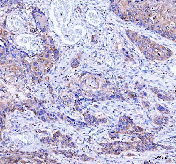
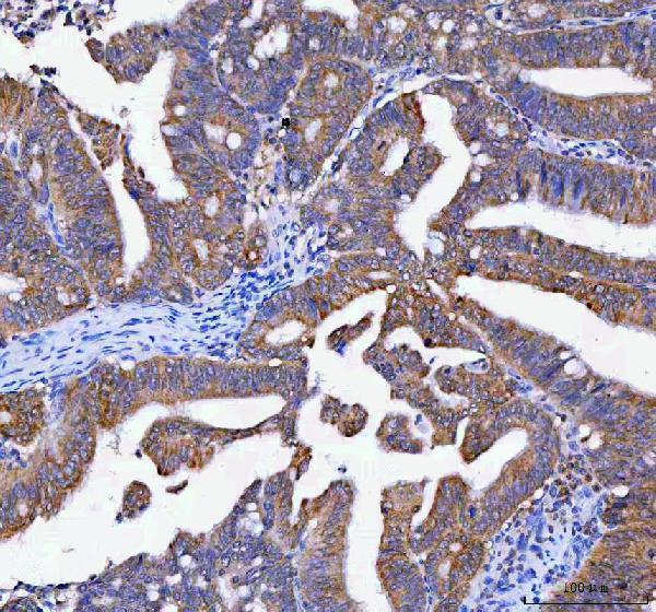
Plan LYPLA1 staining in paraffin sections using cytoplasmic glandular staining as a reference pattern (HPA tissue IHC). Compare the catalog antibody’s IHC conditions with positive and negative tissue controls (datasheet A07249-2; HPA tissue IHC).

Expected localisation, validated protocols, controls and antibodies — the at-a-glance facts below, then the full design guide.
| Expected localisation | Predominantly cytoplasmic in tissue (HPA tissue IHC) | |
| Staining pattern | Cytoplasmic staining in glandular cells of several tissues (HPA tissue IHC) | |
| Antigen retrieval | EDTA pH 8.0 HIER, heat-mediated (datasheet A07249-2) | |
| Positive control | Colon+4 more · see all | |
| Negative control | Adipose tissue+4 more · see all |
| Fixation | Keep fixation consistent across sections (standard IHC practice; not target-specific) | |
| Caveat | Esophageal carcinoma stains despite negative normal squamous cells (datasheet A07249-2; HPA tissue IHC) | |
| Regulation | No specific expression regulator reported (UniProt) | |
| Isoform / epitope | 2 isoforms; check antibody epitope coverage (UniProt) |
The catalog antibody’s IHC-P protocol is accompanied by published LYPLA1 staining protocols for head and neck biopsies, kidney sections, and breast cancer tissue microarrays (PMC7367650; PMC9676193; PMC10463390).
| Sample | Paraffin-embedded human esophageal squamous carcinoma tissue; fixative not specified (datasheet A07249-2) |
| Fixation | Image fixative and duration unreported (datasheet A07249-2); verify before use. |
| Sectioning | 4–5 µm sections on charged slides (standard) |
| Deparaffinisation | Xylene, graded ethanol series to water (standard) |
| Antigen retrieval | Heat retrieval: EDTA pH 8.0 (datasheet A07249-2); 20 min, 95–100 °C (standard) |
| Peroxidase block | 3% H2O2, 10 min, room temperature (standard) |
| Blocking | 10% goat serum (datasheet A07249-2) |
| Primary antibody | Rabbit anti-LYPLA1, 2-5 μg/ml (datasheet A07249-2) |
| Primary incubation | Overnight at 4 °C (datasheet A07249-2) |
| Detection | HRP-conjugated secondary, DAB chromogen (datasheet A07249-2) |
| Counterstain | Hematoxylin, blue, dehydrate and mount (standard) |
| Expected result | LYPLA1-positive staining in glandular cells of colon (HPA tissue IHC: High). HPA tissue profile: Cytoplasmic expression in several tissues. No signal in the no-primary control. |
LYPLA1 should appear mainly in the cytoplasm, with weak cell, nuclear and endoplasmic reticulum membrane localization possible (UniProt O75608 localization). It has no transmembrane segment (UniProt O75608 topology). In paraffin sections, look first at glandular cells of the colon, duodenum, gallbladder, rectum, small intestine or stomach, where staining is high (HPA: tissue IHC). HPA calls the tissue staining reliability Enhanced but reports only medium consistency with RNA expression (HPA: tissue IHC).
| Glandular cells show clear cytoplasmic chromogen, especially in colon, duodenum, gallbladder, rectum, small intestine or stomach (HPA: High in glandular cells). | This fits the reported tissue pattern and predominant cytoplasmic localization (HPA: tissue IHC; UniProt O75608 localization). Judge intensity within the identified cell type; a generally dark section alone does not establish specific staining (general IHC practice). |
| Strong, isolated nuclear or sharply membrane-only staining dominates, with little glandular cytoplasm stained. | That distribution conflicts with the predominant cytoplasmic pattern (UniProt O75608 localization; HPA: tissue IHC). Weak nuclear or cell membrane signal remains plausible (UniProt O75608 localization), so check controls and antibody behavior before calling the dominant pattern an artefact (general IHC practice). |
| Adipocytes or esophageal squamous epithelial cells stain prominently while glandular cells do not. | Those cell types were not detected in the HPA tissue survey, whereas several glandular cell populations were high (HPA: tissue IHC). The mismatch warrants checks for cross-reactivity or endogenous chromogenic activity; neither cause is established by HPA alone (general IHC practice). |
| Chromogen is spread across the section, including spaces between cells or the negative control. | A diffuse deposit cannot be scored as LYPLA1's reported cellular pattern (HPA: cytoplasmic tissue expression). Review blocking, washes, detection reagents and the negative control for background sources (general IHC practice); the record does not identify a LYPLA1-specific cause. |
| A well-preserved glandular area in a reported high-staining tissue has no detectable signal. | This disagrees with the reported HPA pattern for that cell type (HPA: High in glandular cells). First confirm cell identity and that a control section developed; then review retrieval and detection conditions as general IHC troubleshooting, without assuming LYPLA1-specific fixation sensitivity (general IHC practice). |
| Compartment and topology | Predominantly cytoplasmic localization guides scoring; weak cell, nuclear and endoplasmic reticulum membrane signal can occur (UniProt O75608 localization). Absence of a transmembrane segment does not rule out the reported weak membrane association (UniProt O75608 topology and localization). |
| Cell type within a tissue | HPA reports high staining in glandular cells at six named sites, medium in adrenal gland and appendix glandular cells, and undetected staining in selected adipocytes, squamous cells and other cell types (HPA: tissue IHC). Score the stated cell population rather than treating an entire organ as uniformly positive or negative. |
| Strength of supporting evidence | HPA050941 has Enhanced IHC validation, while the tissue profile has Enhanced reliability with medium antibody–RNA consistency (HPA: antibody validation; HPA: tissue IHC). These labels support the observed pattern but do not validate every antibody, section or unexpected compartment. |
| Isoforms and processing | UniProt lists two LYPLA1 isoforms and a chain spanning residues 1–230, with no signal peptide or propeptide reported (UniProt O75608 isoforms and processing). The supplied sources do not map the IHC antibody epitope or show whether isoforms stain differently; do not infer isoform identity from the slide. |
| Situation | Likely cause | Next action |
|---|---|---|
| High-staining glandular cells are blank (HPA: tissue IHC). | The expected cellular signal is missing; the supplied sources do not establish a LYPLA1-specific fixation or retrieval effect. | Verify cell identity and a working positive control, then review retrieval, primary antibody and chromogen steps (general IHC practice). |
| The section has widespread faint brown haze. | Diffuse signal lacks the reported cytoplasmic cell pattern (HPA: tissue IHC); background or endogenous detection activity is possible (general IHC practice). | Compare a primary-omission control and review blocking, washes and detection chemistry (general IHC practice). |
| Only nuclei are strongly stained. | Predominantly cytoplasmic localization is expected; HPA's uncertain nucleoplasmic IF finding does not establish a nuclear-dominant IHC pattern (UniProt O75608 localization; HPA: subcellular ICC-IF). | Check the negative control and repeat scoring against glandular cytoplasm; investigate persistent nuclear-dominant signal (general IHC practice). |
| Adipocytes or esophageal squamous cells stain more than nearby glandular cells. | HPA reports those particular cell types as not detected, while several glandular populations are high (HPA: tissue IHC). | Confirm cell identification, inspect controls for nonspecific or endogenous signal, and avoid calling those cells LYPLA1-positive on intensity alone (general IHC practice). |
| A weak membrane rim is visible alongside cytoplasm. | Weak cell and nuclear membrane localization is reported, despite no transmembrane segment (UniProt O75608 localization and topology). | Score the accompanying cytoplasmic pattern and cell type; investigate a membrane-only result with controls (HPA: tissue IHC; general IHC practice). |
| What should IF/ICC show? | HPA reports supported cytosol and uncertain nucleoplasm in ICC-IF, with a caution that the finding uses antibodies targeting proteins from multiple genes (HPA: subcellular ICC-IF). | Expect cytosolic signal as the better-supported comparison; treat nucleoplasmic signal cautiously (HPA: subcellular ICC-IF). Use the separate IF/ICC guide for experimental conditions. |


Comprehensive Human Protein Atlas IHC scoring per tissue (reliability: Enhanced — Medium consistency between antibody staining and RNA expression data.). Rows are taken directly from the HPA tissue chart — click any row's HPA link to view the source.
| Tissue | Cell type | Level | Evidence | Source |
|---|---|---|---|---|
| Colon | Glandular cells | High | Protein (IHC) | HPA → |
| Duodenum | Glandular cells | High | Protein (IHC) | HPA → |
| Gallbladder | Glandular cells | High | Protein (IHC) | HPA → |
| Rectum | Glandular cells | High | Protein (IHC) | HPA → |
| Small intestine | Glandular cells | High | Protein (IHC) | HPA → |
| Tissue | Cell type | Level | Evidence | Source |
|---|---|---|---|---|
| Adipose tissue | Adipocytes | Not detected | Protein (IHC) | HPA → |
| Bone marrow | Hematopoietic cells | Not detected | Protein (IHC) | HPA → |
| Breast | Adipocytes | Not detected | Protein (IHC) | HPA → |
| Cerebellum | Cells in granular layer | Not detected | Protein (IHC) | HPA → |
| Esophagus | Squamous epithelial cells | Not detected | Protein (IHC) | HPA → |
Troubleshoot LYPLA1 staining in paraffin sections by checking retrieval, cellular compartment, controls and scoring before interpreting chromogenic signal.
A07249-2 has IHC images from human paraffin sections (catalog IHC captions); M07249-1 lists ICC/IF for human, mouse and rat, with no IF image supplied (catalog applications, reactivity and IF image alts).
A07249-2 lists IHC and shows LYPLA1 staining in paraffin sections of human esophageal squamous carcinoma, rectum adenocarcinoma and lung cancer (catalog applications and IHC captions). M07249-1 lists ICC/IF and human, mouse and rat reactivity, but supplies no IF image (catalog applications, reactivity and IF image alts).
Which to pick: Choose A07249-2 for tissue IHC: its own captions document human paraffin sections, EDTA retrieval at pH 8.0 and 2 μg/ml primary antibody; the fixative is unreported (catalog A07249-2 IHC captions). Choose M07249-1 for IF/ICC based on its listed application, rabbit monoclonal clone 21L68 and 1:50 IF dilution; no IF image is supplied (catalog M07249-1 applications, clone, dilution and IF image alts). For cross-species work, both list human, mouse and rat reactivity, but the supplied IHC images show human sections only and no IF images are supplied (catalog reactivity and image alts).