MACROH2A1 / Core histone macro-H2A.1 · IHC design guide

Design Immunohistochemistry for MACROH2A1

Plan MACROH2A1 paraffin-section IHC using the catalog antibody at 1–2 μg/ml (datasheet A04635-3). Assess nuclear staining against the reported ubiquitous tissue profile, while accounting for the caution that staining may reflect proteins from more than one gene (HPA tissue IHC).

Evidence assembled Oct 2026 · For research use; verify linked source records and product datasheet before use
Immunohistochemistry protocol sheet for MACROH2A1 (IHC for MACROH2A1): expected localisation Nuclear staining in tissue (HPA tissue IHC), antibody A04635-3, validated IHC image, and IHC protocol steps
Printable MACROH2A1 IHC protocol sheet — expected localisation Nuclear staining in tissue (HPA tissue IHC), antibody A04635-3, controls and protocol steps. Open the full MACROH2A1 IHC guide →

MACROH2A1 Immunohistochemistry Experimental Design Guide

Expected localisation, validated protocols, controls and antibodies — the at-a-glance facts below, then the full design guide.

Must know before staining
Expected localisation Nuclear staining in tissue (HPA tissue IHC)
Staining pattern Nuclear staining across diverse tissue cell types (HPA tissue IHC)
Antigen retrieval EDTA pH 8.0 HIER, heat-mediated (datasheet A04635-3)
Positive control ⓘ Bone marrow+4 more · see all
Negative control ⓘ None in HPA (detected in all 45 tissues); use no-primary + isotype controls
Important caveats
Reasons your staining may differ from the expected pattern.
Fixation Keep fixation consistent across paraffin sections. (standard IHC practice; not target-specific)
Caveat Staining may reflect proteins from more than one gene (HPA tissue IHC)
Regulation Low tissue specificity (HPA tissue RNA)
Isoform / epitope 3 isoforms; epitope coverage is unspecified (UniProt)
Section 1

Recommended MACROH2A1 IHC & IF Protocols

The catalog antibody protocol is paired with one published macroH2A1 IHC protocol for deparaffinized sections (datasheet; PMC7471871).

Recommended immunohistochemistry (IHC-P) protocol parameters
SampleParaffin-embedded human colon adenocarcinoma tissue; fixative not specified (datasheet A04635-3)
FixationImage fixative and duration unreported (datasheet A04635-3); verify before use.
Sectioning4–5 µm sections on charged slides (standard)
DeparaffinisationXylene, graded ethanol series to water (standard)
Antigen retrievalHeat retrieval: EDTA pH 8.0 (datasheet A04635-3); 20 min, 95–100 °C (standard)
Peroxidase block3% H2O2, 10 min, room temperature (standard)
Blocking10% goat serum (datasheet A04635-3)
Primary antibodyRabbit anti-MACROH2A1, 1-2 μg/ml (datasheet A04635-3)
Primary incubationOvernight at 4 °C (datasheet A04635-3)
DetectionHRP-conjugated secondary, DAB chromogen (datasheet A04635-3)
CounterstainHematoxylin, blue, dehydrate and mount (standard)
Expected resultMACROH2A1-positive staining in hematopoietic cells of bone marrow (HPA tissue IHC: High). HPA tissue profile: Ubiquitous nuclear expression. No signal in the no-primary control.
💡Decision noteStart with heat-mediated EDTA pH 8.0 retrieval for the catalog antibody (datasheet: A04635-3); the published protocol specifies microwave retrieval without a buffer (PMC7471871).
Section 2

What Is the Expected MACROH2A1 Staining Pattern?

MACROH2A1 is a nuclear chromatin protein without a transmembrane segment (UniProt O75367: location and topology). Expect staining in nuclei across many tissues, including hematopoietic cells in bone marrow and glial cells in brain (HPA: ubiquitous nuclear expression; High in those cells). HPA rates its tissue IHC profile Supported but cautions that the staining may detect protein from more than one gene (HPA: reliability description).

What am I looking at on my slide?
Distinct nuclear chromogen in bone marrow hematopoietic cells or brain glial cells.This matches HPA's High staining in these cell populations and its ubiquitous nuclear profile (HPA: tissue IHC). Judge signal within identified cells, since nearby cell types need not have the same reported intensity (HPA: cell-specific tissue observations).
Predominantly cytoplasmic, membranous, or extracellular color with little nuclear signal.That compartment conflicts with the nuclear and chromosome location (UniProt O75367: subcellular location) and nucleoplasmic IF signal (HPA: subcellular). Check morphology and controls before calling it MACROH2A1; off-target binding or detection artefact is possible (general IHC practice).
Strong color appears in an unexpected cell population while expected nuclei remain weak.Compare cell identity with HPA's cell-level observations; hepatocytes and some glandular cells are reported Low, not negative (HPA: tissue IHC). Cross-reactivity or endogenous chromogenic detection activity may explain disproportionate staining (general IHC practice).
Color spreads across stroma, tissue edges, or the whole section without clear nuclear contrast.This is background rather than the expected nuclear pattern (UniProt O75367: location; HPA: ubiquitous nuclear expression). Inspect secondary-only and reagent controls, blocking, washing, and detection development to locate nonspecific signal (general IHC practice).
No nuclear signal appears in bone marrow hematopoietic cells or another HPA High population.Treat this as a failed or uninformative run until tissue integrity and detection controls pass; these cells are expected to stain strongly (HPA: High in hematopoietic cells). Review the catalog antibody's IHC-P instructions for the assay conditions (general IHC practice).
💡Expected MACROH2A1 appearanceCall positive when identifiable nuclei show clear chromogenic staining in HPA High cell populations; predominantly extranuclear color or color without cellular boundaries is suspect (HPA: tissue IHC; UniProt O75367: nuclear location; general IHC practice).
How each factor affects the staining
Tissue and cell selectionHPA reports High staining in bone marrow hematopoietic cells, brain glia, duodenal glandular cells, and lung alveolar cells (HPA: tissue IHC). Liver hepatocytes and several glandular populations are Low; HPA lists no negative tissue (HPA: tissue IHC).
Antibody evidenceHPA041189 is IHC Supported; HPA050962 has no listed IHC status (HPA: antibody validation). HPA also cautions that its tissue staining may detect protein from more than one gene, so a matching pattern alone cannot prove MACROH2A1 specificity (HPA: reliability description).
Target forms and epitopeUniProt lists three isoforms, histone H2A and macro domains, and modified residues (UniProt O75367: isoforms, domains, PTMs). The supplied record does not map the catalog antibody's epitope, so these features cannot predict its staining differences or retrieval response.
IF/ICC: where should signal appear?The corresponding IF/ICC expectation is nucleoplasmic signal (HPA: supported nucleoplasm location). That observation supports a compartment check, but ICC validation does not substitute for antibody-specific IHC evidence (HPA: subcellular and antibody validation).
Why is my staining missing, weak or wrong?
SituationLikely causeNext action
Expected nuclei are blank in a known HPA High tissue.The run may have lost usable antigen or detection signal; the section alone cannot identify which step failed (general IHC practice).Check tissue morphology and assay controls, then review the catalog antibody's IHC-P instructions for retrieval, dilution, and detection (general IHC practice; HPA: High cell populations).
Cytoplasm or membrane stains more strongly than nuclei.The distribution disagrees with the target's nuclear location (UniProt O75367: subcellular location). Nonspecific binding or misread tissue pigment may be involved (general IHC practice).Inspect the matching negative reagent control and compare staining with a nuclear counterstain before scoring cells positive (general IHC practice).
Diffuse brown background obscures cell boundaries.Nonspecific reagent binding, incomplete washing, or excessive chromogen development can reduce contrast (general IHC practice).Check reagent controls and review blocking, wash, and development steps under the established IHC-P workflow (general IHC practice).
Signal is strong in a population reported Low by HPA.Low does not mean absent (HPA: tissue IHC). Cross-reactivity or endogenous detection activity is another possibility if the signal is disproportionate (general IHC practice).Verify cell identity and compare negative reagent controls; avoid declaring specificity from intensity alone (general IHC practice; HPA: specificity caution).
Two antibodies give different nuclear patterns.HPA assigns IHC Supported to HPA041189 but lists no IHC status for HPA050962; their IHC evidence is unequal (HPA: antibody validation).Interpret each result against its own IHC evidence and controls; investigate discordance before combining scores (general IHC practice).
IF/ICC and paraffin IHC seem inconsistent.HPA's nucleoplasmic ICC-IF observation and tissue IHC observations come from different assay contexts (HPA: subcellular and tissue IHC).Use the IF result as a location check, then assess the paraffin section with its own antibody-specific IHC controls (general IHC practice).

Sample controls for MACROH2A1 IHC & IF

🧪Run bone marrow first and score nuclear staining in hematopoietic cells (HPA: High in hematopoietic cells; UniProt O75367: nucleus). HPA detects MACROH2A1 in all 45 scored tissues, so no negative tissue is designated; use no-primary and isotype controls, and expect cells without specific nuclear staining on the positive slide to show counterstain only (HPA: no negative tissue rows; standard IHC practice).
Positive control tissue: Bone marrow (Hematopoietic cells, HPA High)
Negative control tissue: None in HPA: MACROH2A1 is detected in all 45 scored tissues. Use a no-primary (secondary-only) and an isotype control instead.
ICC-IF cell lines (HPA subcellular resource): HPA ICC-IF images show MACROH2A1 in A-431, U-251MG, U2OS, NIH 3T3, with annotated localisation: Nucleoplasm (supported) (HPA subcellular).
Technical controls: Include a no-primary, secondary-only slide and a concentration-matched rabbit IgG isotype control for the rabbit catalog antibody; if available, use MACROH2A1 knockout material as a biological specificity control (selected-SKU caption: rabbit primary; standard IHC practice). Block endogenous peroxidase in bone marrow before HRP/DAB detection and compare background with the no-primary slide (HPA: bone marrow positive; selected-SKU caption: HRP/DAB; standard IHC practice).
⚠️Feasibility: Paraffin-section IHC is documented with heat-mediated EDTA retrieval at pH 8.0, but that caption does not establish that retrieval is required for every specimen (selected-SKU A04635-3 caption). A target-specific fixation window or fixation effect is unreported, and the selected-SKU caption does not state the fixative; the supplied evidence also does not establish whether frozen sections or IF are easier (selected-SKU A04635-3 caption; HPA subcellular: nucleoplasm in ICC-IF). In bone marrow, endogenous peroxidase can produce chromogenic background, so assess it with the secondary-only control (HPA: bone marrow positive; standard IHC practice).

HPA tissue IHC evidence for MACROH2A1

Comprehensive Human Protein Atlas IHC scoring per tissue (reliability: Supported — High consistency between antibody staining and RNA expression data. Caution, targets protein from more than one gene.). Rows are taken directly from the HPA tissue chart — click any row's HPA link to view the source.

Positive expression · recommended positive controls

TissueCell typeLevelEvidenceSource
Bone marrow Hematopoietic cells High Protein (IHC) HPA →
Caudate Glial cells High Protein (IHC) HPA →
Cerebellum Cells in granular layer High Protein (IHC) HPA →
Cerebral cortex Glial cells High Protein (IHC) HPA →
Duodenum Glandular cells High Protein (IHC) HPA →

Undetected expression · recommended negative controls

TissueCell typeLevelEvidenceSource
None in HPA: MACROH2A1 is detected in all 45 scored tissues. Use a no-primary (secondary-only) and an isotype control instead.
Section 3

Advanced MACROH2A1 IHC Tips

Troubleshoot MACROH2A1 staining by checking nuclear localisation, retrieval conditions, antibody specificity, and tissue controls before comparing chromogenic IHC results (UniProt O75367; HPA tissue IHC).

Which retrieval conditions should I start with for weak MACROH2A1 staining?
Start with heat-mediated retrieval in EDTA at pH 8.0 for paraffin sections (datasheet A04635-3). This is the retrieval used for the catalog antibody’s human colon adenocarcinoma IHC image, followed by 2 μg/ml primary antibody overnight at 4°C (datasheet A04635-3). If nuclear staining remains weak, compare a modest increase in heating time on matched sections while monitoring tissue damage and background (standard IHC practice). Keep antibody concentration and detection conditions fixed during that comparison, then judge retrieval by nuclear signal in intact cells rather than by DAB at tissue edges (UniProt O75367 localisation; standard IHC practice).
Could fixation explain weak or uneven nuclear staining?
The selected paraffin-section caption does not state a fixative, so target-specific fixation sensitivity for MACROH2A1 is unknown (datasheet A04635-3). Record the fixative, fixation duration, section thickness, and processing history for each specimen before comparing staining intensity (standard IHC practice). On matched sections, check whether weak nuclear signal tracks with poorly preserved morphology or varies between the tissue edge and centre, then repeat retrieval under the documented EDTA pH 8.0 condition (datasheet A04635-3; standard IHC practice). Treat any improvement after changing fixation or retrieval as an assay observation requiring matched controls, rather than as an established MACROH2A1-specific fixation effect (standard IHC practice).
What staining pattern should count as plausible MACROH2A1 IHC?
Score staining primarily within nuclei: MACROH2A1 is a chromatin-associated histone, and HPA reports ubiquitous nuclear expression (UniProt O75367 localisation and function; HPA tissue IHC). Nuclear signal may appear uneven because the protein is enriched in inactive X chromosome chromatin and senescence-associated heterochromatin, but routine chromogenic sections cannot establish either structure from DAB pattern alone (UniProt O75367 localisation; standard IHC practice). Compare intact cells within the same section and check a hematoxylin counterstain before classifying apparent extranuclear deposits (standard IHC practice). Predominantly cytoplasmic or extracellular staining should prompt review of background, tissue integrity, and antibody specificity rather than immediate biological interpretation (UniProt O75367 localisation; standard IHC practice).
Can this stain distinguish MACROH2A1 isoforms or epitope modifications?
Do not assign isoform identity from this chromogenic stain alone: UniProt lists 3 MACROH2A1 isoforms, while the supplied IHC caption does not map the catalog antibody’s epitope (UniProt O75367 isoforms; datasheet A04635-3). The protein contains an H2A domain at residues 2–117 and a macro domain at 184–367, so epitope location would matter for interpreting domain-specific recognition (UniProt O75367 domains). Report staining as MACROH2A1-compatible nuclear immunoreactivity unless independent antibody documentation establishes isoform selectivity (standard IHC practice). Modified residues include methylated and acetylated sites, but no supplied evidence shows that these modifications alter this antibody’s staining; investigate apparent condition-dependent loss with orthogonal controls (UniProt O75367 modified residues; standard IHC practice).
How should I check this nuclear pattern by IF alongside cell markers?
For a separate IF experiment, pair MACROH2A1 with a validated marker of the cell population being assessed, then inspect whether its signal falls inside that population’s counterstained nuclei (UniProt O75367 localisation; standard IF practice). Choose a fluorophore channel with low tissue autofluorescence, and examine an unstained or secondary-only section before interpreting faint nuclear signal (standard IF practice). Because MACROH2A1 is nuclear and has no transmembrane segment, use permeabilisation compatible with access to nuclear chromatin, then check that nuclei remain intact (UniProt O75367 topology and localisation; standard IF practice). Optimise IF fixation and permeabilisation independently; the selected paraffin IHC caption supplies no IF fixation condition or IF validation for this antibody (datasheet A04635-3).
How do I separate specific nuclear DAB signal from background?
First compare intact nuclei with a no-primary control processed through the same secondary, peroxidase, and DAB steps; residual colour reveals detection-system background (standard IHC practice). Include an endogenous-peroxidase block before HRP detection and review whether DAB collects at damaged tissue edges or within necrotic areas (standard IHC practice). The selected image used 10% goat serum blocking, a peroxidase-conjugated anti-rabbit secondary, and DAB, but these conditions do not establish specificity in every specimen (datasheet A04635-3). If diffuse colour persists, reassess blocking, antibody concentration, washing, and detection exposure while retaining a nuclear reference pattern (UniProt O75367 localisation; standard IHC practice).
How should I quantify MACROH2A1 across paraffin sections? ⚠ ANSWER MARKED FOR VERIFICATION
Define the scored cell population and analyse intact nuclei within comparable tissue regions, excluding folds, edges, and necrosis before calculating a result (standard IHC practice). For graded nuclear DAB, record the percentage of cells at each intensity and calculate an H-score; alternatively report the percentage of positive nuclei with a prespecified threshold (standard IHC practice). Normalise counts to the number of eligible nuclei, or report positive-cell density per mm² when cell abundance itself is the outcome (standard IHC practice). Keep retrieval, staining run, image settings, and scoring thresholds consistent, since HPA describes broadly distributed nuclear staining rather than a universally negative tissue baseline (HPA tissue IHC; standard IHC practice).
When is an apparent positive result biologically credible?
A credible result shows staining in morphologically intact nuclei with low signal in no-primary controls, consistent with MACROH2A1’s chromatin localisation (UniProt O75367 localisation; standard IHC practice). Do not interpret cytoplasmic deposits, edge accentuation, necrotic debris, or colour persisting after primary omission as evidence of MACROH2A1 expression (UniProt O75367 localisation; standard IHC practice). HPA reports ubiquitous nuclear expression and cautions that its tissue staining targets protein from more than one gene, so its patterns cannot independently prove this catalog antibody’s specificity (HPA tissue IHC). Compare the observed cell population and nuclear distribution with morphology, then seek an orthogonal specificity control before making isoform-level or mechanistic claims (standard IHC practice).
Boster reagents

Best MACROH2A1 / Core histone macro-H2A.1 IHC Antibodies

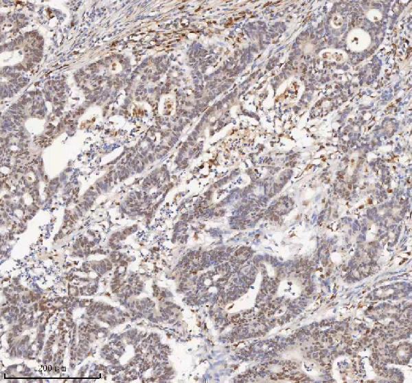
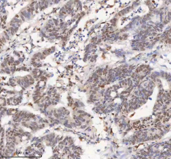
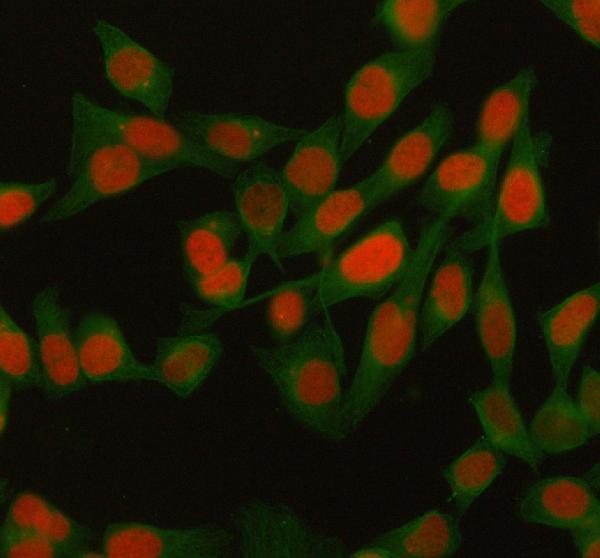
A04635-3 has IHC images from paraffin sections of human colon adenocarcinoma and liver cancer, plus an IF/ICC image from HELA cells (catalog image captions: A04635-3).

Real IHC data IHC analysis of H2AFY/MACROH2A1 using anti-H2AFY/MACROH2A1 antibody (A04635-3). H2AFY/MACROH2A1 was detected in a paraffin-embedded section of human colon adenocarcinoma tissue. Heat mediated antigen retrieval was performed in EDTA buffer (pH 8.0, epitope retrieval solution). The tissue section was blocked with 10% goat serum. The tissue section was then incubated with 2 μg/ml rabbit anti-H2AFY/MACROH2A1 Antibody (A04635-3) overnight at 4°C. Peroxidase Conjugated Goat Anti-rabbit IgG was used as secondary antibody and incubated for 30 minutes at 37°C. The tissue section was developed using HRP Conjugated Rabbit IgG Super Vision Assay Kit (Catalog # SV0002) with DAB as the chromogen.
Anti-H2AFY/MACROH2A1 Antibody ®
Cat # A04635-3

A04635-3 is listed for IHC and IF/ICC; its IHC images show human colon adenocarcinoma and liver cancer paraffin sections (catalog applications and IHC captions: A04635-3). Its IF/ICC image shows HELA cells (catalog IF caption: A04635-3).

Which to pick: Choose A04635-3 for paraffin-section IHC: its caption documents EDTA retrieval at pH 8.0 and 2 μg/ml primary antibody; the fixative is unreported (catalog IHC caption: A04635-3). Choose the same SKU for IF/ICC because its application list includes both and its IF image shows HELA cells (catalog applications and IF caption: A04635-3). For mouse or rat samples, A04635-3 lists species reactivity, but the supplied IHC and IF images show human samples only; it is a rabbit antibody with clonality unreported (catalog reactivity, host and image captions: A04635-3).

Each figure is that product's own IHC / IF validation image from its datasheet.

References

  1. UniProt Consortium. UniProt entry O75367 (H2AY_HUMAN, Core histone macro-H2A.1).
  2. Human Protein Atlas. MACROH2A1 tissue IHC expression (reliability: Supported).
  3. Human Protein Atlas. MACROH2A1 subcellular location (ICC-IF): Localized to the nucleoplasm..
  4. Human Protein Atlas. MACROH2A1 antibody validation summary (2 antibodies).
  5. MacroH2A1 Immunoexpression in Breast Cancer. Frontiers in oncology 2020 — PMC7471871.
  6. Loss of macroH2A1 decreases mitochondrial metabolism and reduces the aggressiveness of uveal melanoma cells. Aging 2020 — PMC7288915.
  7. The histone variant MacroH2A regulates Ca(2+) influx through TRPC3 and TRPC6 channels. Oncogenesis 2013 — PMC3816217.
  8. Loss of histone macroH2A1 in hepatocellular carcinoma cells promotes paracrine-mediated chemoresistance and CD4(+)CD25(+)FoxP3(+) regulatory T cells activation. Theranostics 2020 — PMC6929991.
  9. PubMed PMID:9714746 — UniProt-cited evidence.
  10. PubMed PMID:9653160 — UniProt-cited evidence.
  11. PubMed PMID:14702039 — UniProt-cited evidence.