This website uses cookies to ensure you get the best experience on our website.


- Table of Contents
Plan MAD2L2 IHC in paraffin sections using the catalog antibody at 2–5 μg/ml (datasheet A02357-4). Expect cytoplasmic staining in most tissue types, and compare signal within the same cell type when scoring (HPA tissue IHC).

Expected localisation, validated protocols, controls and antibodies — the at-a-glance facts below, then the full design guide.
| Expected localisation | Cytoplasmic staining in most tissue types (HPA tissue IHC) | |
| Staining pattern | Cytoplasmic staining across most tissue types (HPA tissue IHC) | |
| Antigen retrieval | EDTA pH 8.0 HIER, heat-mediated (datasheet A02357-4) | |
| Positive control | Bone marrow+4 more · see all | |
| Negative control | Breast+2 more · see all |
| Fixation | Keep fixation consistent across sections. (standard IHC practice; not target-specific) | |
| Caveat | Lung macrophages stain strongly; score cell types separately (HPA tissue IHC) | |
| Regulation | No stimulus-linked regulation specified (UniProt) | |
| Isoform / epitope | One 1–211 chain; no listed isoforms or processing (UniProt) |
Compare the catalog antibody’s IHC-P protocol (datasheet A02357-4) with two published MAD2L2 IHC workflows (PMC10956193; PMC6090455).
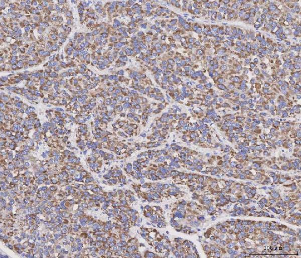
| Sample | Paraffin-embedded human liver cancer tissue; fixative not specified (datasheet A02357-4) |
| Fixation | Image fixative and duration unreported (datasheet A02357-4); verify before use. |
| Sectioning | 4–5 µm sections on charged slides (standard) |
| Deparaffinisation | Xylene, graded ethanol series to water (standard) |
| Antigen retrieval | Heat retrieval: EDTA pH 8.0 (datasheet A02357-4); 20 min, 95–100 °C (standard) |
| Peroxidase block | 3% H2O2, 10 min, room temperature (standard) |
| Blocking | 10% goat serum (datasheet A02357-4) |
| Primary antibody | Rabbit anti-MAD2L2, 2-5 μg/ml (datasheet A02357-4) |
| Primary incubation | Overnight at 4 °C (datasheet A02357-4) |
| Detection | HRP-conjugated secondary, DAB chromogen (datasheet A02357-4) |
| Counterstain | Hematoxylin, blue, dehydrate and mount (standard) |
| Expected result | MAD2L2-positive staining in hematopoietic cells of bone marrow (HPA tissue IHC: High). HPA tissue profile: Cytoplasmic expression in most tissue types. No signal in the no-primary control. |
MAD2L2 is found in the cytoplasm and nucleus, with no transmembrane segment (UniProt Q9UI95). In paraffin section IHC, expect mainly cytoplasmic staining across many tissues, especially bone marrow hematopoietic cells, lung macrophages and testis Leydig cells (HPA tissue IHC). HPA rates the tissue IHC evidence Approved, with medium consistency between staining and RNA expression; use its cell level observations as guides rather than universal cutoffs (HPA tissue IHC).
| Cytoplasmic signal in bone marrow hematopoietic cells, lung macrophages or testis Leydig cells. | This matches the High staining reported in each cell population and the mainly cytoplasmic tissue profile (HPA tissue IHC). Assess the relevant cells within each section; staining elsewhere in the tissue does not replace that comparison. |
| Predominantly membranous or extracellular staining, without the expected cytoplasmic pattern. | Treat this as a possible artefact: MAD2L2 has no transmembrane segment or signal peptide, and HPA describes cytoplasmic tissue IHC (UniProt Q9UI95; HPA tissue IHC). Nuclear signal alone needs separate assessment because nuclear localisation is also reported (UniProt Q9UI95). |
| Strong signal in a cell population reported as undetected, such as skeletal muscle myocytes. | Consider cross-reactivity or endogenous detection activity and check controls before scoring it as MAD2L2. The undetected call applies to the named cells in the sampled tissue; it does not establish that every cell there must be negative (HPA tissue IHC). |
| Diffuse chromogen across cells and surrounding tissue, obscuring cell boundaries. | The distribution cannot support a reliable cell level call. Check the no-primary control and detection background, then reassess whether distinct cytoplasmic signal remains in an HPA High population (general IHC practice; HPA tissue IHC). |
| No signal in bone marrow hematopoietic cells, lung macrophages or testis Leydig cells. | A blank result in a reported High population raises concern about the staining run or antibody performance (HPA tissue IHC). Verify tissue identity, section quality and a working positive control before interpreting other samples as negative (general IHC practice). |
| Tissue and cell selection | Expression is ubiquitous by UniProt, but HPA reports different IHC levels by cell population: High in three listed positive populations and undetected in breast adipocytes, hippocampal glia and skeletal muscle myocytes (UniProt Q9UI95; HPA tissue IHC). Choose controls by named cell type, not organ name alone. |
| IF/ICC Q: Should nuclear staining be expected? | Yes. HPA reports supported nucleoplasmic localisation in ICC-IF, while its tissue IHC summary is cytoplasmic; UniProt also lists nucleus and cytoplasm (HPA subcellular; HPA tissue IHC; UniProt Q9UI95). Interpret each assay against its own observed pattern. |
| Antibody validation | CAB008110 is Approved for IHC, while HPA003882 is Supported for ICC-IF; those labels apply to different antibodies and applications (HPA antibodies). HPA's tissue IHC reliability is Approved with medium staining versus RNA consistency, so an unexpected slide still needs controls (HPA tissue IHC). |
| Topology and processing | The annotated protein is a single 1–211 chain with no signal peptide, propeptide, transmembrane segment or glycosylation sites (UniProt Q9UI95). These annotations support scrutiny of membrane or extracellular staining; they do not establish epitope access or fixation sensitivity. |
| Situation | Likely cause | Next action |
|---|---|---|
| The reported High cells are blank. | A failed staining run, unsuitable section or ineffective antigen retrieval is possible; no MAD2L2-specific retrieval requirement is supplied (HPA tissue IHC; general IHC practice). | Check tissue identity and a run positive control, then review the antibody's validated IHC conditions and the run's retrieval and detection steps (general IHC practice). |
| Macrophages or hematopoietic cells stain heavily in an HRP chromogen run. | These are HPA High populations, but endogenous peroxidase can also contribute chromogen in some cells (HPA tissue IHC; general IHC practice). | Compare a no-primary control and confirm the peroxidase blocking step before attributing all signal to MAD2L2 (general IHC practice). |
| A skeletal muscle myocyte appears strongly positive. | HPA reports myocytes as undetected; cross-reactivity, background or cell misidentification could explain the mismatch (HPA tissue IHC). | Confirm the cell type and staining compartment, inspect controls, and repeat with an independently validated IHC antibody if the finding matters (general IHC practice). |
| The section shows mainly membrane or extracellular deposits. | That pattern conflicts with the HPA cytoplasmic IHC profile and UniProt's lack of a transmembrane segment or signal peptide (HPA tissue IHC; UniProt Q9UI95). | Inspect the no-primary control and chromogen precipitate, then score only cell associated staining that remains credible after background review (general IHC practice). |
| Staining is faint in liver hepatocytes or bronchial respiratory epithelial cells. | HPA reports Low staining in both populations, so weak signal there alone does not establish a failed assay (HPA tissue IHC). | Compare with a named High population in the same run before changing the assay or calling the sample negative (HPA tissue IHC; general IHC practice). |
| IHC looks cytoplasmic, but an ICC-IF image looks nuclear. | The observations come from different assays: HPA tissue IHC is chiefly cytoplasmic, and HPA ICC-IF supports nucleoplasmic localisation (HPA tissue IHC; HPA subcellular). | Check each result against its assay specific evidence and antibody validation; do not use the ICC-IF localisation alone to reject a controlled cytoplasmic IHC result (HPA antibodies; HPA tissue IHC; HPA subcellular). |
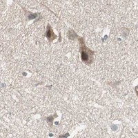


Comprehensive Human Protein Atlas IHC scoring per tissue (reliability: Approved — Medium consistency between antibody staining and RNA expression data.). Rows are taken directly from the HPA tissue chart — click any row's HPA link to view the source.
| Tissue | Cell type | Level | Evidence | Source |
|---|---|---|---|---|
| Bone marrow | Hematopoietic cells | High | Protein (IHC) | HPA → |
| Lung | Macrophages | High | Protein (IHC) | HPA → |
| Testis | Leydig cells | High | Protein (IHC) | HPA → |
| Adipose tissue | Adipocytes | Medium | Protein (IHC) | HPA → |
| Adrenal gland | Glandular cells | Medium | Protein (IHC) | HPA → |
Troubleshoot MAD2L2 staining in paraffin sections by checking retrieval, compartment, cell identity and controls before comparing chromogenic signal across samples.
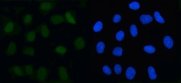
A02357-4 has IHC data from human liver cancer paraffin sections and IF data from U2OS cells (A02357-4 image captions). M02357 lists human, mouse and rat reactivity (M02357 catalog).
A02357-4 will render with its IHC figure from human liver cancer paraffin tissue; its separate IF caption shows U2OS cells (A02357-4 image captions). M02357 will render with listed IHC and IF/ICC applications and human, mouse and rat reactivity, but no IHC or IF figure is supplied (M02357 catalog).
Which to pick: For tissue IHC, pick A02357-4: its own caption shows staining in a human liver cancer paraffin section at 2 μg/ml after EDTA retrieval at pH 8.0; the fixative is unreported (A02357-4 IHC image caption). For IF/ICC, pick A02357-4 because its IF caption shows U2OS cells stained at 5 μg/ml (A02357-4 IF image caption). For mouse or rat work, consider M02357, a rabbit monoclonal listed for IHC and IF/ICC in human, mouse and rat, with no image evidence supplied here (M02357 catalog).