This website uses cookies to ensure you get the best experience on our website.


- Table of Contents
Plan chromogenic IHC for MAGED4 in paraffin sections with the catalog antibody at 2–5 μg/ml (datasheet A16674-1). Evaluate cytoplasmic staining in selected tissues while accounting for reported off-target binding (HPA tissue IHC).

Expected localisation, validated protocols, controls and antibodies — the at-a-glance facts below, then the full design guide.
| Expected localisation | Cytoplasmic in selected tissues (HPA tissue IHC) | |
| Staining pattern | Cytoplasmic staining in selected glandular and stromal cells (HPA tissue IHC) | |
| Antigen retrieval | EDTA pH 8.0 HIER, heat-mediated (datasheet A16674-1) | |
| Positive control | Adrenal gland+3 more · see all | |
| Negative control | Adipose tissue+4 more · see all |
| Fixation | Keep fixation consistent across sections. (standard IHC practice; not target-specific) | |
| Caveat | Off-target staining reported; antibody targets >1 gene (HPA tissue IHC) | |
| Regulation | Isoforms 1–2 occur in glioma cells (UniProt) | |
| Isoform / epitope | 4 isoforms; check epitope coverage across variants (UniProt) |
Compare the catalog antibody’s IHC-P protocol using EDTA pH 8.0 retrieval (datasheet A16674-1) with the published oral squamous cell carcinoma protocol (PMC12732915).
| Sample | Paraffin-embedded human adrenocortical adenoma tissue; fixative not specified (datasheet A16674-1) |
| Fixation | Image fixative and duration unreported (datasheet A16674-1); verify before use. |
| Sectioning | 4–5 µm sections on charged slides (standard) |
| Deparaffinisation | Xylene, graded ethanol series to water (standard) |
| Antigen retrieval | Heat retrieval: EDTA pH 8.0 (datasheet A16674-1); 20 min, 95–100 °C (standard) |
| Peroxidase block | 3% H2O2, 10 min, room temperature (standard) |
| Blocking | 10% goat serum (datasheet A16674-1) |
| Primary antibody | Rabbit anti-MAGED4, 2-5 μg/ml (datasheet A16674-1) |
| Primary incubation | Overnight at 4 °C (datasheet A16674-1) |
| Detection | HRP-conjugated secondary, DAB chromogen (datasheet A16674-1) |
| Counterstain | Hematoxylin, blue, dehydrate and mount (standard) |
| Expected result | MAGED4-positive staining in glandular cells of adrenal gland (HPA tissue IHC: Medium). HPA tissue profile: Cytoplasmic expression in selected tissues. No signal in the no-primary control. |
In paraffin-section IHC, expect cytoplasmic MAGED4 staining in selected cells: adrenal glandular cells, breast adipocytes, endometrial stromal cells and placental trophoblastic cells show medium staining (HPA tissue IHC). HPA rates the tissue pattern Approved, with medium consistency against RNA data and a warning about presumed off-target binding (HPA tissue IHC). UniProt assigns no subcellular location and reports no transmembrane segment (UniProt Q96JG8).
| Distinct cytoplasmic stain in the listed cell populations, with limited surrounding stain. | This matches HPA's selected-tissue cytoplasmic pattern. The reported level is Medium in each listed positive population; compare cells within the same section before judging intensity (HPA tissue IHC). |
| Predominantly nuclear or sharply membrane-restricted stain in an otherwise interpretable IHC section. | Neither is the reported tissue-IHC pattern (HPA tissue IHC). Check morphology, counterstain and detection controls before calling it MAGED4. HPA's separate ICC-IF summary says Membrane, but supplies no cell-line images or ICC validation for HPA003554 (HPA subcellular; HPA antibodies). |
| Strong stain in colon or appendix glandular cells, bronchial respiratory epithelium, or bone-marrow hematopoietic cells. | These populations were Not detected in the HPA tissue set (HPA tissue IHC). Investigate antibody cross-reactivity or endogenous chromogenic activity; unexpected staining alone cannot establish expression, especially given HPA's off-target warning (HPA tissue IHC). |
| Uniform color across tissue, stroma and empty areas, obscuring cell boundaries. | This is background rather than a readable cell-specific pattern (general IHC practice). Review blocking, washes, primary-antibody concentration and chromogen exposure; use a no-primary control to distinguish detection background from primary-dependent staining (general IHC practice). |
| No stain in a section selected as a positive control. | First confirm that the section contains the HPA-positive cell population, since positivity is cell-specific (HPA tissue IHC). If it does, inspect tissue preservation and the staining run with independent controls before interpreting a test section as negative (general IHC practice). |
| Tissue and cell context | HPA reports Medium stain in breast adipocytes but Not detected in adipose-tissue adipocytes; identify the sampled tissue before using adipocytes as controls (HPA tissue IHC). |
| Evidence strength and specificity | IHC is Approved, with medium RNA agreement; HPA also cautions that the target spans more than one gene and presumed off-target binding was disregarded in its profile (HPA tissue IHC). |
| IHC versus IF/ICC: what should be expected? | Use the cytoplasmic tissue-IHC pattern for this assay. HPA's ICC-IF summary says Membrane, yet lists no cell-line images; HPA003554 has IHC Approved and no ICC status (HPA tissue IHC; HPA subcellular; HPA antibodies). |
| Isoforms and protein architecture | UniProt lists four isoforms, one chain spanning residues 1–741, and no signal peptide, propeptide or transmembrane segment. These facts do not identify the antibody epitope or prove equal detection of all isoforms (UniProt Q96JG8). |
| Antigen retrieval | Retrieval is a routine paraffin-IHC variable, but target-specific fixation sensitivity and an optimal retrieval condition are unreported here; assess retrieval changes against tissue and detection controls (general IHC practice). |
| Situation | Likely cause | Next action |
|---|---|---|
| Positive-control slide is blank. | The chosen area may lack the reported positive cell type, or the staining run may have failed (HPA tissue IHC; general IHC practice). | Confirm the cell population in the section; then review a working assay control and detection reagents before changing target interpretation (general IHC practice). |
| Signal appears mainly nuclear. | This conflicts with HPA's cytoplasmic tissue-IHC profile; nonspecific binding or difficult-to-read counterstain is possible (HPA tissue IHC; general IHC practice). | Check morphology and a no-primary control; repeat with adjusted detection conditions if background obscures the cytoplasm (general IHC practice). |
| Unexpected cell types stain strongly. | Cross-reactivity is plausible because HPA notes presumed off-target binding and a target spanning more than one gene (HPA tissue IHC). | Compare the exact cell type with HPA positives and negatives, then seek independent antibody or orthogonal support before assigning MAGED4 expression (HPA tissue IHC; general IHC practice). |
| Color is widespread or follows tissue edges. | Background or excessive chromogen development can obscure cell-restricted staining (general IHC practice). | Inspect no-primary and detection controls; refine blocking, washes, primary concentration or development time one variable at a time (general IHC practice). |
| Brain-region section is negative despite expected brain expression. | RNA or broad tissue statements do not ensure detectable protein in every cell: HPA records Not detected in caudate glia and cerebellar granular-layer cells (HPA tissue IHC; UniProt Q96JG8). | Score the sampled cell population against its own HPA entry; use an HPA Medium-positive population to check the run (HPA tissue IHC). |
| An IF/ICC result looks membrane-localized. | HPA labels its ICC-IF summary Membrane, but supplies no cell-line images and no ICC validation for HPA003554 (HPA subcellular; HPA antibodies). | Interpret that result with the separate IF/ICC guide and appropriate controls; do not substitute its localization for the tissue-IHC scoring pattern (HPA tissue IHC; general IF practice). |


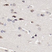
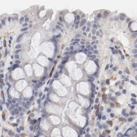
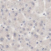
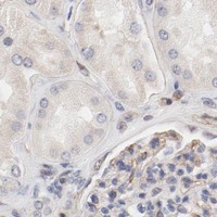
Comprehensive Human Protein Atlas IHC scoring per tissue (reliability: Approved — Medium consistency between antibody staining and RNA expression data. Caution, targets protein from more than one gene. Presumed off target binding observed and disregarded.). Rows are taken directly from the HPA tissue chart — click any row's HPA link to view the source.
| Tissue | Cell type | Level | Evidence | Source |
|---|---|---|---|---|
| Adrenal gland | Glandular cells | Medium | Protein (IHC) | HPA → |
| Breast | Adipocytes | Medium | Protein (IHC) | HPA → |
| Endometrium | Cells in endometrial stroma | Medium | Protein (IHC) | HPA → |
| Placenta | Trophoblastic cells | Medium | Protein (IHC) | HPA → |
| Tissue | Cell type | Level | Evidence | Source |
|---|---|---|---|---|
| Adipose tissue | Adipocytes | Not detected | Protein (IHC) | HPA → |
| Appendix | Glandular cells | Not detected | Protein (IHC) | HPA → |
| Bone marrow | Hematopoietic cells | Not detected | Protein (IHC) | HPA → |
| Bronchus | Respiratory epithelial cells | Not detected | Protein (IHC) | HPA → |
| Caudate | Glial cells | Not detected | Protein (IHC) | HPA → |
Troubleshoot MAGED4 staining in paraffin sections by checking retrieval, cellular distribution, antibody specificity, and scoring against matched controls.
A16674-1 has human paraffin-section IHC data in adrenocortical adenoma and bladder urothelial carcinoma, plus IF/ICC data in A549 cells (catalog image captions).
A16674-1 is listed for human IHC and shown on paraffin sections of adrenocortical adenoma and bladder urothelial carcinoma (catalog applications, reactivity and IHC image captions). The same SKU is listed for IF/ICC and shown in A549 cells (catalog applications and IF image caption).
Which to pick: For tissue IHC, choose A16674-1: its rabbit antibody is shown on paraffin sections after EDTA retrieval at pH 8.0, using 2 μg/ml primary antibody (catalog host and IHC image captions). For IF/ICC, choose A16674-1: it is listed for both applications and shown in A549 cells at 5 μg/ml (catalog applications and IF image caption). No cross-species option is documented, clonality is unreported, and the paraffin-section captions do not report a fixative.