This website uses cookies to ensure you get the best experience on our website.


- Table of Contents
Plan MAML3 chromogenic IHC in paraffin sections using the catalog antibody’s documented conditions. Compare cytoplasmic and nuclear staining with HPA tissue observations, while treating nuclear speckles as a molecular expectation from UniProt.

Expected localisation, validated protocols, controls and antibodies — the at-a-glance facts below, then the full design guide.
| Expected localisation | Cytoplasmic and nuclear staining observed (HPA tissue IHC) | |
| Staining pattern | High in adipocytes; overall cytoplasmic/nuclear staining (HPA tissue IHC) | |
| Antigen retrieval | EDTA pH 8.0 HIER, heat-mediated (datasheet A07975-1) | |
| Positive control | Adipose tissue+4 more · see all | |
| Negative control | None in HPA (detected in all 44 tissues); use no-primary + isotype controls |
| Fixation | Keep fixation conditions consistent across samples. (standard IHC practice; not target-specific) | |
| Caveat | Staining has medium consistency with RNA expression (HPA tissue IHC) | |
| Regulation | Expression regulation not annotated (UniProt) | |
| Isoform / epitope | No isoforms or processing variants annotated (UniProt) |
The catalog antibody protocol and two published MAML3 IHC protocols provide starting conditions for paraffin sections (datasheet A07975-1; PMC12398435; PMC8419063).
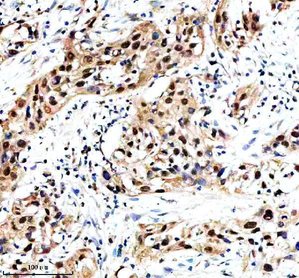
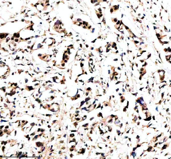
| Sample | Paraffin-embedded human bladder cancer tissue; fixative not specified (datasheet A07975-1) |
| Fixation | Image fixative and duration unreported (datasheet A07975-1); verify before use. |
| Sectioning | 4–5 µm sections on charged slides (standard) |
| Deparaffinisation | Xylene, graded ethanol series to water (standard) |
| Antigen retrieval | Heat retrieval: EDTA pH 8.0 (datasheet A07975-1); 20 min, 95–100 °C (standard) |
| Peroxidase block | 3% H2O2, 10 min, room temperature (standard) |
| Blocking | 10% goat serum (datasheet A07975-1) |
| Primary antibody | Rabbit anti-MAML3, 2-5 μg/ml (datasheet A07975-1) |
| Primary incubation | Overnight at 4 °C (datasheet A07975-1) |
| Detection | HRP-conjugated secondary, DAB chromogen (datasheet A07975-1) |
| Counterstain | Hematoxylin, blue, dehydrate and mount (standard) |
| Expected result | MAML3-positive staining in adipocytes of adipose tissue (HPA tissue IHC: High). HPA tissue profile: General cytoplasmic and nuclear expression. No signal in the no-primary control. |
MAML3 is a nuclear transcriptional coactivator with punctate localization in nuclear speckles (UniProt Q96JK9; HPA: nuclear speckles supported). In paraffin section IHC, look for staining in the cell populations HPA reports as high, including adipocytes, hematopoietic cells, endothelial cells, and selected glandular and squamous epithelial cells (HPA: tissue IHC). HPA also describes general cytoplasmic and nuclear expression; its IHC reliability is Approved, with medium consistency against RNA data (HPA: tissue IHC). MAML3 has no transmembrane segment (UniProt Q96JK9 topology).
| Punctate nuclear staining in an HPA high cell population. | This fits MAML3 localization to nuclear speckles (UniProt Q96JK9; HPA: nuclear speckles supported). High staining is reported in adipocytes in adipose tissue and breast, hematopoietic cells in bone marrow, and endothelial cells in colon (HPA: tissue IHC). Judge the identified cell population and compartment together; a dark region alone does not establish a positive result (General IHC interpretation). |
| Predominant membrane or extracellular staining, without a convincing nuclear component. | That pattern is inconsistent with the annotated nuclear localization and absence of a transmembrane segment (UniProt Q96JK9). Treat it as suspect and compare an adjacent section stained without primary antibody (General IHC practice). Cytoplasmic staining alone needs care: HPA describes general cytoplasmic and nuclear expression, so cytoplasm is not automatically an artefact (HPA: tissue IHC). |
| Strong staining in a cell population reported as low by HPA. | Compare cell identity and staining distribution before calling it MAML3. HPA reports low staining in caudate glial cells, parathyroid glandular cells, soft tissue fibroblasts, and spleen red pulp cells (HPA: tissue IHC). An unexpected strong result warrants checks for antibody cross-reactivity or endogenous detection activity (General IHC practice); low does not mean absent. |
| Diffuse color across tissue, including spaces or many unrelated structures. | A widespread deposit that obscures cell borders and nuclei is difficult to score as specific IHC staining (General IHC interpretation). Compare the no-primary control and inspect blocking, washing, and detection steps for background (General IHC practice). Do not use diffuse color as evidence for MAML3 nuclear speckles, which are the supported subcellular location (HPA: subcellular ICC-IF). |
| No discernible signal in a section containing HPA high cells. | First confirm that the expected cells are present: duodenal and gallbladder glandular cells and esophageal and oral mucosal squamous cells are reported high (HPA: tissue IHC). A blank result may reflect an assay failure or sample variation (General IHC interpretation). HPA rates the IHC pattern Approved but notes medium consistency with RNA, so one negative section does not settle target absence (HPA: tissue IHC). |
| Cell population and tissue context | HPA reports high IHC staining in specific cells within adipose tissue, bone marrow, breast, colon, duodenum, esophagus, gallbladder, and oral mucosa, while RNA has low tissue specificity (HPA: tissue IHC). Select a control by its reported cell population, then verify that those cells are present in the section (General IHC practice). The supplied HPA list contains no designated negative tissue (HPA: tissue IHC). |
| Subcellular evidence and antibody validation | UniProt places MAML3 in the nucleus in a punctate manner, and HPA supports nuclear speckles by ICC-IF (UniProt Q96JK9; HPA: subcellular ICC-IF). HPA calls antibody HPA037717 IHC Approved and ICC Supported; the supplied record does not assign IHC Enhanced status (HPA: antibody validation). Interpret mixed cytoplasmic and nuclear tissue staining within that stated evidence limit (HPA: tissue IHC). |
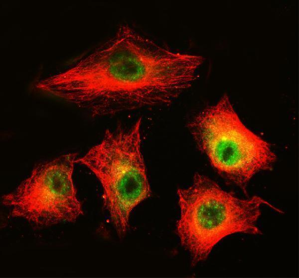
| IF/ICC Q: What should a positive cell look like? | A: Expect nuclear speckles, supported by HPA ICC-IF and consistent with UniProt's punctate nuclear annotation (HPA: subcellular ICC-IF; UniProt Q96JK9). HPA lists ICC-IF images from A-431, U-251MG, and U2OS and marks HPA037717 ICC Supported (HPA: subcellular ICC-IF; HPA: antibody validation). This is a localization answer, not an IHC protocol recommendation. |
| Situation | Likely cause | Next action |
|---|---|---|
| Nuclear signal is weak in an HPA high cell population. | The expected cells may be sparse, or IHC detection may be underperforming (General IHC interpretation). HPA high denotes an observed staining level in named cells, not guaranteed intensity in every section (HPA: tissue IHC). | Confirm cell identity and section quality; then review the catalog antibody's IHC-P instructions for retrieval and detection and check a concurrently processed control (General IHC practice). Do not infer MAML3 specific fixation sensitivity from these records. |
| Predominant membrane staining appears. | A membrane dominant pattern conflicts with nuclear speckles and no transmembrane segment (HPA: subcellular ICC-IF; UniProt Q96JK9 topology). | Compare the no-primary control and inspect the chromogenic deposit for nonspecific edge or tissue staining (General IHC practice). Report membrane only staining as unconfirmed until a nuclear pattern can be reproduced (General IHC interpretation). |
| Strong color appears in HPA low cells. | Cross-reactivity or endogenous detection activity may contribute (General IHC practice). HPA reports low staining in caudate glial cells, parathyroid glandular cells, soft tissue fibroblasts, and spleen red pulp cells (HPA: tissue IHC). | Verify cell identity, run a no-primary control, and assess the detection system's endogenous activity controls (General IHC practice). Treat HPA low as a comparison point, not proof that any staining is false. |
| Cytoplasmic color dominates a candidate positive cell. | HPA describes general cytoplasmic and nuclear tissue expression, whereas its supported ICC-IF location is nuclear speckles (HPA: tissue IHC; HPA: subcellular ICC-IF). | Score nuclear and cytoplasmic compartments separately and check whether identifiable positive cells retain nuclear staining (General IHC interpretation). Avoid calling cytoplasm alone definitive without additional validation. |
| Color is diffuse across the section. | Broad background may arise from blocking, washing, or detection conditions (General IHC practice); it cannot resolve the supported speckled nuclear location (HPA: subcellular ICC-IF). | Compare no-primary and tissue controls, then review blocking, wash steps, and detection timing under the chosen IHC-P workflow (General IHC practice). Reassess cell specific staining only after background is controlled. |
| A control section shows no staining anywhere. | The control may lack the named HPA high cells, or an assay step may have failed (HPA: tissue IHC; General IHC practice). HPA supplies no designated negative tissue in this record (HPA: tissue IHC). | Check morphology for the expected cell population, confirm that the antibody and detection steps ran, and repeat with a documented HPA high cell population (General IHC practice; HPA: tissue IHC). Record a blank result as inconclusive until the control works. |
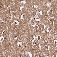


Comprehensive Human Protein Atlas IHC scoring per tissue (reliability: Approved — Medium consistency between antibody staining and RNA expression data.). Rows are taken directly from the HPA tissue chart — click any row's HPA link to view the source.
| Tissue | Cell type | Level | Evidence | Source |
|---|---|---|---|---|
| Adipose tissue | Adipocytes | High | Protein (IHC) | HPA → |
| Bone marrow | Hematopoietic cells | High | Protein (IHC) | HPA → |
| Breast | Adipocytes | High | Protein (IHC) | HPA → |
| Colon | Endothelial cells | High | Protein (IHC) | HPA → |
| Duodenum | Glandular cells | High | Protein (IHC) | HPA → |
| Tissue | Cell type | Level | Evidence | Source |
|---|---|---|---|---|
| None in HPA: MAML3 is detected in all 44 scored tissues. Use a no-primary (secondary-only) and an isotype control instead. | ||||
Troubleshoot chromogenic MAML3 IHC in paraffin sections using the catalog antibody’s tissue example and the reported nuclear speckle localisation (datasheet A07975-1; UniProt Q96JK9).
The catalog antibody has IHC images from human paraffin sections of bladder and breast cancer and IF images from those tissues and A549 cells (catalog image captions).
A07975-1 is listed for human IHC and illustrated on paraffin sections of bladder and breast cancer (catalog: applications and reactivity; A07975-1 IHC captions). The same SKU is listed for IF/ICC, with images from A549 cells and paraffin sections of bladder and breast cancer (catalog: applications; A07975-1 IF captions).
Which to pick: Choose A07975-1 for human paraffin-section IHC; its own captions show staining in bladder and breast cancer tissue at 2 μg/ml after EDTA retrieval (A07975-1 IHC captions). For IF/ICC, the same rabbit antibody has images from A549 cells and human paraffin sections at 5 μg/ml (catalog: host and applications; A07975-1 IF captions). No cross-species option is listed because A07975-1 is specified for human reactivity only; its paraffin-section IHC captions do not report the fixative (catalog: reactivity; A07975-1 IHC captions).