MAP1B / Microtubule-associated protein 1B · IHC design guide

Design Immunohistochemistry for MAP1B

Plan chromogenic MAP1B IHC in paraffin sections using the reported cytoplasmic staining in CNS, peripheral nerve, glomeruli and pancreatic islets (HPA tissue IHC). Compare cell types within each tissue, since neuronal staining can be high while glial staining is low or undetected (HPA tissue IHC).

Evidence assembled Oct 2026 · For research use; verify linked source records and product datasheet before use
Immunohistochemistry protocol sheet for MAP1B (IHC for MAP1B): expected localisation Cytoplasmic in CNS, peripheral nerve and glomeruli (HPA tissue IHC), antibody A01760-1, validated IHC image, and IHC protocol steps
Printable MAP1B IHC protocol sheet — expected localisation Cytoplasmic in CNS, peripheral nerve and glomeruli (HPA tissue IHC), antibody A01760-1, controls and protocol steps. Open the full MAP1B IHC guide →

MAP1B Immunohistochemistry Experimental Design Guide

Expected localisation, validated protocols, controls and antibodies — the at-a-glance facts below, then the full design guide.

Must know before staining
Expected localisation Cytoplasmic in CNS, peripheral nerve and glomeruli (HPA tissue IHC)
Staining pattern Cytoplasmic CNS, peripheral nerve, glomerular and islet cells (HPA tissue IHC)
Antigen retrieval EDTA pH 8.0 HIER, heat-mediated (datasheet A01760-1)
Positive control ⓘ Cerebellum+4 more · see all
Negative control ⓘ Adipose tissue+4 more · see all
Important caveats
Reasons your staining may differ from the expected pattern.
Fixation Keep fixation consistent across sections. (standard IHC practice; not target-specific)
Caveat Glial staining can be low or absent; score cell types separately (HPA tissue IHC)
Regulation Expression regulation is not annotated (UniProt)
Isoform / epitope No annotated isoforms; cleavage separates heavy-chain and LC1 epitopes (UniProt)
Section 1

Recommended MAP1B IHC & IF Protocols

The catalog antibody’s IHC-P protocol is accompanied by published MAP1B IHC methods for breast, urothelial and lung tumors (PMC10866687; PMC7139768; PMC12885910).

Recommended immunohistochemistry (IHC-P) protocol parameters
SampleParaffin-embedded mouse brain tissue; fixative not specified (datasheet A01760-1)
FixationImage fixative and duration unreported (datasheet A01760-1); verify before use.
Sectioning4–5 µm sections on charged slides (standard)
DeparaffinisationXylene, graded ethanol series to water (standard)
Antigen retrievalHeat retrieval: EDTA pH 8.0 (datasheet A01760-1); 20 min, 95–100 °C (standard)
Peroxidase block3% H2O2, 10 min, room temperature (standard)
Blocking10% goat serum (datasheet A01760-1)
Primary antibodyRabbit anti-MAP1B, 2-5 μg/ml (datasheet A01760-1)
Primary incubationOvernight at 4 °C (datasheet A01760-1)
DetectionHRP-conjugated secondary, DAB chromogen (datasheet A01760-1)
CounterstainHematoxylin, blue, dehydrate and mount (standard)
Expected resultMAP1B-positive staining in gLUC cells - cytoplasm/membrane of cerebellum (HPA tissue IHC: High). HPA tissue profile: Cytoplasmic expression in the CNS, peripheral nerve, renal glomeruli, pancreatic islets and seminiferous duct in testis. No signal in the no-primary control.
💡Decision noteStart with heat-mediated EDTA pH 8.0 retrieval for the catalog antibody (datasheet A01760-1); the lung tumor study used citrate buffer at 95 °C for 5 minutes (PMC12885910).
Section 2

What Is the Expected MAP1B Staining Pattern?

MAP1B is a cytoplasmic cytoskeletal protein associated with microtubules, synapses and dendritic spines; it has no transmembrane segment (UniProt P46821). In paraffin section IHC, expect prominent staining in cerebral cortical neurons and cerebellar GLUC cells, with additional staining in selected peripheral and non-neural cells (HPA: tissue IHC, Enhanced reliability). Read the signal against cell identity and compartment, since the HPA pattern varies by tissue and cell type.

What am I looking at on my slide?
Strong cytoplasmic staining in cerebral cortical neurons or cerebellar GLUC cells, with tissue structure preserved.This fits HPA's High IHC staining in those cells and MAP1B's cytoplasmic cytoskeletal location (HPA: tissue IHC; UniProt P46821). Cerebellar GLUC cells are annotated as cytoplasm/membrane; that observation does not imply MAP1B is a transmembrane protein (HPA: cerebellum; UniProt P46821 topology).
Predominantly nuclear staining, or a uniform sharp membrane rim across many cells.Treat this as a compartment mismatch requiring control review: UniProt places MAP1B in the cytoplasm and cytoskeleton and reports no transmembrane segment (UniProt P46821). HPA's cytoplasm/membrane label for cerebellar GLUC cells should not be generalized to every tissue (HPA: cerebellum).
Strong staining in adipocytes or bone marrow hematopoietic cells.These cell populations are listed as Not detected, so reproducible staining warrants a check for cross-reactivity or endogenous detection activity (HPA: adipose tissue; HPA: bone marrow; standard IHC practice). A tissue-level judgment must follow the identified cells: HPA also reports staining in other cell types and tissues (HPA: tissue IHC).
Diffuse chromogen over cells and surrounding tissue, with little distinction between compartments.The pattern cannot establish MAP1B localization. Compare a no-primary control and inspect blocking, washes and detection background; these are general IHC checks, not reported MAP1B-specific effects (standard IHC practice). Score only a distinguishable cellular pattern against the HPA reference (HPA: tissue IHC).
No discernible signal in cerebral cortical neurons despite an interpretable section.Cortical neuronal staining is High in the HPA reference, making this a useful positive-control failure to investigate (HPA: cerebral cortex). Confirm tissue identity and control performance, then review the antibody and staining workflow; absence in one section alone does not establish absent MAP1B (standard IHC practice).
💡Expected MAP1B appearanceA convincing positive is prominent cytoplasmic staining in cerebral cortical neurons or cerebellar GLUC cells, reported as High by HPA; dominant nuclear staining or strong signal in HPA Not detected cell populations calls for control review (HPA: tissue IHC; UniProt P46821).
How each factor affects the staining
Compartment and topologyMAP1B is cytoplasmic and cytoskeletal, with synaptic and dendritic-spine annotations and no transmembrane segment (UniProt P46821). Interpret chromogen within the cell's anatomy; do not require a continuous surface-membrane outline (UniProt P46821; standard IHC practice).
Tissue and cell selectionHPA reports High staining in cortical neurons and cerebellar GLUC cells, Medium staining in peripheral nerve, glomerular cells and pancreatic endocrine cells, and Not detected staining in adipocytes (HPA: tissue IHC). These are cell-specific observations, not whole-tissue guarantees.
Antibody evidenceTwo listed antibodies, HPA022275 and CAB009792, each have Enhanced IHC validation (HPA: antibody validation). HPA describes Enhanced tissue reliability as high consistency between antibody staining and RNA expression data (HPA: tissue IHC). Validation supports pattern comparison but does not resolve every unexpected signal.
Processing and modified residuesUniProt annotates MAP1B heavy chain at residues 2–2206, light chain LC1 at 2207–2468, and multiple modified residues including phosphosites (UniProt P46821). The payload gives no antibody epitope or target-specific fixation sensitivity; therefore these annotations cannot predict which stained cells or compartments a given reagent will detect.
IF/ICC Q&A: where should signal localize?HPA reports mainly cytosolic MAP1B in ICC-IF, with an additional approved equatorial-segment localization; its main cytosol location is supported (HPA: subcellular ICC-IF). Use that as an IF localization reference, while interpreting paraffin-section chromogen against the tissue IHC observations (HPA: tissue IHC).
Why is my staining missing, weak or wrong?
SituationLikely causeNext action
The known-positive cortical neuronal control is blank.The cause is unresolved; the expected neuronal staining is High in HPA (HPA: cerebral cortex).Check tissue identity, section integrity, reagent performance and detection controls before interpreting the study samples (standard IHC practice).
Cerebellar signal appears only as a crisp outline around every cell.A universal surface-rim pattern is not established by MAP1B's cytoskeletal annotation or HPA's GLUC cytoplasm/membrane label (UniProt P46821; HPA: cerebellum).Compare cell morphology and a no-primary control; reassess the signal's intracellular distribution (standard IHC practice).
Adipocytes or bone marrow hematopoietic cells stain strongly.Those specific cells are Not detected in HPA; cross-reactivity or endogenous detection activity is possible, not proven (HPA: adipose tissue; HPA: bone marrow; standard IHC practice).Compare the positive tissue and no-primary control; investigate reagent specificity and detection background before assigning MAP1B positivity (standard IHC practice).
Chromogen is widespread and obscures cell boundaries.Background staining prevents a reliable compartment call; the payload does not identify a MAP1B-specific cause (standard IHC practice).Review blocking, washes and the detection control, then judge whether cytoplasmic staining remains distinguishable (standard IHC practice; UniProt P46821).
Glomerular or pancreatic endocrine staining looks weaker than cortical neuronal staining.HPA grades glomerular cells and pancreatic endocrine cells Medium, while cortical neurons are High (HPA: kidney; HPA: pancreas; HPA: cerebral cortex).Evaluate each named cell population against its own HPA reference; avoid applying the cortical intensity threshold to every tissue (HPA: tissue IHC).
A run shows staining, but its cell type or compartment varies between sections.The observation alone cannot separate biological distribution from staining variability; HPA reports distinct cell-specific patterns (HPA: tissue IHC).Compare matched positive and no-primary controls, confirm the cell types, and repeat interpretation with consistent scoring criteria (standard IHC practice).

Sample controls for MAP1B IHC & IF

🧪Run cerebellum first and score cytoplasmic staining in GLUC cells (HPA: High in cerebellar GLUC cells). Use adipose tissue as the negative comparator, scoring adipocytes (HPA: Not detected in adipocytes); other cells on the cerebellum slide should lack comparable staining, but their MAP1B-negative status is unverified by the supplied HPA row (HPA: High in cerebellar GLUC cells).
Positive control tissue: Cerebellum (GLUC cells - cytoplasm/membrane, HPA High)
Negative control tissue: Adipose tissue (HPA Not detected)
ICC-IF cell lines (HPA subcellular resource): HPA ICC-IF images show MAP1B in U-251MG, U2OS, A-549, ASC52telo, Sperm, with annotated localisation: Cytosol (supported) (HPA subcellular).
Technical controls: Include a no-primary, secondary-only control and a host- and immunoglobulin-matched rabbit IgG isotype control (selected-SKU tissue-IHC caption: rabbit primary and anti-rabbit secondary; standard IHC practice). Use MAP1B knockout material, if available, or competition with the cognate immunizing peptide as a biological specificity control; quench endogenous peroxidase before HRP/DAB detection in brain sections (standard IHC practice; selected-SKU tissue-IHC caption: HRP/DAB detection).
⚠️Feasibility: A target-specific fixation window or fixation effect is unreported, and the selected A01760-1 paraffin mouse-brain caption does not state a fixative (selected-SKU tissue-IHC caption). The caption uses heat-mediated EDTA retrieval at pH 8.0, which is a starting condition to validate for cerebellum rather than an established requirement for every specimen; the supplied evidence does not establish whether frozen sections or IF are easier (selected-SKU tissue-IHC caption). Neural-tissue lipofuscin can add autofluorescence if the experiment uses IF (standard IF practice).

HPA tissue IHC evidence for MAP1B

Comprehensive Human Protein Atlas IHC scoring per tissue (reliability: Enhanced — High consistency between antibody staining and RNA expression data.). Rows are taken directly from the HPA tissue chart — click any row's HPA link to view the source.

Positive expression · recommended positive controls

TissueCell typeLevelEvidenceSource
Cerebellum GLUC cells - cytoplasm/membrane High Protein (IHC) HPA →
Cerebral cortex Neuronal cells High Protein (IHC) HPA →
Placenta Decidual cells High Protein (IHC) HPA →
Soft tissue Peripheral nerve Medium Protein (IHC) HPA →
Bronchus Ciliated cells (cell body) Medium Protein (IHC) HPA →

Undetected expression · recommended negative controls

TissueCell typeLevelEvidenceSource
Adipose tissue Adipocytes Not detected Protein (IHC) HPA →
Appendix Endocrine cells Not detected Protein (IHC) HPA →
Bone marrow Hematopoietic cells Not detected Protein (IHC) HPA →
Breast Adipocytes Not detected Protein (IHC) HPA →
Caudate Glial cells Not detected Protein (IHC) HPA →
Section 3

Advanced MAP1B IHC Tips

Use the catalog antibody’s paraffin-section example and MAP1B’s documented localisation to troubleshoot chromogenic IHC; assess IF separately.

What retrieval should I use when MAP1B staining is weak in paraffin sections?
Start with heat-mediated retrieval in EDTA at pH 8.0 (datasheet A01760-1). The catalog antibody’s mouse brain paraffin-section example pairs that retrieval with 2 μg/ml primary antibody overnight at 4°C, so compare retrieval changes against that documented starting condition (datasheet A01760-1). If staining remains weak, test an alternative retrieval buffer on adjacent sections while keeping antibody concentration and detection constant, then compare signal with tissue damage and background (standard IHC practice). Include cerebral cortex neuronal cells as a positive reference and assess the expected cytoplasmic pattern, since these cells show high MAP1B staining (HPA tissue IHC: cerebral cortex; UniProt P46821 localisation).
Can I change fixation to improve weak MAP1B IHC staining?
The selected antibody example identifies a paraffin-embedded mouse brain section but does not state its fixative, so MAP1B-specific fixation sensitivity is unknown here (datasheet A01760-1). Do not attribute weak staining to a particular fixative from this evidence; instead, document the fixative, fixation interval, section handling and retrieval conditions for each specimen (datasheet A01760-1; standard IHC practice). Compare matched sections processed under controlled conditions, using the documented EDTA pH 8.0 retrieval as a reference (datasheet A01760-1; standard IHC practice). Judge any improvement by preserved morphology and cytoplasmic staining in a positive reference such as cerebral cortex neuronal cells, alongside a negative reagent control (HPA tissue IHC: cerebral cortex; standard IHC practice).
Which staining pattern fits MAP1B, and how should unexpected nuclear signal be checked?
MAP1B is associated with cytoplasm and cytoskeleton, including synapses and dendritic spines; it has no annotated transmembrane segment (UniProt P46821 localisation and topology). In tissue IHC, HPA describes cytoplasmic expression in the CNS and peripheral nerve, with high staining in cerebral cortex neuronal cells (HPA tissue IHC). Score staining within identifiable cells and processes, recording cytoplasmic signal separately from any nuclear or diffuse extracellular colour (UniProt P46821 localisation; standard IHC practice). If nuclei dominate, review counterstain, peroxidase blocking and primary-omission controls before treating that pattern as MAP1B; compare the same run with a positive neuronal reference (UniProt P46821 localisation; HPA tissue IHC: cerebral cortex; standard IHC practice).
Could MAP1B processing or phosphorylation change what this antibody detects in sections?
MAP1B is processed into a heavy chain spanning residues 2–2206 and light chain LC1 spanning 2207–2468; no isoforms are annotated in this record (UniProt P46821 processing and isoforms). The record also lists numerous modified residues, including phosphoserines at 336, 339 and 343, but the antibody epitope is not supplied here (UniProt P46821 modified residues; datasheet A01760-1). Therefore, do not assign staining to either chain or a phosphorylation state without epitope mapping or direct validation (UniProt P46821 processing; standard IHC practice). Compare consistently processed sections and report the antibody identity and retrieval conditions when interpreting regional differences, because an unmapped epitope limits molecular attribution (datasheet A01760-1; standard IHC practice).
How should I investigate MAP1B by IF alongside this chromogenic IHC guide?
For a separate IF experiment, pair MAP1B with a validated marker of the expected cell type, such as a neuronal marker when examining cerebral cortex, where neuronal staining is high (HPA tissue IHC: cerebral cortex; standard IF practice). Choose spectrally separated fluorophores and inspect unstained tissue in each channel to select detection away from tissue autofluorescence (standard IF practice). MAP1B has no annotated transmembrane segment and is mainly cytosolic, so optimise permeabilisation for an intracellular epitope while preserving morphology; the antibody’s precise epitope is not specified (UniProt P46821 topology; HPA subcellular; datasheet A01760-1; standard IF practice). Establish IF fixation and antibody conditions directly for the IF assay, since the selected paraffin-section IHC caption supplies no IF fixation or permeabilisation conditions (datasheet A01760-1; standard IF practice).
How can I separate MAP1B staining from DAB background?
The documented paraffin-section workflow uses a rabbit primary, a peroxidase-conjugated secondary and DAB development (datasheet A01760-1). Include a primary-omission section to assess secondary and detection background, and apply a peroxidase-blocking step to assess endogenous enzyme activity (standard IHC practice). If diffuse colour persists, review serum blocking, wash stringency and DAB development while changing one condition at a time; the documented example used 10% goat serum and 2 μg/ml primary antibody (datasheet A01760-1; standard IHC practice). Compare the result with identifiable neuronal cytoplasm in cerebral cortex and with areas expected to show little staining, rather than accepting uniform colour as specific MAP1B signal (HPA tissue IHC: cerebral cortex; standard IHC practice).
What should I score when comparing MAP1B IHC across sections? ⚠ ANSWER MARKED FOR VERIFICATION
Define the anatomical region and cell population before scoring, because HPA reports high staining in cerebral cortex neuronal cells but no detectable staining in caudate glial cells (HPA tissue IHC). For cell-based comparisons, record the percentage of positive target cells and an H-score based on cytoplasmic intensity; report the denominator as all eligible cells in the same annotated population (UniProt P46821 localisation; standard IHC practice). If processes dominate, measure MAP1B-positive area or process density per mm² of comparable tissue area and state the image threshold used (UniProt P46821 localisation; standard image analysis practice). Keep retrieval, DAB development, imaging and region selection consistent, and exclude damaged or folded areas from the denominator (standard IHC practice).
When should apparent MAP1B positivity be treated as an artefact?
A credible signal follows identifiable cytoplasm or neuronal processes, consistent with MAP1B’s cytoskeletal localisation and high staining in cerebral cortex neuronal cells (UniProt P46821 localisation; HPA tissue IHC). Treat isolated nuclear colour, extracellular precipitate or widespread staining in an unexpected cell population as unresolved until controls and morphology support it (UniProt P46821 localisation; HPA tissue IHC; standard IHC practice). Examine section edges and necrotic areas for local staining artefacts, and check primary-omission and peroxidase-blocked controls for detection-related colour (standard IHC practice). HPA’s enhanced tissue-IHC reliability supports its reported pattern, but an individual section still needs appropriate controls and cell-level interpretation (HPA tissue IHC: reliability; standard IHC practice).
Boster reagents

Best MAP1B / Microtubule-associated protein 1B IHC Antibodies

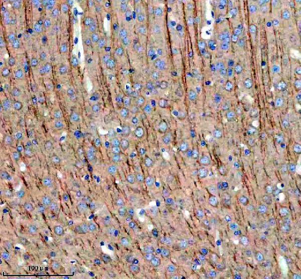
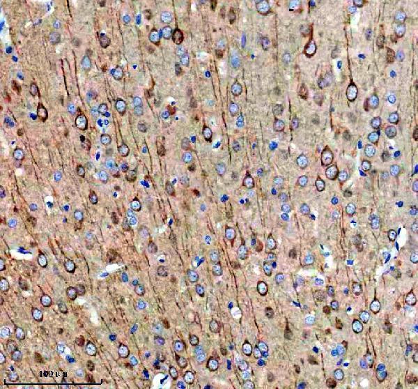
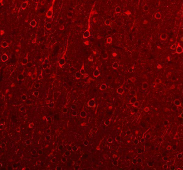
A01760-1 has real MAP1B IHC and IF images from mouse and rat paraffin brain sections (catalog image captions). Human reactivity is listed (catalog reactivity).

Real IHC data IHC analysis of MAP1B using anti-MAP1B antibody (A01760-1). MAP1B was detected in a paraffin-embedded section of mouse brain tissue. Heat mediated antigen retrieval was performed in EDTA buffer (pH 8.0, epitope retrieval solution). The tissue section was blocked with 10% goat serum. The tissue section was then incubated with 2 μg/ml rabbit anti-MAP1B Antibody (A01760-1) overnight at 4°C. Peroxidase Conjugated Goat Anti-rabbit IgG was used as secondary antibody and incubated for 30 minutes at 37°C. The tissue section was developed using HRP Conjugated Rabbit IgG Super Vision Assay Kit (Catalog # SV0002) with DAB as the chromogen.
Anti-MAP1B Antibody ®
Cat # A01760-1

A01760-1 is listed for IHC and IF with human, mouse, and rat reactivity (catalog applications; catalog reactivity). Its images show IHC and IF in mouse and rat paraffin brain sections (catalog IHC and IF image captions).

Which to pick: Choose A01760-1 for tissue IHC in mouse or rat paraffin sections; its IHC captions document EDTA retrieval at pH 8.0 and 2 μg/ml primary antibody (A01760-1 IHC image captions). For IF, A01760-1 has mouse and rat paraffin brain images; ICC is not listed or pictured (A01760-1 applications; A01760-1 IF image captions). For cross-species work, it is a rabbit antibody with listed human, mouse, and rat reactivity, while the pictured tissue data cover mouse and rat; the IHC captions do not report the fixative (A01760-1 catalog host and reactivity; A01760-1 IHC image captions).

Each figure is that product's own IHC / IF validation image from its datasheet.

References

  1. UniProt Consortium. UniProt entry P46821 (MAP1B_HUMAN, Microtubule-associated protein 1B).
  2. Human Protein Atlas. MAP1B tissue IHC expression (reliability: Enhanced).
  3. Human Protein Atlas. MAP1B subcellular location (ICC-IF): Mainly localized to the cytosol. In addition localized to the equatorial segment..
  4. Human Protein Atlas. MAP1B antibody validation summary (2 antibodies).
  5. A MAP1B-cortactin-Tks5 axis regulates TNBC invasion and tumorigenesis. The Journal of cell biology 2024 — PMC10866687.
  6. Role of Microtubule-Associated Protein 1b in Urothelial Carcinoma: Overexpression Predicts Poor Prognosis. Cancers 2020 — PMC7139768.
  7. Genes Related to Motility in an Ionizing Radiation and Estrogen Breast Cancer Model. Biology 2024 — PMC11591951.
  8. Single-patient single-cell RNA sequencing reveals neuroendocrine predominance and immunosuppression in small-cell lung cancer. Translational cancer research 2026 — PMC12885910.
  9. PubMed PMID:7806212 — UniProt-cited evidence.
  10. PubMed PMID:12684070 — UniProt-cited evidence.
  11. PubMed PMID:15372022 — UniProt-cited evidence.