This website uses cookies to ensure you get the best experience on our website.


- Table of Contents
Plan MAP1S chromogenic IHC in paraffin sections using the IHC-validated antibody A04308 at 2–5 μg/ml (datasheet A04308). This guide covers the cytoplasmic tissue pattern (HPA tissue IHC) and the heavy and light chains relevant to epitope selection (UniProt).

Expected localisation, validated protocols, controls and antibodies — the at-a-glance facts below, then the full design guide.
| Expected localisation | Cytoplasmic in most tissues (HPA tissue IHC); nuclear location annotated (UniProt) | |
| Staining pattern | Cytoplasmic in most tissues; high in nasopharyngeal ciliated cells (HPA tissue IHC) | |
| Antigen retrieval | EDTA pH 8.0 HIER, heat-mediated (datasheet A04308) | |
| Positive control | Nasopharynx+4 more · see all | |
| Negative control | Adipose tissue+4 more · see all |
| Fixation | Keep fixation consistent across sections (standard IHC practice; not target-specific) | |
| Caveat | Antibody staining has medium agreement with RNA data (HPA tissue IHC) | |
| Regulation | Staining intensity regulation is unreported (UniProt) | |
| Isoform / epitope | Two isoforms; heavy and light chains affect epitope coverage (UniProt) |
The catalog antibody protocol uses EDTA retrieval (datasheet: A04308). Two published MAP1S tissue staining protocols provide citrate retrieval and chromogenic detection details (PMC9240238; PMC4646605).
| Sample | Paraffin-embedded mouse brain tissue; fixative not specified (datasheet A04308) |
| Fixation | Image fixative and duration unreported (datasheet A04308); verify before use. |
| Sectioning | 4–5 µm sections on charged slides (standard) |
| Deparaffinisation | Xylene, graded ethanol series to water (standard) |
| Antigen retrieval | Heat retrieval: EDTA pH 8.0 (datasheet A04308); 20 min, 95–100 °C (standard) |
| Peroxidase block | 3% H2O2, 10 min, room temperature (standard) |
| Blocking | 10% goat serum (datasheet A04308) |
| Primary antibody | Rabbit anti-MAP1S, 2-5 μg/ml (datasheet A04308) |
| Primary incubation | Overnight at 4 °C (datasheet A04308) |
| Detection | HRP-conjugated secondary, DAB chromogen (datasheet A04308) |
| Counterstain | Hematoxylin, blue, dehydrate and mount (standard) |
| Expected result | MAP1S-positive staining in ciliated cells (cell body) of nasopharynx (HPA tissue IHC: High). HPA tissue profile: Cytoplasmic expression in most tissues. No signal in the no-primary control. |
MAP1S is chiefly cytoplasmic in tissue IHC, with expression reported in most tissues (HPA tissue IHC: Approved; medium consistency with RNA; pending external verification). Expect staining in nasopharyngeal ciliated cell bodies and in glandular or basal cells in the specified positive tissues (HPA tissue IHC). Cytoskeletal and context-dependent nuclear localisation are also reported (UniProt Q66K74 localisation); MAP1S has no transmembrane segment (UniProt Q66K74 topology).
| Cytoplasmic staining in nasopharyngeal ciliated cell bodies, with glandular or basal cell staining in other positive tissues. | This fits the reported tissue pattern: nasopharyngeal ciliated cell bodies are High; appendix, breast, colon, duodenum and endometrium glandular cells, and bronchial basal cells, are Medium (HPA tissue IHC). Judge the named cell population and compartment together; a tissue name alone does not establish that every cell should stain (HPA tissue IHC). |
| Predominantly nuclear staining in an otherwise routine tissue section, with little cytoplasmic staining. | Recheck the compartment assignment and controls before calling this MAP1S: tissue IHC is described as cytoplasmic in most tissues (HPA tissue IHC). Nuclear localisation can occur, including in cells exhibiting apoptosis (UniProt Q66K74 localisation), and nucleoplasmic and nucleolar signals are additional ICC-IF locations (HPA subcellular); nuclear colour alone is therefore not proof of artefact. |
| Strong staining in an HPA-listed cell population reported as Not detected, such as adipocytes or bone-marrow hematopoietic cells. | That result conflicts with the specified cell-level observations (HPA tissue IHC: adipocytes and hematopoietic cells Not detected). Consider antibody cross-reactivity or endogenous detection activity, then compare the named cells with a matched control section (general IHC practice). A negative designation for one cell population does not establish that its whole tissue must be blank (HPA tissue IHC). |
| Diffuse colour across tissue, extracellular space or the slide, obscuring cell borders. | This is difficult to score as the reported cell-body or cytoplasmic pattern (HPA tissue IHC). Assess background with a primary-antibody-omission control and check blocking, washes and detection reagents (general IHC practice). Do not interpret widespread colour as broader MAP1S expression until the cell-specific pattern is visible. |
| No detectable signal in nasopharyngeal ciliated cell bodies or another stated positive cell population. | A blank result conflicts with High staining in nasopharyngeal ciliated cell bodies or Medium staining in the specified positive populations (HPA tissue IHC). It does not, by itself, establish MAP1S absence: check section quality, detection controls, antibody working conditions and the identity of the scored cells (general IHC practice). HPA reports medium consistency with RNA and pending external verification (HPA tissue IHC). |
| Cell population and tissue selection (HPA tissue IHC). | HPA describes cytoplasmic expression in most tissues, but the listed intensities belong to specific cells: High in nasopharyngeal ciliated cell bodies; Medium in several glandular populations and bronchial basal cells; Not detected in specified populations including adipocytes and bone-marrow hematopoietic cells (HPA tissue IHC). Select and score controls at cell level. |
| Compartment and cellular state (UniProt Q66K74 localisation; HPA subcellular). | Diffuse cytoplasmic staining with partial microtubule localisation is reported in interphase cells, and spindle microtubule localisation during mitosis (UniProt Q66K74 localisation). UniProt also reports a perinuclear punctate network and nuclear detection in cells exhibiting apoptosis; HPA ICC-IF lists cytosol as a main supported location and nucleoplasm and nucleoli as additional supported locations (HPA subcellular). |
| Antibody evidence (HPA antibodies; HPA tissue IHC). | HPA050934 and HPA054637 each have IHC Approved status; HPA050934 has ICC Supported status (HPA antibodies). The tissue IHC profile has medium consistency with RNA and awaits external verification (HPA tissue IHC). These labels support a working expectation, while leaving an unexpected compartment or cell population to be resolved with controls. |
| Protein processing and isoforms (UniProt Q66K74). | UniProt lists a heavy chain spanning residues 1–829, a light chain spanning 830–1059, and 2 isoforms (UniProt Q66K74 processing; isoforms). These facts do not identify the IHC antibody's epitope or predict which processed form a section will display; interpret any proposed chain-specific pattern only if the antibody's epitope is established. |
| Membrane assignment and retrieval limits (UniProt Q66K74 topology; HPA tissue IHC). | No transmembrane segment is annotated (UniProt Q66K74 topology), so a membrane-only outline would need scrutiny against the reported cytoplasmic tissue pattern (HPA tissue IHC). Neither source supplies target-specific fixation sensitivity or an antigen-retrieval condition; optimize retrieval as a general IHC variable without treating an HPA staining level as evidence of a fixation effect (general IHC practice). |
| Situation | Likely cause | Next action |
|---|---|---|
| The named positive cells are blank or much weaker than expected (HPA tissue IHC). | Target-specific fixation effects are not established by the supplied assay evidence. Verify with a matched IHC source before attributing a result to fixation. | Confirm the scored cell type, run a stated positive tissue alongside the sample, and check the detection control. If needed, compare retrieval and antibody working conditions systematically (general IHC practice); no MAP1S-specific retrieval setting is supplied. |
| Brown signal spreads across the section and obscures cell boundaries (general IHC practice). | Nonspecific antibody binding, endogenous detection activity or excessive development can produce background (general IHC practice). | Inspect a primary-antibody-omission control; review blocking, washes and chromogen development, then rescore only cell-associated signal (general IHC practice). Compare the resulting pattern with HPA's cytoplasmic tissue profile (HPA tissue IHC). |
| A listed Not detected cell population stains strongly (HPA tissue IHC). | Cross-reactivity or endogenous detection activity is possible (general IHC practice); HPA's negative calls apply to the named cell populations, not automatically to every cell in those tissues (HPA tissue IHC). | Verify cell identity against morphology and counterstain, inspect omission and positive controls, and compare another IHC-approved antibody if available (general IHC practice; HPA antibodies: HPA050934 and HPA054637 IHC Approved). |
| Signal appears exclusively nuclear in a routine tissue section (HPA tissue IHC). | The pattern differs from HPA's usual cytoplasmic tissue profile; genuine nuclear MAP1S is context dependent in UniProt, and HPA reports additional nuclear ICC-IF locations (HPA tissue IHC; UniProt Q66K74 localisation; HPA subcellular). | Check whether the signal is confined to morphologically apoptotic cells, compare cytoplasmic positive cells on the same run, and inspect controls before assigning a localisation or artefact (UniProt Q66K74 localisation; general IHC practice). |
| Staining varies sharply between adjacent areas of one section (general IHC practice). | Uneven reagent coverage, washing or development can create spatial variation (general IHC practice); the supplied sources provide no MAP1S-specific fixation-effect evidence. | Inspect section integrity and reagent coverage, repeat with consistent processing, and compare like cell populations across areas (general IHC practice). Avoid inferring a MAP1S fixation effect from this pattern alone. |
| IF/ICC Q&A: Where should MAP1S fluorescence appear (HPA subcellular)? | ICC-IF compartment evidence addresses a different preparation from paraffin tissue IHC (HPA subcellular; HPA tissue IHC). | Expect mainly cytosol and sperm principal piece, with additional nucleoplasm and nucleoli (HPA subcellular). UniProt also reports microtubule-associated and context-dependent nuclear localisation (UniProt Q66K74 localisation); use the separate IF/ICC guide for its procedure. |
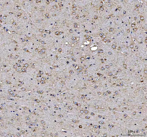
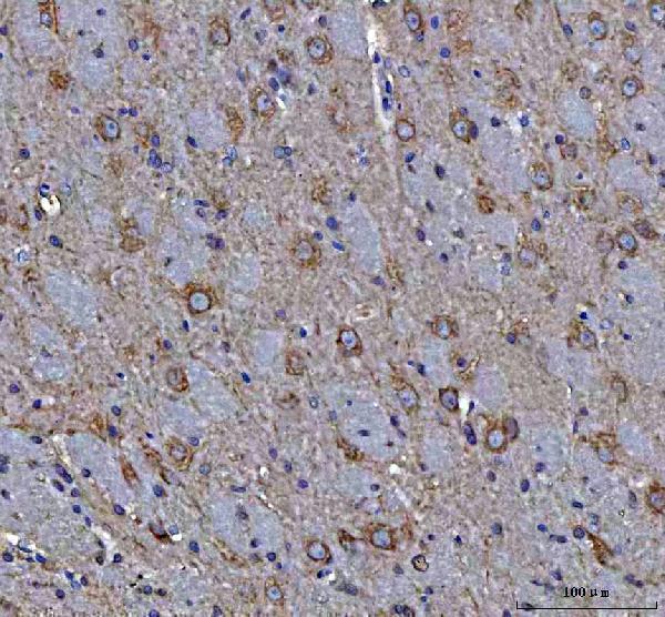


Comprehensive Human Protein Atlas IHC scoring per tissue (reliability: Approved — Medium consistency between antibody staining and RNA expression data. Pending external verification.). Rows are taken directly from the HPA tissue chart — click any row's HPA link to view the source.
| Tissue | Cell type | Level | Evidence | Source |
|---|---|---|---|---|
| Nasopharynx | Ciliated cells (cell body) | High | Protein (IHC) | HPA → |
| Appendix | Glandular cells | Medium | Protein (IHC) | HPA → |
| Breast | Glandular cells | Medium | Protein (IHC) | HPA → |
| Bronchus | Basal cells | Medium | Protein (IHC) | HPA → |
| Colon | Glandular cells | Medium | Protein (IHC) | HPA → |
| Tissue | Cell type | Level | Evidence | Source |
|---|---|---|---|---|
| Adipose tissue | Adipocytes | Not detected | Protein (IHC) | HPA → |
| Adrenal gland | Glandular cells | Not detected | Protein (IHC) | HPA → |
| Bone marrow | Hematopoietic cells | Not detected | Protein (IHC) | HPA → |
| Caudate | Glial cells | Not detected | Protein (IHC) | HPA → |
| Cerebellum | Cells in granular layer | Not detected | Protein (IHC) | HPA → |
Troubleshoot MAP1S staining in paraffin sections by checking retrieval, cellular localisation, and controls before comparing signal across specimens.
A04308 has IHC images from paraffin-embedded mouse and rat brain sections (catalog image captions); human, mouse and rat reactivity is listed (catalog), but no IF/ICC data are supplied (catalog).
A04308 is the only SKU rendered and lists IHC among its applications (catalog: A04308 applications). Its images show staining of paraffin-embedded mouse and rat brain sections after EDTA pH 8.0 retrieval, using 2 μg/ml primary antibody (catalog: A04308 IHC image captions).
Which to pick: Choose A04308 for paraffin-section IHC using the captioned mouse or rat brain conditions; the fixative is unreported (catalog: A04308 IHC image captions). For cross-species planning, A04308 lists human, mouse and rat reactivity, though the supplied IHC images cover mouse and rat only (catalog: A04308 reactivity and IHC image captions). No IF/ICC application or image is supplied, so there is no payload-supported IF/ICC pick; A04308 is a rabbit antibody with no clone reported (catalog: A04308 applications, images, host and clone).