This website uses cookies to ensure you get the best experience on our website.


- Table of Contents
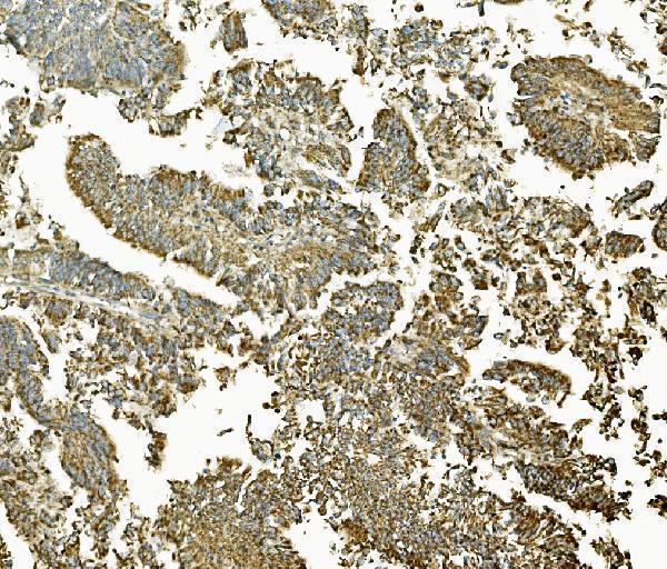
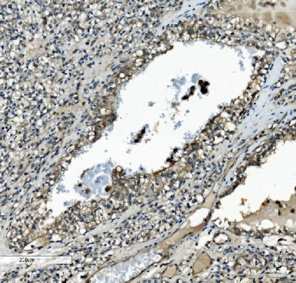
Plan MAP2K2 paraffin IHC around cytoplasmic staining, with high signal reported in colon glandular cells (HPA tissue IHC). In a paraffin-section example, catalog antibody A00996-2 was applied at 2 μg/mL overnight at 4°C (datasheet: A00996-2).

Expected localisation, validated protocols, controls and antibodies — the at-a-glance facts below, then the full design guide.
| Expected localisation | General cytoplasmic tissue staining (HPA tissue IHC) | |
| Staining pattern | Cytoplasmic staining in glandular and hematopoietic cells (HPA tissue IHC) | |
| Antigen retrieval | EDTA pH 8.0 HIER, heat-mediated (datasheet A00996-2) | |
| Positive control | Appendix+4 more · see all | |
| Negative control | Heart muscle+2 more · see all |
| Fixation | Keep fixation conditions consistent across sections (standard IHC practice; not target-specific) | |
| Caveat | Bone marrow endogenous peroxidase can mimic DAB signal (HPA tissue IHC; standard IHC practice) | |
| Regulation | Expression regulation unreported (UniProt) | |
| Isoform / epitope | No isoforms or cleavage annotated to alter the epitope map (UniProt) |
The catalog antibody’s IHC-P protocol is accompanied by four published MAP2K2 IHC protocols covering head and neck, pancreatic, breast, and prostate tumors (PMC10741104; PMC10563513; PMC10342177; PMC10126012).
| Sample | Paraffin-embedded human rectal cancer tissue; fixative not specified (datasheet A00996-2) |
| Fixation | Image fixative and duration unreported (datasheet A00996-2); verify before use. |
| Sectioning | 4–5 µm sections on charged slides (standard) |
| Deparaffinisation | Xylene, graded ethanol series to water (standard) |
| Antigen retrieval | Heat retrieval: EDTA pH 8.0 (datasheet A00996-2); 20 min, 95–100 °C (standard) |
| Peroxidase block | 3% H2O2, 10 min, room temperature (standard) |
| Blocking | 10% goat serum (datasheet A00996-2) |
| Primary antibody | Rabbit anti-MAP2K2, 2-5μg/ml (datasheet A00996-2) |
| Primary incubation | Overnight at 4 °C (datasheet A00996-2) |
| Detection | Streptavidin-biotin complex (SABC), DAB chromogen (datasheet A00996-2) |
| Counterstain | Hematoxylin, blue, dehydrate and mount (standard) |
| Expected result | MAP2K2-positive staining in glandular cells of appendix (HPA tissue IHC: High). HPA tissue profile: General cytoplasmic expression. No signal in the no-primary control. |
In paraffin-section IHC, expect mainly cytoplasmic MAP2K2 staining, particularly in glandular cells of the colon, appendix and duodenum, and in cerebellar Purkinje cells (HPA tissue IHC: High; general cytoplasmic expression). Membrane association may occur through KSR1 interaction, although MAP2K2 has no transmembrane segment (UniProt P36507: subcellular location and topology). HPA rates the tissue pattern Supported, with medium consistency between staining and RNA expression (HPA tissue IHC: reliability).
| Cytoplasmic staining in glandular epithelium or Purkinje cells. | This fits the reported compartment and cell distribution (HPA tissue IHC: general cytoplasmic expression; High in glandular and Purkinje cells). Judge staining within the named cell population rather than treating every cell in a positive tissue as equally positive (HPA tissue IHC: cell-specific levels). |
| Predominantly nuclear staining, with little cytoplasmic signal. | A nuclear-only pattern conflicts with the reported cytoplasmic IHC profile and cytosolic IF location (HPA tissue IHC: profile; HPA subcellular: Cytosol). Investigate nonspecific primary binding, detection background and compartment identification before assigning that signal to MAP2K2 (general IHC practice). |
| Strong staining in cardiomyocytes, skeletal myocytes or smooth muscle cells. | HPA reports MAP2K2 as Not detected in these respective cell populations (HPA tissue IHC: heart, skeletal muscle and smooth muscle). Unexpected signal warrants checks for cross-reactivity or endogenous detection activity; it does not, by itself, establish either cause (general IHC practice). |
| Uniform haze across cells, stroma and tissue-free areas. | A diffuse field obscures the cell and compartment pattern needed to interpret MAP2K2 (HPA tissue IHC: cell-specific cytoplasmic profile). Compare a section processed without primary antibody and review blocking, washes and detection chemistry for background (general IHC practice). |
| No staining in a section expected to contain HPA-high cells. | First confirm that the relevant cells are present: HPA names glandular cells in the colon, appendix and duodenum, and Purkinje cells in the cerebellum (HPA tissue IHC: High). If present, review antibody suitability and the IHC workflow before interpreting the absence as biological (general IHC practice). |
| Tissue and cell selection | HPA reports High staining in several glandular populations, hematopoietic cells and Purkinje cells, but Not detected staining in the listed muscle cell populations (HPA tissue IHC: cell levels). Use the specified cells when comparing sections; low tissue specificity of RNA does not make all cell types equivalent IHC controls (HPA tissue IHC: RNA specificity and staining). |
| Compartment and membrane association | Cytoplasm is the reported IHC pattern, while UniProt lists cytoplasm and membrane and says membrane localization is probably regulated by KSR1 interaction (HPA tissue IHC: profile; UniProt P36507: subcellular location). With no transmembrane segment, a strict cell-surface outline is not the default expected pattern (UniProt P36507: topology). |
| Strength of reference evidence | HPA calls tissue IHC Supported and describes medium consistency with RNA data; its 2 listed antibodies each have Supported IHC status (HPA tissue IHC: reliability; HPA antibodies: HPA051993 and CAB003835). These observations guide pattern checks but do not establish that every sample or staining run must have the same intensity (general IHC practice). |
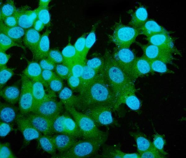
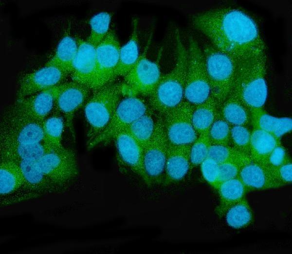
| IF/ICC Q&A: where should MAP2K2 appear? | Mainly in the cytosol; HPA additionally reports signal in the sperm mid piece, principal piece and end piece (HPA subcellular: enhanced cytosol; approved additional locations). This localization answers the IF/ICC interpretation question and supplies no IF/ICC protocol conditions (HPA subcellular: localization record). |
| Fixation and retrieval evidence | Target-specific fixation effects are not established by the supplied assay evidence. Verify with a matched IHC source before attributing a result to fixation. |
| Situation | Likely cause | Next action |
|---|---|---|
| Known-positive glandular cells show no chromogenic signal. | The expected cells may be absent from the section, or staining and detection may have failed (HPA tissue IHC: High in specified glandular cells; general IHC practice). | Verify cell identity on the counterstain, then review IHC validation, retrieval, primary incubation and detection with a positive control section (general IHC practice). |
| Colour appears equally in negative-control and test sections. | Primary-independent background, including endogenous enzyme activity when enzyme detection is used, is possible (general IHC practice). | Compare a no-primary control, check the detection reagent and apply the appropriate endogenous-activity block for that detection system (general IHC practice). |
| Strong nuclear colour dominates a stained tissue. | This conflicts with reported cytoplasmic IHC and cytosolic IF localization (HPA tissue IHC: profile; HPA subcellular: Cytosol). | Check nuclear boundaries with the counterstain, compare the no-primary control and reassess antibody specificity before scoring MAP2K2 (general IHC practice). |
| Muscle cells stain strongly despite a clean negative control. | HPA reports Not detected staining in cardiomyocytes, skeletal myocytes and smooth muscle cells; the discrepancy needs investigation (HPA tissue IHC: muscle entries). | Confirm cell identity and compare staining with an HPA-high cell population; evaluate primary-antibody specificity if the discrepancy persists (general IHC practice). |
| Signal spreads through stroma and obscures cell borders. | Diffuse background can prevent assessment of the reported cell-specific cytoplasmic pattern (HPA tissue IHC: profile; general IHC practice). | Inspect a no-primary section and review blocking, wash steps, antibody concentration and chromogen development as general IHC practice. |
| A faint membrane outline appears alongside cytoplasmic staining. | Membrane association is plausible through KSR1, but MAP2K2 lacks a transmembrane segment (UniProt P36507: subcellular location and topology). | Score the predominant cytoplasmic pattern and compare controls before treating the outline as specific membrane localization (HPA tissue IHC: profile; general IHC practice). |

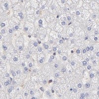


Comprehensive Human Protein Atlas IHC scoring per tissue (reliability: Supported — Medium consistency between antibody staining and RNA expression data.). Rows are taken directly from the HPA tissue chart — click any row's HPA link to view the source.
| Tissue | Cell type | Level | Evidence | Source |
|---|---|---|---|---|
| Appendix | Glandular cells | High | Protein (IHC) | HPA → |
| Bone marrow | Hematopoietic cells | High | Protein (IHC) | HPA → |
| Cerebellum | Purkinje cells | High | Protein (IHC) | HPA → |
| Colon | Glandular cells | High | Protein (IHC) | HPA → |
| Duodenum | Glandular cells | High | Protein (IHC) | HPA → |
Troubleshoot MAP2K2 staining in paraffin sections by checking retrieval, cell compartment, controls, and scoring before interpreting differences in signal.
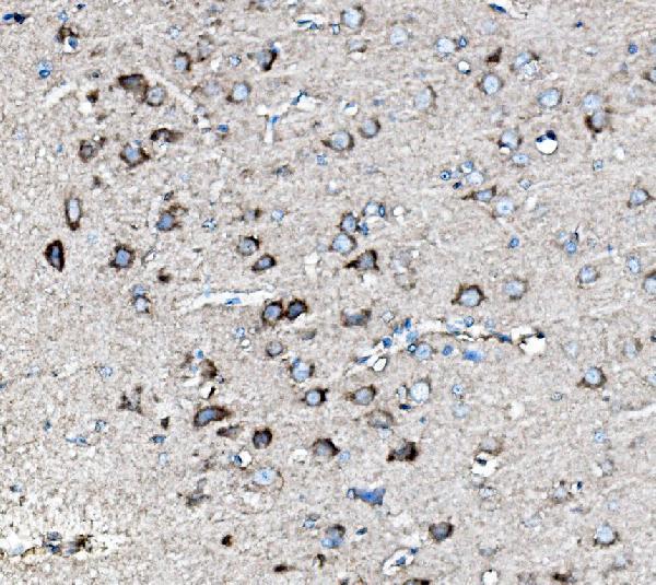
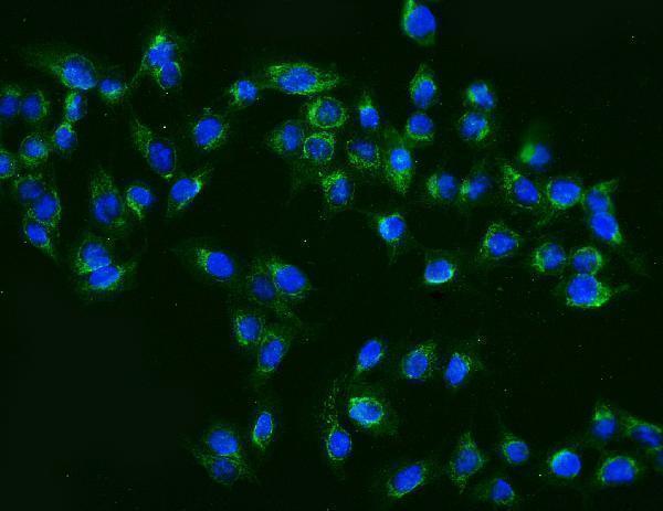
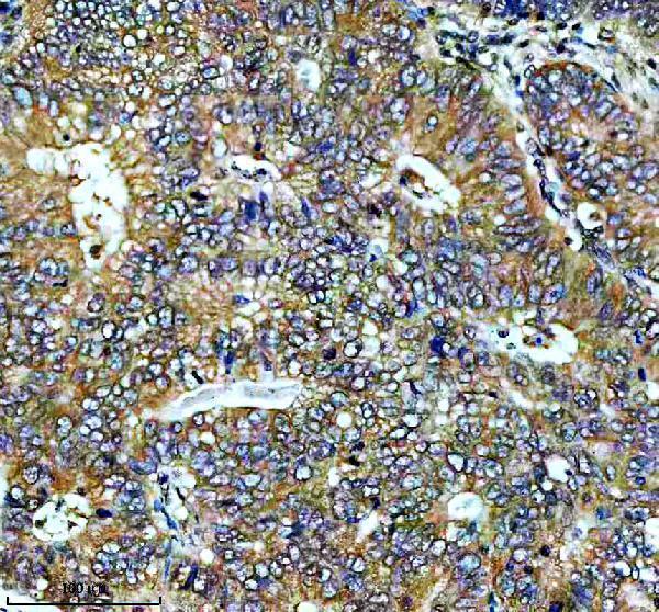
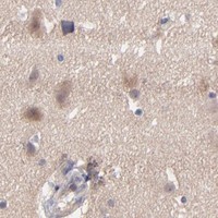
Catalog images document paraffin-section IHC in human tumors and mouse and rat brain, plus IF/ICC in U20S, K562, and MCF-7 cells (catalog image captions).
A00996-2 has IHC images from human rectal cancer and mouse and rat brain, plus IF/ICC images from U20S and K562 cells (A00996-2 image captions); M00996 has an IHC image from human stomach cancer (M00996 IHC caption). M00996-3 has an IHC image from human renal clear cell carcinoma and an IF/ICC image from MCF-7 cells (M00996-3 image captions); M00996-2 has an IF/ICC image from MCF-7 cells and does not list IHC as an application (M00996-2 image caption; catalog applications).
Which to pick: For paraffin-section IHC, choose A00996-2 for human rectal cancer or mouse and rat brain (2–5 μg/mL; A00996-2 catalog dilution and IHC captions), M00996 for human stomach cancer (1:50; M00996 catalog dilution and IHC caption), or mouse monoclonal M00996-3 for human renal clear cell carcinoma (2 μg/mL in its IHC caption; M00996-3 catalog clone and IHC caption). For IF/ICC, mouse monoclonals M00996-2 and M00996-3 have MCF-7 images, while A00996-2 has U20S and K562 images (catalog clones and IF captions). For cross-species tissue IHC, A00996-2 has human, mouse, and rat paraffin-section images (A00996-2 IHC captions); the fixative is unreported in the paraffin-section captions (catalog IHC captions).