This website uses cookies to ensure you get the best experience on our website.


- Table of Contents
Plan MAP2K7 chromogenic IHC in paraffin sections using the observed variable cytoplasmic tissue pattern and high spermatogonial staining as references (HPA tissue IHC). The guide covers fixation consistency, antibody dilution and interpretation alongside the four annotated isoforms (datasheet: PB9764; UniProt).

Expected localisation, validated protocols, controls and antibodies — the at-a-glance facts below, then the full design guide.
| Expected localisation | Cytoplasm in tissue; nucleus also annotated (HPA tissue IHC; UniProt) | |
| Staining pattern | High in spermatogonia; variable cytoplasmic staining in most tissues (HPA tissue IHC) | |
| Antigen retrieval | EDTA pH 8.0 HIER, heat-mediated (datasheet PB9764) | |
| Positive control | Testis+4 more · see all | |
| Negative control | Caudate+2 more · see all |
| Fixation | Matched tissue-IHC evidence does not establish the fixation claim. Validate the specimen-specific method before use. (selected-SKU IHC image PB9764) | |
| Caveat | Skeletal myocytes may lack detectable staining (HPA tissue IHC) | |
| Regulation | Expression changes are unreported (UniProt) | |
| Isoform / epitope | Four isoforms; antibody epitope coverage is unspecified (UniProt) |
The catalog antibody’s IHC-P protocol (datasheet: PB9764) is accompanied by one published MAP2K7 tissue staining protocol (PMC4863692).
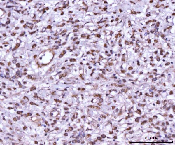
| Sample | Paraffin-embedded human testicular cancer tissue; fixative not specified (datasheet PB9764) |
| Fixation | Image fixative and duration unreported (datasheet PB9764); verify before use. |
| Sectioning | 4–5 µm sections on charged slides (standard) |
| Deparaffinisation | Xylene, graded ethanol series to water (standard) |
| Antigen retrieval | Heat retrieval: EDTA pH 8.0 (datasheet PB9764); 20 min, 95–100 °C (standard) |
| Peroxidase block | 3% H2O2, 10 min, room temperature (standard) |
| Blocking | 10% goat serum (datasheet PB9764) |
| Primary antibody | Rabbit anti-MAP2K7, 1-2μg/ml (datasheet PB9764) |
| Primary incubation | Overnight at 4 °C (datasheet PB9764) |
| Detection | Streptavidin-biotin complex (SABC), DAB chromogen (datasheet PB9764) |
| Counterstain | Hematoxylin, blue, dehydrate and mount (standard) |
| Expected result | MAP2K7-positive staining in spermatogonia cells of testis (HPA tissue IHC: High). HPA tissue profile: Cytoplasmic expression in most tissues at variable levels. No signal in the no-primary control. |
MAP2K7 is expected mainly in the cytoplasm across many tissues, with variable staining intensity (HPA tissue IHC: Supported; medium consistency with RNA). Testis spermatogonia show high staining; several epithelial and hematopoietic populations show medium staining (HPA tissue IHC). UniProt also lists nuclear localization, while its topology shows no transmembrane segment (UniProt O14733: subcellular location and topology). Interpret the compartment and the stained cell type together.
| Cytoplasmic staining in spermatogonia; weaker staining in other listed cell populations. | This matches the observed pattern: high in testis spermatogonia and medium in appendix lymphoid tissue, bone marrow hematopoietic cells, and several epithelial populations (HPA tissue IHC). Score the named cells separately from neighboring cells (general IHC practice). |
| Predominantly nuclear staining with little cytoplasmic signal. | Check the result against the expected cytoplasmic tissue pattern (HPA tissue IHC). Nuclear localization is listed by UniProt, so nuclear signal alone is not proof of an artefact (UniProt O14733: subcellular location). Review controls before assigning it to MAP2K7 (general IHC practice). |
| Strong staining in oral mucosa squamous cells, caudate glia, or skeletal muscle myocytes. | Those specific cell populations were not detected in the supplied tissue observations (HPA tissue IHC). Confirm their identity and inspect controls; unexpected staining may reflect cross-reactivity or detection background (general IHC practice). A tissue-wide label cannot substitute for a cell-specific comparison. |
| Diffuse color across tissue, extracellular spaces, or the negative control. | A non-cellular or control-matched pattern does not establish MAP2K7 localization (general IHC practice). Check blocking, washes, chromogen development, and endogenous detection activity before interpreting stained cells (general IHC practice). |
| No signal in testis spermatogonia. | This conflicts with the reported high staining in that cell population (HPA tissue IHC). First verify that spermatogonia are present in the examined section; then review antibody, detection, and run controls (general IHC practice). A blank slide alone cannot establish biological absence. |
| Cell and tissue context | Compare like cells: HPA reports high staining in spermatogonia, medium staining in selected lymphoid, hematopoietic, myoepithelial, respiratory epithelial, and glandular cells, and variable cytoplasmic expression overall (HPA tissue IHC). |
| RNA and protein agreement | The tissue IHC assessment is Supported, with medium consistency between antibody staining and RNA expression (HPA tissue IHC). Treat an isolated unexpected result as a prompt for controls, rather than as a new reference pattern (general IHC practice). |
| Skeletal muscle discrepancy | UniProt describes highest overall expression in skeletal muscle, whereas HPA reports no staining in skeletal muscle myocytes (UniProt O14733: tissue specificity; HPA tissue IHC: myocytes not detected). Keep those distinct observations visible when choosing a control. |
| Isoforms and antibody recognition | Four isoforms are listed, and isoform 3 is low in placenta, fetal liver, and skeletal muscle (UniProt O14733: isoforms and tissue specificity). The supplied evidence does not map an antibody epitope to an isoform; do not infer isoform-specific IHC staining. |
| Topology and processing | MAP2K7 has no transmembrane segment or signal peptide and has a listed chain spanning residues 2–419 (UniProt O14733: topology and processing). These facts support caution about assigning extracellular color to the target; they do not establish an antigen retrieval condition. |
| Antibody validation | The supplied IHC status is Supported for HPA001633 and CAB004262 (HPA antibodies). That status supports use of their reported patterns as comparisons; it does not show that every antibody or every specimen yields the same intensity. |
| IF/ICC Q&A: Should membrane signal define a positive IHC result? | No. HPA places IF/ICC signal in cytosol and plasma membrane, both uncertain, while tissue IHC describes cytoplasmic staining (HPA subcellular ICC-IF; HPA tissue IHC). Assess IHC against its tissue pattern and controls (general IHC practice). |
| Situation | Likely cause | Next action |
|---|---|---|
| Expected spermatogonia are blank while the rest of the slide is readable. | The reported high-staining cell population may be absent from the section, or the assay may have missed it (HPA tissue IHC: high in spermatogonia; general IHC practice). | Confirm cell identity on the counterstain; compare an appropriate positive section and the run controls before changing conditions (general IHC practice). |
| Color appears in a negative control or broadly between cells. | Background from detection chemistry or insufficient blocking or washing can mimic cell staining (general IHC practice). | Review the negative control, endogenous activity blocking, washes, and chromogen development; accept only a cell-associated pattern that separates from background (general IHC practice). |
| Staining is mainly nuclear. | It differs from the observed cytoplasmic tissue profile, although UniProt also lists the nucleus (HPA tissue IHC; UniProt O14733: subcellular location). | Check section morphology and controls, then compare the same cell type with the HPA tissue pattern before scoring nuclear signal as target-specific (general IHC practice). |
| Skeletal muscle myocytes stain strongly. | This conflicts with the supplied myocyte IHC observation despite UniProt's broader skeletal muscle expression statement (HPA tissue IHC: myocytes not detected; UniProt O14733: tissue specificity). | Recheck cell identification and control staining; report the discrepancy at cell level instead of treating skeletal muscle as an assured IHC positive control (general IHC practice). |
| A listed medium-staining population appears weak or patchy. | Intensity varies across tissues, and the IHC-to-RNA consistency is medium (HPA tissue IHC: profile and reliability). Technical variation can also affect a run (general IHC practice). | Compare the same cell type across sections, inspect run controls, and adjust scoring thresholds only with consistent control performance (general IHC practice). |
| Signal is weak in the positive comparison section. | Antibody dilution, retrieval, or detection conditions can limit IHC signal (general IHC practice); the supplied sources give no MAP2K7-specific fixation or retrieval effect. | Verify the antibody's IHC instructions and run controls, then optimize one general IHC condition at a time; do not claim a MAP2K7-specific retrieval requirement from these sources (general IHC practice). |



Comprehensive Human Protein Atlas IHC scoring per tissue (reliability: Supported — Medium consistency between antibody staining and RNA expression data.). Rows are taken directly from the HPA tissue chart — click any row's HPA link to view the source.
| Tissue | Cell type | Level | Evidence | Source |
|---|---|---|---|---|
| Testis | Spermatogonia cells | High | Protein (IHC) | HPA → |
| Appendix | Lymphoid tissue | Medium | Protein (IHC) | HPA → |
| Bone marrow | Hematopoietic cells | Medium | Protein (IHC) | HPA → |
| Breast | Myoepithelial cells | Medium | Protein (IHC) | HPA → |
| Bronchus | Respiratory epithelial cells | Medium | Protein (IHC) | HPA → |
Troubleshoot MAP2K7 staining in paraffin sections by checking retrieval, compartment, cell type and controls before interpreting chromogenic signal.
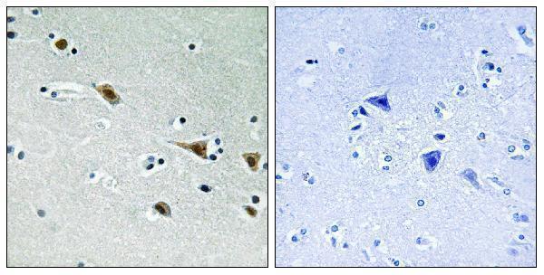
IHC data show MAP2K7 staining in paraffin sections of human testicular cancer and brain; IF data show staining in PC-3 cells (PB9764 and A02713S271-1 image captions).
PB9764 has human testicular cancer paraffin-section IHC data and PC-3 cell IF data (PB9764 image captions). A02713S271-1 has human brain paraffin-section IHC data with phospho-peptide blocking (A02713S271-1 IHC image caption).
Which to pick: For tissue IHC, choose PB9764 for MAP2K7 in human testicular cancer sections (PB9764 IHC image caption), or A02713S271-1 when the target is phosphorylated Ser271 in human brain sections (A02713S271-1 title and IHC image caption); neither caption reports the fixative. For IF/ICC, PB9764 has a PC-3 cell IF example (PB9764 IF image caption). For cross-species planning, PB9764 lists human, monkey, mouse and rat reactivity, while A02713S271-1 lists human, mouse and rat; their illustrated IHC examples are human only (catalog reactivity; PB9764 and A02713S271-1 IHC image captions).