This website uses cookies to ensure you get the best experience on our website.


- Table of Contents
Plan chromogenic paraffin IHC for MAP4 using its general cytoplasmic tissue pattern (HPA tissue IHC). Lung endothelial cells show high staining, while adipocytes are undetected; use these as cell-specific references when scoring (HPA tissue IHC).

Expected localisation, validated protocols, controls and antibodies — the at-a-glance facts below, then the full design guide.
| Expected localisation | Cytoplasm (HPA tissue IHC); cytoskeleton/MTOC (UniProt) | |
| Staining pattern | General cytoplasmic staining; distinct in nerves and endothelium (HPA tissue IHC) | |
| Antigen retrieval | EDTA pH 8.0 HIER, heat-mediated (datasheet A00948-2) | |
| Positive control | Caudate+4 more · see all | |
| Negative control | Adipose tissue+4 more · see all |
| Fixation | Keep fixation consistent across paraffin sections (standard IHC practice; not target-specific) | |
| Caveat | Microtubule polyglutamylation inhibits MAP4 recruitment (UniProt) | |
| Regulation | Expression regulation not annotated (UniProt) | |
| Isoform / epitope | 7 isoforms; antibody epitope coverage needs validation (UniProt) |
The catalog antibody protocol (datasheet A00948-2) is followed by four published MAP4 IHC protocols (PMC10858930; PMC5878896; PMC6435630; PMC10054043).
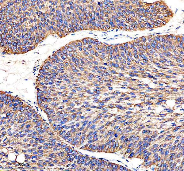
| Sample | Paraffin-embedded human ovarian cancer tissue; fixative not specified (datasheet A00948-2) |
| Fixation | Image fixative and duration unreported (datasheet A00948-2); verify before use. |
| Sectioning | 4–5 µm sections on charged slides (standard) |
| Deparaffinisation | Xylene, graded ethanol series to water (standard) |
| Antigen retrieval | Heat retrieval: EDTA pH 8.0 (datasheet A00948-2); 20 min, 95–100 °C (standard) |
| Peroxidase block | 3% H2O2, 10 min, room temperature (standard) |
| Blocking | 10% goat serum (datasheet A00948-2) |
| Primary antibody | Rabbit anti-MAP4, 1:50 recommended; image 1:100 (datasheet A00948-2) |
| Primary incubation | Overnight at 4 °C (datasheet A00948-2) |
| Detection | HRP-conjugated secondary, DAB chromogen (datasheet A00948-2) |
| Counterstain | Hematoxylin, blue, dehydrate and mount (standard) |
| Expected result | MAP4-positive staining in glial cells of caudate (HPA tissue IHC: High). HPA tissue profile: General cytoplasmic expression. Distinct expression in peripheral nerves and endothelial cells. No signal in the no-primary control. |
MAP4 is a non-neuronal microtubule-associated protein that localizes to the cytoplasm, cytoskeleton and microtubule-organizing center; it has no transmembrane segment (UniProt P27816). In tissue IHC, expect general cytoplasmic staining, with distinct staining in peripheral nerves and endothelial cells (HPA tissue IHC). HPA rates the tissue pattern Enhanced, while noting medium consistency between antibody staining and RNA expression (HPA tissue IHC).
| Cytoplasmic staining in lung endothelial cells, caudate glial cells or endometrial stromal cells. | This fits HPA's High staining in those cell populations and MAP4's cytoskeletal location (HPA tissue IHC; UniProt P27816). Judge the named cells within their tissue context; a positive field alone does not establish that every stained cell is a specific positive. |
| Predominantly nuclear staining, with little cytoplasmic staining in an expected-positive cell population. | A predominantly nuclear result conflicts with the supplied cytoplasmic and cytoskeletal localization (UniProt P27816; HPA tissue IHC). Check antibody specificity and detection controls before scoring it as MAP4; nuclear color alone is insufficient evidence of a true positive (standard IHC practice). |
| Strong staining in adipocytes or bone-marrow hematopoietic cells, especially without staining in an expected-positive control. | HPA reports MAP4 as Not detected in these specified cell populations, so review cell identification and possible cross-reactivity or endogenous detection activity (HPA tissue IHC; standard IHC practice). A negative HPA observation is a comparator, not proof that every specimen must be negative. |
| Diffuse color across cells and extracellular spaces, with poor distinction between cytoplasm and background. | This does not match HPA's cell-resolved tissue pattern (HPA tissue IHC). Run an appropriate detection-only control and review blocking, washing and chromogen development; diffuse deposit may reflect assay background rather than MAP4 localization (standard IHC practice). |
| No staining in lung endothelial cells or another documented High cell population. | The absent expected signal makes that run difficult to interpret (HPA tissue IHC). First check the positive-control section, tissue preservation, retrieval conditions, antibody dilution and detection reagents (standard IHC practice); the supplied sources establish no MAP4-specific fixation sensitivity. |
| Cell population and tissue context | HPA reports High staining in Bergmann glia, cervical squamous epithelium, kidney Bowman's capsule and lung endothelium, but Not detected staining in adipocytes and several other specified populations (HPA tissue IHC). Compare like cells within like tissues; do not score a whole organ as uniformly positive or negative. |
| Strength of the tissue evidence | The HPA tissue profile is rated Enhanced, with medium consistency between antibody staining and RNA expression; both listed antibodies have Enhanced IHC validation (HPA tissue IHC; HPA antibodies). These ratings support the reported pattern but do not validate a different antibody or every individual specimen. |
| Intracellular distribution | UniProt places MAP4 in the cytoskeleton and microtubule-organizing center; HPA describes general cytoplasmic tissue staining (UniProt P27816; HPA tissue IHC). Interpret compartment at the resolution of chromogenic paraffin IHC; fine microtubule structures may not be individually resolved (standard IHC practice). |
| Isoform and epitope coverage | UniProt lists seven MAP4 isoforms (UniProt P27816). If an antibody recognizes an isoform-dependent epitope, coverage could vary; no epitope map or isoform-specific tissue pattern is supplied, so the sources cannot assign a particular staining difference to isoform choice. |
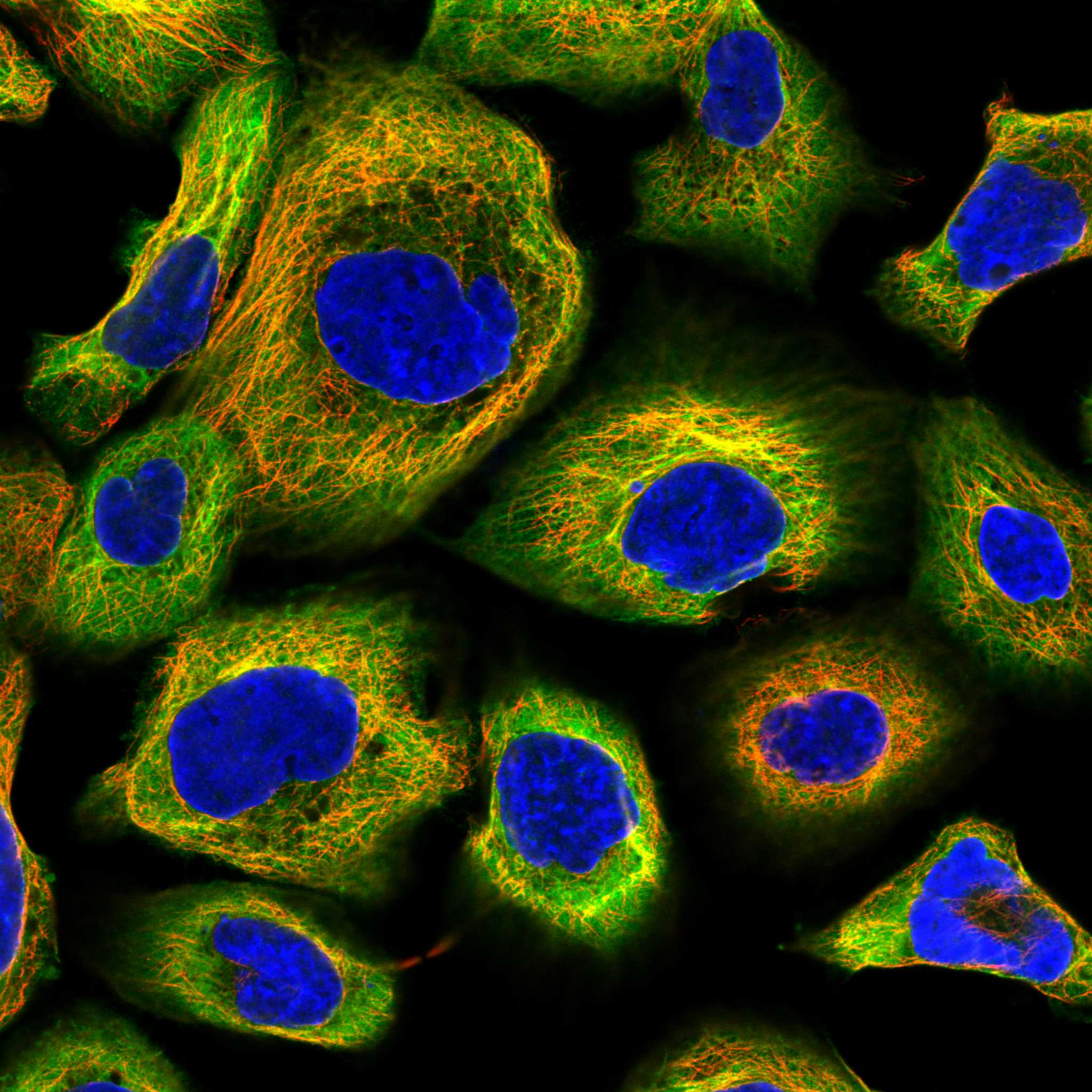
| IF/ICC Q: What localization is reported? | A: HPA supports microtubules, primary cilium and basal body in ICC-IF; it labels plasma membrane and cytosol uncertain, and also reports approved acrosome and equatorial-segment locations (HPA subcellular). This cell-imaging evidence helps interpret IF/ICC but does not establish those fine structures in chromogenic tissue IHC. |
| Situation | Likely cause | Next action |
|---|---|---|
| Expected-positive tissue is blank. | The assay may have missed antigen or failed at detection; HPA reports High staining in several defined cell populations (HPA tissue IHC; standard IHC practice). | Check an HPA-documented positive cell population on the same run, then review retrieval, antibody dilution, reagent activity and detection timing (HPA tissue IHC; standard IHC practice). No MAP4-specific retrieval condition is supplied. |
| The section is uniformly brown. | Excess detection activity, inadequate washing or prolonged chromogen development can obscure cell-specific staining (standard IHC practice). | Inspect a detection-only control; adjust blocking, washing or development using standard IHC optimization, then reassess cytoplasmic staining in named positive cells (standard IHC practice; HPA tissue IHC). |
| Nuclei dominate the signal. | Predominantly nuclear color does not fit MAP4's reported cytoskeletal location (UniProt P27816). | Compare with a known-positive cell population and appropriate negative detection control; review specificity before assigning nuclear color to MAP4 (HPA tissue IHC; standard IHC practice). |
| Adipocytes or marrow hematopoietic cells stain strongly. | That result differs from HPA's Not detected observations for those cells; cross-reactivity or endogenous activity is possible (HPA tissue IHC; standard IHC practice). | Confirm cell identity and compare a detection-only control with a documented positive population in the same run (HPA tissue IHC; standard IHC practice). Investigate the discrepancy before changing the expected pattern. |
| A reported positive tissue looks patchy. | HPA reports staining for named cell populations, not uniform positivity throughout an organ; its tissue profile has medium antibody–RNA consistency (HPA tissue IHC). | Score identifiable cells separately, compare multiple fields and record which compartments carry signal (HPA tissue IHC; standard IHC practice). Do not turn an unstained neighboring cell into a failed control. |
| The IF/ICC image and tissue IHC slide appear different. | HPA's supported cilium and microtubule assignments come from ICC-IF, whereas its tissue IHC profile describes broader cytoplasmic staining (HPA subcellular; HPA tissue IHC). | Interpret each image at its assay's spatial resolution; use the tissue IHC profile for paraffin-section scoring and consult the separate IF/ICC guide for IF workflow (HPA tissue IHC; standard IHC practice). |



Comprehensive Human Protein Atlas IHC scoring per tissue (reliability: Enhanced — Medium consistency between antibody staining and RNA expression data.). Rows are taken directly from the HPA tissue chart — click any row's HPA link to view the source.
| Tissue | Cell type | Level | Evidence | Source |
|---|---|---|---|---|
| Caudate | Glial cells | High | Protein (IHC) | HPA → |
| Cerebellum | Bergmann glia - cytoplasm/membrane | High | Protein (IHC) | HPA → |
| Cervix | Squamous epithelial cells | High | Protein (IHC) | HPA → |
| Endometrium | Cells in endometrial stroma | High | Protein (IHC) | HPA → |
| Epididymis | Glandular cells | High | Protein (IHC) | HPA → |
| Tissue | Cell type | Level | Evidence | Source |
|---|---|---|---|---|
| Adipose tissue | Adipocytes | Not detected | Protein (IHC) | HPA → |
| Adrenal gland | Glandular cells | Not detected | Protein (IHC) | HPA → |
| Bone marrow | Hematopoietic cells | Not detected | Protein (IHC) | HPA → |
| Nasopharynx | Respiratory epithelial cells | Not detected | Protein (IHC) | HPA → |
| Parathyroid gland | Glandular cells | Not detected | Protein (IHC) | HPA → |
Troubleshoot MAP4 staining in paraffin section chromogenic IHC, using the catalog antibody’s tissue image and independent localisation evidence as reference points.
A00948-2 has real IHC data from a paraffin-embedded human ovarian cancer section (IHC image caption). IF/ICC is listed for human, mouse, and rat (catalog applications and reactivity).
A00948-2 was shown by IHC in a paraffin-embedded human ovarian cancer section (IHC image caption). Its listed applications include IHC, IF, and ICC for human, mouse, and rat, but no IF image is supplied (catalog applications, reactivity, and IF image alts).
Which to pick: For tissue IHC, choose A00948-2: its image caption documents EDTA retrieval at pH 8.0 and DAB detection in a paraffin-embedded human section; the fixative is unreported (A00948-2 IHC image caption). For IF/ICC, A00948-2 is the listed option, though no IF image or IF dilution is supplied (catalog applications, IF image alts, and dilution fields). For cross-species work, A00948-2 is listed as a rabbit polyclonal antibody reactive with human, mouse, and rat; its pictured IHC evidence is human only (catalog host, dilution_raw, and reactivity; A00948-2 IHC image caption).