This website uses cookies to ensure you get the best experience on our website.


- Table of Contents
Plan MAP4K3 IHC in paraffin sections using the catalog antibody at 2–5 μg/ml (datasheet A07188-2). Compare cytoplasmic staining across tissues (HPA tissue IHC) and include controls when interpreting variable staining (HPA tissue IHC: medium staining–RNA consistency).

Expected localisation, validated protocols, controls and antibodies — the at-a-glance facts below, then the full design guide.
| Expected localisation | Cytoplasmic staining in several tissues (HPA tissue IHC) | |
| Staining pattern | High staining in fallopian tube ciliary rootlets (HPA tissue IHC) | |
| Antigen retrieval | EDTA pH 8.0 HIER, heat-mediated (datasheet A07188-2) | |
| Positive control | Fallopian tube+4 more · see all | |
| Negative control | Adipose tissue+4 more · see all |
| Fixation | Matched tissue-IHC evidence does not establish the fixation claim. Validate the specimen-specific method before use. (selected-SKU IHC image A07188-2) | |
| Caveat | Staining and RNA show medium consistency (HPA tissue IHC) | |
| Regulation | Expression regulation unreported (UniProt) | |
| Isoform / epitope | 3 isoforms; check epitope coverage (UniProt) |
The catalog antibody’s IHC-P protocol (datasheet A07188-2) is paired with one published MAP4K3 IHC protocol (PMC4467118).
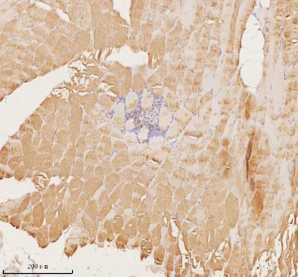
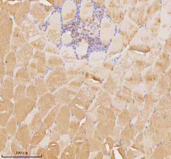
| Sample | Paraffin-embedded human skeletal muscle tissue; fixative not specified (datasheet A07188-2) |
| Fixation | Image fixative and duration unreported (datasheet A07188-2); verify before use. |
| Sectioning | 4–5 µm sections on charged slides (standard) |
| Deparaffinisation | Xylene, graded ethanol series to water (standard) |
| Antigen retrieval | Heat retrieval: EDTA pH 8.0 (datasheet A07188-2); 20 min, 95–100 °C (standard) |
| Peroxidase block | 3% H2O2, 10 min, room temperature (standard) |
| Blocking | 10% goat serum (datasheet A07188-2) |
| Primary antibody | Rabbit anti-MAP4K3, 2-5 μg/ml (datasheet A07188-2) |
| Primary incubation | Overnight at 4 °C (datasheet A07188-2) |
| Detection | HRP-conjugated secondary, DAB chromogen (datasheet A07188-2) |
| Counterstain | Hematoxylin, blue, dehydrate and mount (standard) |
| Expected result | MAP4K3-positive staining in ciliated cells (ciliary rootlets) of fallopian tube (HPA tissue IHC: High). HPA tissue profile: Cytoplasmic expression in several tissues. No signal in the no-primary control. |
In paraffin section IHC, expect cytoplasmic MAP4K3 staining in selected cell populations, especially fallopian tube ciliated cell rootlets, with moderate staining reported in several other tissues (HPA: tissue IHC). MAP4K3 has no annotated transmembrane segment (UniProt Q8IVH8: topology). The tissue IHC profile is Approved, with medium consistency against RNA data and external verification pending (HPA: tissue IHC reliability).
| Strong, localized staining at ciliary rootlets in fallopian tube ciliated cells. | This matches the highest reported tissue IHC signal and provides a useful positive reference. Assess staining in identified ciliated cells rather than treating the entire section as uniformly positive (HPA: High in fallopian tube ciliated cell rootlets). |
| Cytoplasmic staining in glomerular cells, lung macrophages, pancreatic exocrine cells, placental trophoblasts, or skeletal myocytes. | Moderate staining in these specified cell populations is compatible with the atlas pattern. Confirm cell identity from section morphology before scoring; an isolated stained structure should not define the whole tissue as positive (HPA: Medium in these cell populations). |
| Predominantly nuclear or sharply membranous staining without the expected cytoplasmic pattern. | Treat the compartment mismatch as a reason to investigate assay background or nonspecific binding. Tissue IHC reports cytoplasmic expression, while UniProt annotates no transmembrane segment; neither source alone proves that every unexpected signal is artefactual (HPA: cytoplasmic expression; UniProt Q8IVH8: topology). |
| Strong staining in cells reported as undetected, or widespread staining across unrelated cell types. | For example, adipocytes and adrenal glandular cells are reported as not detected. Check cell identification, primary antibody controls, and chromogen background before calling these cells MAP4K3 positive; an atlas negative is a reference observation, not proof of universal absence (HPA: adipocytes and adrenal glandular cells Not detected; standard IHC practice). |
| Diffuse color across tissue, including areas without a plausible cell pattern. | A diffuse result does not reproduce the reported cell and compartment distribution. Review the no primary control, blocking, washes, and chromogenic detection background before interpreting intensity as MAP4K3 expression (HPA: tissue IHC pattern; standard IHC practice). |
| Cell population and tissue selection | Signal differs within and between tissues: fallopian tube ciliated cell rootlets are High, whereas lung macrophages and pancreatic exocrine cells are Medium. Select and score the named cells rather than assuming all cells in a positive tissue stain alike (HPA: tissue IHC). |
| Strength of the tissue IHC evidence | The profile is Approved but has medium consistency with RNA expression and awaits external verification. Use the listed pattern as an interpretation benchmark and investigate unexpected staining with assay controls (HPA: tissue IHC reliability; standard IHC practice). |
| Antibody validation | The listed rabbit polyclonal HPA030380 has IHC Approved status. The supplied record does not label its IHC result Enhanced, so agreement with an independently validated reagent cannot be assumed (HPA: antibody validation). |
| Protein topology and isoforms | MAP4K3 has no signal peptide or transmembrane segment and has 3 listed isoforms. These facts support an intracellular interpretation but do not identify which isoform an IHC antibody detects or establish an epitope location (UniProt Q8IVH8: topology and isoforms). |
| Chromogenic detection background | Endogenous activity relevant to the chosen detection system can produce color unrelated to the primary antibody. Interpret staining alongside a no primary control and apply an appropriate detection activity block when indicated (standard IHC practice). |
| IF/ICC Q&A: Where should MAP4K3 appear? | HPA reports a mainly centriolar satellite location in ICC-IF, with images from A-431, U-251MG, and U2OS. Use that as an IF localization reference; paraffin section IHC is described more broadly as cytoplasmic (HPA: subcellular ICC-IF and tissue IHC). |
| Situation | Likely cause | Next action |
|---|---|---|
| No staining in fallopian tube ciliated cells. | The strongest listed positive reference is missing; assay failure or section quality may contribute, but the image alone cannot identify the cause (HPA: High in ciliated cell rootlets; standard IHC practice). | Check that ciliated cells are present and identifiable, then review primary antibody application, retrieval conditions, and detection controls against the validated IHC workflow (standard IHC practice). |
| Little or no staining in lung parenchyma despite an otherwise usable section. | The reported lung signal is in macrophages at Medium intensity, so a field lacking identifiable macrophages may appear negative (HPA: Medium in lung macrophages). | Inspect fields containing morphologically identifiable macrophages and compare with a named positive reference tissue; avoid scoring all lung cells as the expected positive population (HPA: lung macrophages; standard IHC practice). |
| Strong color appears in adipocytes or adrenal glandular cells. | Both populations are listed as Not detected, so the result conflicts with the atlas pattern; nonspecific signal or misidentified cells are possibilities (HPA: tissue IHC; standard IHC practice). | Recheck morphology and a no primary control, then review blocking and detection background before assigning MAP4K3 positivity (standard IHC practice). |
| Most cells show uniform, weak brown haze. | Diffuse chromogen can obscure the reported cell selective cytoplasmic pattern (HPA: tissue IHC; standard IHC practice). | Compare with the no primary control and review washing, blocking, and the chosen detection system. Score only cellular staining that remains interpretable above background (standard IHC practice). |
| Signal is predominantly nuclear or outlines plasma membranes. | The dominant compartment differs from the reported cytoplasmic IHC profile; no transmembrane segment is annotated (HPA: cytoplasmic expression; UniProt Q8IVH8: topology). | Check the positive reference and controls, then reassess antibody specificity and detection background before accepting the unexpected localization (standard IHC practice). |
| Weak staining in a cell population expected to be moderately positive. | HPA intensity categories describe observed patterns, and its Approved profile has medium RNA consistency with external verification pending; a single weak field is inconclusive (HPA: tissue IHC reliability and intensity). | Confirm cell identity, assess additional suitable fields, and compare the run with its positive and no primary controls before changing the interpretation (standard IHC practice). |


Comprehensive Human Protein Atlas IHC scoring per tissue (reliability: Approved — Medium consistency between antibody staining and RNA expression data. Pending external verification.). Rows are taken directly from the HPA tissue chart — click any row's HPA link to view the source.
| Tissue | Cell type | Level | Evidence | Source |
|---|---|---|---|---|
| Fallopian tube | Ciliated cells (ciliary rootlets) | High | Protein (IHC) | HPA → |
| Bronchus | Ciliated cells (ciliary rootlets) | Medium | Protein (IHC) | HPA → |
| Kidney | Cells in glomeruli | Medium | Protein (IHC) | HPA → |
| Lung | Macrophages | Medium | Protein (IHC) | HPA → |
| Nasopharynx | Ciliated cells (ciliary rootlets) | Medium | Protein (IHC) | HPA → |
| Tissue | Cell type | Level | Evidence | Source |
|---|---|---|---|---|
| Adipose tissue | Adipocytes | Not detected | Protein (IHC) | HPA → |
| Adrenal gland | Glandular cells | Not detected | Protein (IHC) | HPA → |
| Appendix | Glandular cells | Not detected | Protein (IHC) | HPA → |
| Breast | Adipocytes | Not detected | Protein (IHC) | HPA → |
| Caudate | Glial cells | Not detected | Protein (IHC) | HPA → |
Troubleshoot MAP4K3 staining in paraffin sections by checking retrieval, cellular pattern, controls, and scoring before assigning biological meaning.
The IHC-validated anti-MAP4K3 antibody has an image from a human skeletal muscle paraffin section (catalog IHC caption); its listed species reactivity is human, mouse and rat (catalog reactivity).
A07188-2 will render with an IHC image from a human skeletal muscle paraffin section (catalog IHC caption). Its application list includes IHC, and its listed reactivity covers human, mouse and rat (catalog applications and reactivity).
Which to pick: For tissue IHC, choose A07188-2: its caption documents a human skeletal muscle paraffin section stained at 2 μg/ml after heat retrieval in EDTA, pH 8.0 (catalog IHC caption). The caption does not report the fixative (catalog IHC caption). A07188-2 has no listed IF/ICC application or IF image, so it is not an evidenced IF/ICC choice; for cross-species work, it lists mouse and rat reactivity, while the supplied IHC image documents human tissue only (catalog applications, reactivity and IHC caption).