MAP4K4 / Mitogen-activated protein kinase kinase kinase kinase 4 · IHC design guide

Design Immunohistochemistry for MAP4K4

Plan chromogenic IHC on paraffin sections using cytoplasmic staining as the expected MAP4K4 pattern (HPA tissue IHC). Compare kidney tubules and hepatocytes with reference staining, and interpret intensity cautiously because antibody staining has low consistency with RNA expression (HPA tissue IHC).

Evidence assembled Oct 2026 · For research use; verify linked source records and product datasheet before use
Immunohistochemistry protocol sheet for MAP4K4 (IHC for MAP4K4): expected localisation Cytoplasmic staining in several tissues (HPA tissue IHC), antibody A02829-2, validated IHC image, and IHC protocol steps
Printable MAP4K4 IHC protocol sheet — expected localisation Cytoplasmic staining in several tissues (HPA tissue IHC), antibody A02829-2, controls and protocol steps. Open the full MAP4K4 IHC guide →

MAP4K4 Immunohistochemistry Experimental Design Guide

Expected localisation, validated protocols, controls and antibodies — the at-a-glance facts below, then the full design guide.

Must know before staining
Expected localisation Cytoplasmic staining in several tissues (HPA tissue IHC)
Staining pattern Cytoplasmic staining in kidney tubules and hepatocytes (HPA tissue IHC)
Antigen retrieval EDTA pH 8.0 HIER, heat-mediated (datasheet A02829-2)
Positive control ⓘ Kidney+3 more · see all
Negative control ⓘ Adipose tissue+4 more · see all
Important caveats
Reasons your staining may differ from the expected pattern.
Fixation Keep fixation consistent across sections (standard IHC practice; not target-specific); Matched tissue-IHC evidence does not establish the fixation claim. Validate the specimen-specific method before use. (selected-SKU IHC image A02829-2)
Caveat Staining has low consistency with RNA expression (HPA tissue IHC)
Regulation Brain-enhanced RNA expression (HPA tissue RNA)
Isoform / epitope 6 isoforms; antibody epitope coverage needs verification (UniProt)
Section 1

Recommended MAP4K4 IHC & IF Protocols

Compare the catalog antibody’s IHC-P protocol (datasheet A02829-2) with published MAP4K4 staining methods for endometrial tissue (PMC13592435) and Kaposi sarcoma tissue (PMC3820715).

Recommended immunohistochemistry (IHC-P) protocol parameters
SampleParaffin-embedded human cervical cancer tissue; fixative not specified (datasheet A02829-2)
FixationImage fixative and duration unreported (datasheet A02829-2); verify before use.
Sectioning4–5 µm sections on charged slides (standard)
DeparaffinisationXylene, graded ethanol series to water (standard)
Antigen retrievalHeat retrieval: EDTA pH 8.0 (datasheet A02829-2); 20 min, 95–100 °C (standard)
Peroxidase block3% H2O2, 10 min, room temperature (standard)
Blocking10% goat serum (datasheet A02829-2)
Primary antibodyRabbit anti-MAP4K4, 2-5 μg/ml (datasheet A02829-2)
Primary incubationOvernight at 4 °C (datasheet A02829-2)
DetectionHRP-conjugated secondary, DAB chromogen (datasheet A02829-2)
CounterstainHematoxylin, blue, dehydrate and mount (standard)
Expected resultMAP4K4-positive staining in cells in tubules of kidney (HPA tissue IHC: Medium). HPA tissue profile: Cytoplasmic expression in several different tissues types. No signal in the no-primary control.
💡Decision noteStart with EDTA pH 8.0 heat retrieval for the catalog antibody (datasheet A02829-2); the published endometrial protocol used citrate pH 6.0 (PMC13592435).
Section 2

What Is the Expected MAP4K4 Staining Pattern?

MAP4K4 is a cytoplasmic protein with no transmembrane segment (UniProt O95819: subcellular location and topology). In paraffin-section IHC, expect cytoplasmic staining in kidney tubular cells, hepatocytes, stomach glandular cells, and cells in seminiferous ducts; HPA reports medium staining in each (HPA: tissue IHC). Treat this as a provisional reference pattern: HPA rates the tissue staining Uncertain because antibody staining and RNA expression show low consistency, with a possible splice or transcript discrepancy (HPA: reliability).

What am I looking at on my slide?
Cytoplasmic staining in kidney tubular cells, hepatocytes, stomach glandular cells, or cells in seminiferous ducts.This matches the reported medium IHC pattern (HPA: tissue IHC) and cytoplasmic location (UniProt O95819: subcellular location). Compare cell type and compartment, while keeping HPA's Uncertain reliability in view (HPA: reliability).
Predominantly nuclear or sharply membrane-bound staining, with little cytoplasmic signal.That compartment differs from the recorded cytoplasmic location and lacks support from a transmembrane segment (UniProt O95819: subcellular location and topology). Treat it as a possible staining artefact; review morphology, counterstain, and control slides before assigning it to MAP4K4 (general IHC practice).
Strong staining in a cell type reported as undetected, such as adipocytes in adipose tissue or hematopoietic cells in bone marrow.Those cell-specific results conflict with HPA's Not detected observations (HPA: tissue IHC). Cross-reactivity or endogenous chromogen-generating activity is possible; inspect detection controls and repeat with a separate validated antibody if available (general IHC practice).
Diffuse color across tissue, stroma, and spaces without clear cell boundaries.A non-cellular wash of color does not resemble HPA's cell-specific cytoplasmic pattern (HPA: tissue IHC). Background from detection reagents or insufficient blocking is possible; compare a no-primary control and review washing, blocking, and chromogen development (general IHC practice).
No staining in kidney tubular cells despite preserved morphology.HPA reports medium staining in kidney tubular cells, so an absent result needs a technical check, though the reference pattern is rated Uncertain (HPA: tissue IHC and reliability). Check the assay controls, antigen-retrieval conditions, antibody dilution, and detection reagents (general IHC practice).
💡Expected MAP4K4 appearanceCall a provisional positive when cytoplasmic color is discernible at roughly medium intensity in the reported kidney, liver, stomach, or testis cell types (HPA: tissue IHC; UniProt O95819: subcellular location); isolated nuclear, membrane-like, or diffuse non-cellular color warrants control-based review (general IHC practice).
How each factor affects the staining
Tissue and cell typeHPA reports medium staining in kidney tubular cells, hepatocytes, stomach glandular cells, and cells in seminiferous ducts; it reports low staining in several other specified cell types (HPA: tissue IHC). Score cells within tissue rather than assuming uniform tissue-wide staining (general IHC practice).
Protein location and topologyMAP4K4 is annotated in the cytoplasm and has no transmembrane segment (UniProt O95819: subcellular location and topology). These annotations support cytoplasmic interpretation; they do not establish that every cytoplasmic cell must stain in a given section.
Isoform compositionUniProt lists 6 isoforms, with isoform 5 abundant in brain and isoform 4 predominant in liver, skeletal muscle, and placenta (UniProt O95819: isoforms and tissue specificity). The supplied records do not identify the IHC antibody's epitope, so isoform-specific staining cannot be assigned.
Validation and RNA contextHPA rates the tissue pattern Uncertain and antibody HPA008476 Uncertain for IHC; it notes low staining–RNA consistency and a possible splice or transcript discrepancy (HPA: reliability and antibody validation). Brain-enhanced RNA alone does not predict strong brain IHC: reported staining is low in specified brain cells (HPA: tissue IHC).
Processing and modificationsUniProt reports no signal peptide or propeptide and lists phosphorylation sites and other modified residues (UniProt O95819: processing and modified residues). Without an antibody epitope or target-specific retrieval data, these facts cannot predict staining intensity, retrieval needs, or loss of signal in fixed sections.
Why is my staining missing, weak or wrong?
SituationLikely causeNext action
Expected cytoplasmic signal is absent in kidney tubular cells.A missed assay step or poorly matched retrieval or antibody conditions may suppress staining (general IHC practice); HPA's kidney reference is medium but Uncertain (HPA: tissue IHC and reliability).Confirm a working positive control and detection reagents, then review the antibody's validated IHC-P instructions for retrieval and dilution; compare kidney cells on a repeat section (general IHC practice).
Color is predominantly nuclear or confined to cell borders.The pattern conflicts with cytoplasmic location and absence of a transmembrane segment (UniProt O95819: subcellular location and topology); misread morphology or nonspecific detection is possible (general IHC practice).Check the counterstain and a no-primary control, then assess whether cytoplasmic color remains in correctly identified cells (general IHC practice).
Unexpected strong color appears in HPA-undetected cell types.Adipocytes in adipose tissue and hematopoietic cells in bone marrow are reported Not detected (HPA: tissue IHC); cross-reactivity or endogenous detection activity may explain a discordant result (general IHC practice).Run suitable detection controls, assess cell identity, and seek confirmation with another IHC-validated antibody if available (general IHC practice).
Whole sections show diffuse brown background.Excess chromogen development, inadequate washing, or unblocked endogenous activity can produce background in chromogenic IHC (general IHC practice); this differs from HPA's cell-specific cytoplasmic profile (HPA: tissue IHC).Compare a no-primary control; review wash, blocking, and development conditions while retaining a positive tissue control (general IHC practice).
Brain appears weak despite brain-enhanced RNA.HPA calls RNA tissue enhanced in brain but records low staining in glial cells of cerebral cortex and caudate and Purkinje cells of cerebellum (HPA: tissue IHC). RNA abundance and observed IHC staining are distinct measurements.Interpret the observed cell-specific IHC signal against the low protein-staining reference, and retain HPA's Uncertain reliability caveat (HPA: tissue IHC and reliability).
Can an IF/ICC image settle an ambiguous IHC result?The supplied HPA subcellular record has no main IF location or cell lines with ICC-IF images (HPA: subcellular); it provides no image-based corroboration for this case.Use the cytoplasmic UniProt annotation and the qualified tissue IHC observations for interpretation (UniProt O95819: subcellular location; HPA: tissue IHC and reliability). Consult the separate IF/ICC guide for that application's workflow.

Sample controls for MAP4K4 IHC & IF

🧪Run liver first: hepatocytes should show MAP4K4 staining (HPA: liver hepatocytes, Medium). Use adipose tissue as the negative tissue: adipocytes should lack detectable staining (HPA: adipose adipocytes, Not detected); on the liver slide, internal negative cells should lack specific cytoplasmic DAB staining, but the supplied HPA row identifies no validated internal negative cell type (HPA: liver hepatocytes, Medium; UniProt O95819: cytoplasm).
Positive control tissue: Kidney (Cells in tubules, HPA Medium)
Negative control tissue: Adipose tissue (HPA Not detected)
ICC-IF cell lines (HPA subcellular resource): HPA carries no ICC-IF cell line for MAP4K4; derive a cell-line control from the positive tissue's cell type (Cells in tubules) and confirm it by RNA or western blot first.
Technical controls: Include a no-primary, secondary-only slide and a concentration-matched rabbit IgG isotype control for the rabbit primary, alongside MAP4K4-knockout material or a validated peptide-block control (selected-SKU caption: rabbit anti-MAP4K4; standard IHC practice). Block endogenous peroxidase and check liver pigment for apparent DAB signal (standard chromogenic IHC practice).
⚠️Feasibility: A target-specific fixation window or fixation effect is unreported, and the selected-SKU paraffin-section caption does not state a fixative (selected-SKU tissue-IHC caption). The reported staining used heat retrieval in EDTA, pH 8.0, followed by 2 μg/ml primary overnight at 4°C; the evidence does not establish whether retrieval is required (selected-SKU tissue-IHC caption). Relative ease of frozen-section IHC or IF is unreported, and liver pigment can complicate chromogenic scoring (supplied application evidence; standard IHC practice).

HPA tissue IHC evidence for MAP4K4

Comprehensive Human Protein Atlas IHC scoring per tissue (reliability: Uncertain — Low consistency between antibody staining and RNA expression data. Pending external verification. Caution, Splice and/or transcript discrepancy exists.). Rows are taken directly from the HPA tissue chart — click any row's HPA link to view the source.

Positive expression · recommended positive controls

TissueCell typeLevelEvidenceSource
Kidney Cells in tubules Medium Protein (IHC) HPA →
Liver Hepatocytes Medium Protein (IHC) HPA →
Stomach Glandular cells Medium Protein (IHC) HPA →
Testis Cells in seminiferous ducts Medium Protein (IHC) HPA →

Undetected expression · recommended negative controls

TissueCell typeLevelEvidenceSource
Adipose tissue Adipocytes Not detected Protein (IHC) HPA →
Adrenal gland Glandular cells Not detected Protein (IHC) HPA →
Bone marrow Hematopoietic cells Not detected Protein (IHC) HPA →
Breast Adipocytes Not detected Protein (IHC) HPA →
Bronchus Respiratory epithelial cells Not detected Protein (IHC) HPA →
Section 3

Advanced MAP4K4 IHC Tips

Use the documented MAP4K4 tissue protocol as a starting point, then assess staining by compartment, cell type, and controls.

Which retrieval condition should I try first for MAP4K4 in paraffin sections?
Start with heat-mediated antigen retrieval in EDTA at pH 8.0 (datasheet A02829-2). The documented paraffin-section example used that condition before overnight incubation with 2 μg/ml antibody at 4°C (datasheet A02829-2). Keep heating and cooling conditions consistent across comparison slides, because retrieval variation can change chromogenic staining intensity (standard IHC practice). If staining remains weak, compare a carefully controlled alternative retrieval condition on adjacent sections while retaining the documented EDTA condition as the reference (standard IHC practice). Check tissue integrity and the expected cytoplasmic pattern before treating stronger signal as improved detection (UniProt O95819 localisation; standard IHC practice).
How should I assess fixation when MAP4K4 staining is weak?
The selected paraffin-section caption does not state a fixative, so MAP4K4-specific fixation sensitivity is unknown (datasheet A02829-2). Record each specimen’s fixative and fixation duration, and compare sections processed together before attributing weak staining to antigen loss (standard IHC practice). Excessive or inconsistent fixation can alter antigen accessibility in IHC generally, but no MAP4K4-specific effect is established here (standard IHC practice; datasheet A02829-2). Apply the documented EDTA retrieval at pH 8.0 consistently when comparing specimens (datasheet A02829-2). Include an appropriate positive tissue control and inspect morphology so that processing differences do not masquerade as biological differences (standard IHC practice).
What MAP4K4 staining pattern should count as plausible?
Assess predominantly cytoplasmic staining, consistent with the recorded subcellular location and reported tissue profile (UniProt O95819 localisation; HPA tissue IHC profile). MAP4K4 has no annotated transmembrane segment, so a crisp membrane-only pattern needs independent validation before interpretation (UniProt O95819 topology; standard IHC practice). Kidney tubule cells, hepatocytes, stomach glandular cells, and cells in seminiferous ducts have reported medium staining and can guide compartment checks (HPA tissue IHC). Distinguish positive cells from adjacent unstained cells on the same section, and assess the counterstain and morphology before scoring (standard IHC practice). Treat an exclusively nuclear or diffuse extracellular pattern as a troubleshooting signal rather than proof of a new location (UniProt O95819 localisation; standard IHC practice).
Could isoforms or epitope accessibility explain discordant tissue staining?
MAP4K4 has 6 annotated isoforms, and the supplied antibody caption does not identify its epitope or establish isoform coverage (UniProt O95819 isoforms; datasheet A02829-2). Isoform 5 is abundant in brain, while isoform 4 predominates in liver, skeletal muscle, and placenta (UniProt O95819 tissue specificity). Check the antibody’s epitope documentation before interpreting differences between those tissues as changes in total MAP4K4 (standard IHC practice; UniProt O95819 isoforms). The protein includes a kinase domain at residues 25–289 and a CNH domain at 926–1213, providing landmarks for assessing a documented epitope (UniProt O95819 domains). An unknown epitope cannot support an isoform-specific staining claim (standard IHC practice).
How can I assess MAP4K4 by multiplex IF alongside chromogenic IHC?
Treat IF as a separate assay: the supplied paraffin-section example documents chromogenic detection with DAB, not IF performance (datasheet A02829-2). Pair MAP4K4 with a validated marker for the cell type being assessed, and check each channel separately before interpreting overlap (standard IF practice). Choose fluorophores after examining unstained tissue autofluorescence, favoring channels with a clear signal above that background (standard IF practice). Because MAP4K4 is annotated in the cytoplasm and has no transmembrane segment, use permeabilisation appropriate for access to an intracellular epitope, then verify morphology and background (UniProt O95819 localisation and topology; standard IF practice). Do not transfer the chromogenic antibody concentration of 2 μg/ml to IF without optimisation (datasheet A02829-2; standard IF practice).
What should I change when MAP4K4 DAB staining is widespread?
Separate background from cytoplasmic cell staining by reviewing a no-primary control, tissue morphology, and the counterstain (UniProt O95819 localisation; standard IHC practice). The documented example used 10% goat serum blocking, 2 μg/ml primary antibody overnight at 4°C, and a peroxidase-conjugated secondary for 30 minutes at 37°C (datasheet A02829-2). Check whether serum blocking, primary concentration, washes, or secondary incubation need adjustment using matched sections (standard IHC practice). Include a peroxidase-blocking step when using HRP and DAB, and assess whether pigment or endogenous enzyme activity contributes to apparent signal (standard IHC practice). Preserve the EDTA pH 8.0 retrieval reference while changing one variable at a time (datasheet A02829-2; standard IHC practice).
How should I quantify MAP4K4 staining across tissue samples? ⚠ ANSWER MARKED FOR VERIFICATION
Define the cell population and cytoplasmic scoring compartment before comparing slides (UniProt O95819 localisation; standard IHC practice). For chromogenic sections, record the percentage of positive target cells and staining intensity, then calculate an H-score if an intensity-weighted summary is needed (standard IHC practice). Alternatively, report positive-cell density per mm² of viable tissue when cell abundance itself is the outcome (standard IHC practice). Normalise to the number of evaluable target cells or viable tissue area, and apply the same threshold, imaging settings, and region-selection rules across specimens (standard IHC practice). Report controls and staining variability because HPA rates its MAP4K4 tissue IHC reliability as uncertain (HPA tissue IHC reliability).
How can I distinguish MAP4K4-positive cells from artefact?
Favor staining within intact cytoplasm over isolated nuclear, extracellular, or sharply peripheral signal (UniProt O95819 localisation; standard IHC practice). Confirm that the stained cell type matches the question being tested; HPA reports medium staining in kidney tubule cells and hepatocytes, while its tissue IHC assessment is uncertain (HPA tissue IHC). Exclude section edges, folds, and necrotic areas from scoring when they show disproportionate DAB deposition (standard IHC practice). Use a no-primary control and adequate peroxidase blocking to investigate endogenous enzyme signal (standard IHC practice). The catalog example shows MAP4K4 staining in human cervical cancer, whereas HPA reports no detection in normal cervical glandular cells; interpret that difference within their distinct specimens and assay limits (datasheet A02829-2; HPA tissue IHC).
Boster reagents

Best MAP4K4 / Mitogen-activated protein kinase kinase kinase kinase 4 IHC Antibodies

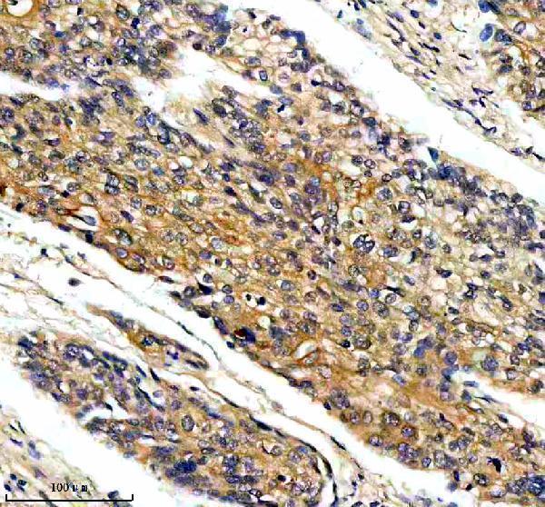
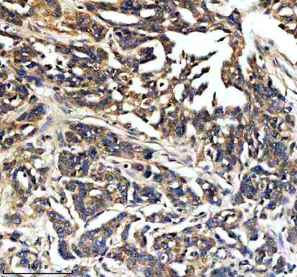
The IHC-validated antibody has images from human cervical and ovarian cancer paraffin sections (catalog image captions). No IF/ICC data are supplied (catalog applications; image payload).

Real IHC data IHC analysis of MAP4K4 using anti-MAP4K4 antibody (A02829-2). MAP4K4 was detected in a paraffin-embedded section of human cervical cancer tissue. Heat mediated antigen retrieval was performed in EDTA buffer (pH 8.0, epitope retrieval solution). The tissue section was blocked with 10% goat serum. The tissue section was then incubated with 2 μg/ml rabbit anti-MAP4K4 Antibody (A02829-2) overnight at 4°C. Peroxidase Conjugated Goat Anti-rabbit IgG was used as secondary antibody and incubated for 30 minutes at 37°C. The tissue section was developed using HRP Conjugated Rabbit IgG Super Vision Assay Kit (Catalog # SV0002) with DAB as the chromogen.
Anti-MAP4K4 Antibody ®
Cat # A02829-2

A02829-2 will render with an IHC figure from a human cervical cancer paraffin section (catalog image caption). Its second catalog image shows a human ovarian cancer paraffin section; the listed reactivity is human, mouse and rat (catalog image caption; catalog reactivity).

Which to pick: Choose A02829-2 for tissue IHC in human paraffin sections: its images document EDTA retrieval at pH 8.0 and primary antibody at 2 μg/ml (catalog image captions); the fixative is unreported (catalog image captions). No SKU can be recommended for IF/ICC because none has IF/ICC listed or an IF image (catalog applications; image payload). A02829-2 lists mouse and rat reactivity, but the supplied tissue IHC images show human samples only (catalog reactivity; catalog image captions).

Each figure is that product's own IHC / IF validation image from its datasheet.

References

  1. UniProt Consortium. UniProt entry O95819 (M4K4_HUMAN, Mitogen-activated protein kinase kinase kinase kinase 4).
  2. Human Protein Atlas. MAP4K4 tissue IHC expression (reliability: Uncertain).
  3. Human Protein Atlas. MAP4K4 subcellular location (ICC-IF): Highest expression in Hep-G2: 324.3 nTPM.
  4. Human Protein Atlas. MAP4K4 antibody validation summary (1 antibodies).
  5. Methodological Pitfalls in Endometriosis Biospecimen Research: Lessons From MAP4K4 Expression Studies. FASEB bioAdvances 2026 — PMC13592435.
  6. The inflammatory kinase MAP4K4 promotes reactivation of Kaposi's sarcoma herpesvirus and enhances the invasiveness of infected endothelial cells. PLoS pathogens 2013 — PMC3820715.
  7. MAP4K4 induces early blood-brain barrier damage in a murine subarachnoid hemorrhage model. Neural regeneration research 2021 — PMC7896238.
  8. A novel pharmacological inhibitor of MAP4K4 activity attenuates metabolic dysfunction-associated steatohepatitis. JHEP reports : innovation in hepatology 2026 — PMC13380734.
  9. PubMed PMID:9890973 — UniProt-cited evidence.
  10. PubMed PMID:12612079 — UniProt-cited evidence.
  11. PubMed PMID:15815621 — UniProt-cited evidence.