This website uses cookies to ensure you get the best experience on our website.


- Table of Contents
Plan MAP4K5 staining in paraffin sections around the reported cytoplasmic tissue pattern, while accounting for its uncertain reliability (HPA tissue IHC). This guide covers tissue controls, fixation consistency and the catalog antibody’s IHC range of 2–5 µg/mL (datasheet A10186-1).

Expected localisation, validated protocols, controls and antibodies — the at-a-glance facts below, then the full design guide.
| Expected localisation | Cytoplasmic staining in a subset of tissues (HPA tissue IHC) | |
| Staining pattern | Cytoplasmic staining, mainly in smooth muscle (HPA tissue IHC) | |
| Antigen retrieval | EDTA pH 8.0 HIER, heat-mediated (datasheet A10186-1) | |
| Positive control | Cerebellum+4 more · see all | |
| Negative control | Adipose tissue+4 more · see all |
| Fixation | Keep fixation consistent across paraffin sections. (standard IHC practice; not target-specific) | |
| Caveat | Tissue staining has uncertain reliability and low RNA concordance (HPA tissue IHC) | |
| Regulation | Higher in ovary, testis and prostate (UniProt) | |
| Isoform / epitope | No listed isoforms; one unprocessed chain (UniProt) |
The catalog antibody protocol uses EDTA pH 8.0 heat retrieval (datasheet A10186-1). The published pancreatic tissue protocol below reports a steamer-based IHC workflow (PMC4811546).
| Sample | Paraffin-embedded human ovarian cancer tissue; fixative not specified (datasheet A10186-1) |
| Fixation | Image fixative and duration unreported (datasheet A10186-1); verify before use. |
| Sectioning | 4–5 µm sections on charged slides (standard) |
| Deparaffinisation | Xylene, graded ethanol series to water (standard) |
| Antigen retrieval | Heat retrieval: EDTA pH 8.0 (datasheet A10186-1); 20 min, 95–100 °C (standard) |
| Peroxidase block | 3% H2O2, 10 min, room temperature (standard) |
| Blocking | 10% goat serum (datasheet A10186-1) |
| Primary antibody | Rabbit anti-MAP4K5, 2μg/ml (datasheet A10186-1) |
| Primary incubation | Overnight at 4 °C (datasheet A10186-1) |
| Detection | Streptavidin-biotin complex (SABC), DAB chromogen (datasheet A10186-1) |
| Counterstain | Hematoxylin, blue, dehydrate and mount (standard) |
| Expected result | MAP4K5-positive staining in purkinje cells of cerebellum (HPA tissue IHC: Medium). HPA tissue profile: Cytoplasmic expression in a subset of tissues, mainly smooth muscle. No signal in the no-primary control. |
MAP4K5 is a cytoplasmic protein with no transmembrane segment (UniProt Q9Y4K4: subcellular location and topology). In paraffin section IHC, expect cytoplasmic staining in selected cells; HPA describes a subset of tissues, mainly smooth muscle, and reports medium staining in several named cell populations (HPA: tissue IHC). Treat these as provisional reference patterns: HPA rates tissue IHC reliability Uncertain because antibody staining and RNA expression show low consistency (HPA: tissue IHC reliability).
| Cytoplasmic staining in smooth muscle or in HPA-listed cells, such as ovarian follicle cells or cerebellar Purkinje cells (HPA: tissue IHC). | This fits the reported compartment and cell distribution (UniProt Q9Y4K4: cytoplasm; HPA: tissue IHC). HPA scores the listed follicle and Purkinje cells Medium, but its tissue IHC reliability is Uncertain (HPA: tissue IHC). |
| Predominantly nuclear staining, or a sharp membrane-only outline, with little cytoplasmic signal. | That conflicts with the reported cytoplasmic IHC profile and UniProt location (HPA: tissue IHC; UniProt Q9Y4K4: cytoplasm). Review controls and imaging before assigning MAP4K5; membrane localization in ICC-IF is itself uncertain (HPA: subcellular). |
| Strong staining in cells HPA reports as not detected, such as adipocytes or cardiomyocytes (HPA: tissue IHC). | Investigate antibody cross-reactivity or endogenous detection activity (standard IHC practice). A discordant cell type is a warning, not proof of either mechanism; HPA's tissue IHC assessment is Uncertain (HPA: tissue IHC reliability). |
| Diffuse colour across many cell types, including areas expected to be unstained. | A widespread haze can obscure cell boundaries and make compartment scoring unreliable (standard IHC practice). Assess a no-primary control and compare staining with HPA's selective cytoplasmic tissue profile (standard IHC practice; HPA: tissue IHC). |
| No cytoplasmic signal in an HPA-listed medium-staining population, such as colon endothelial cells (HPA: tissue IHC). | First check tissue identity, preservation, retrieval, primary incubation and detection controls (standard IHC practice). HPA's Medium rating is an observed pattern, not a guarantee that every section or antibody will stain (HPA: tissue IHC reliability Uncertain). |
| Location and topology | UniProt places MAP4K5 in the cytoplasm and lists no transmembrane segment (UniProt Q9Y4K4: location and topology). Judge chromogenic IHC primarily by cytoplasmic cell staining; do not require a membrane outline to call a positive result (HPA: tissue IHC; standard IHC practice). |
| Tissue and cell selection | HPA reports cytoplasmic staining mainly in smooth muscle and Medium staining in selected cells, including duodenal glandular cells and skeletal myocytes (HPA: tissue IHC). Select controls by the scored cell population within the section, rather than by tissue name alone (standard IHC practice). |
| Expression versus observed staining | UniProt describes expression throughout examined tissues, with higher levels in ovary, testis and prostate (UniProt Q9Y4K4: tissue specificity). HPA reports only a subset of tissues staining and low tissue RNA specificity (HPA: tissue IHC). Do not infer an IHC positive solely from expression information (standard IHC practice). |
| Strength of IHC evidence | HPA calls tissue IHC Uncertain for low antibody-staining and RNA-expression consistency; HPA036696 is also rated Uncertain for IHC (HPA: tissue IHC reliability; HPA: antibody validation). Interpret a new pattern alongside controls rather than treating the reference image as definitive (standard IHC practice). |
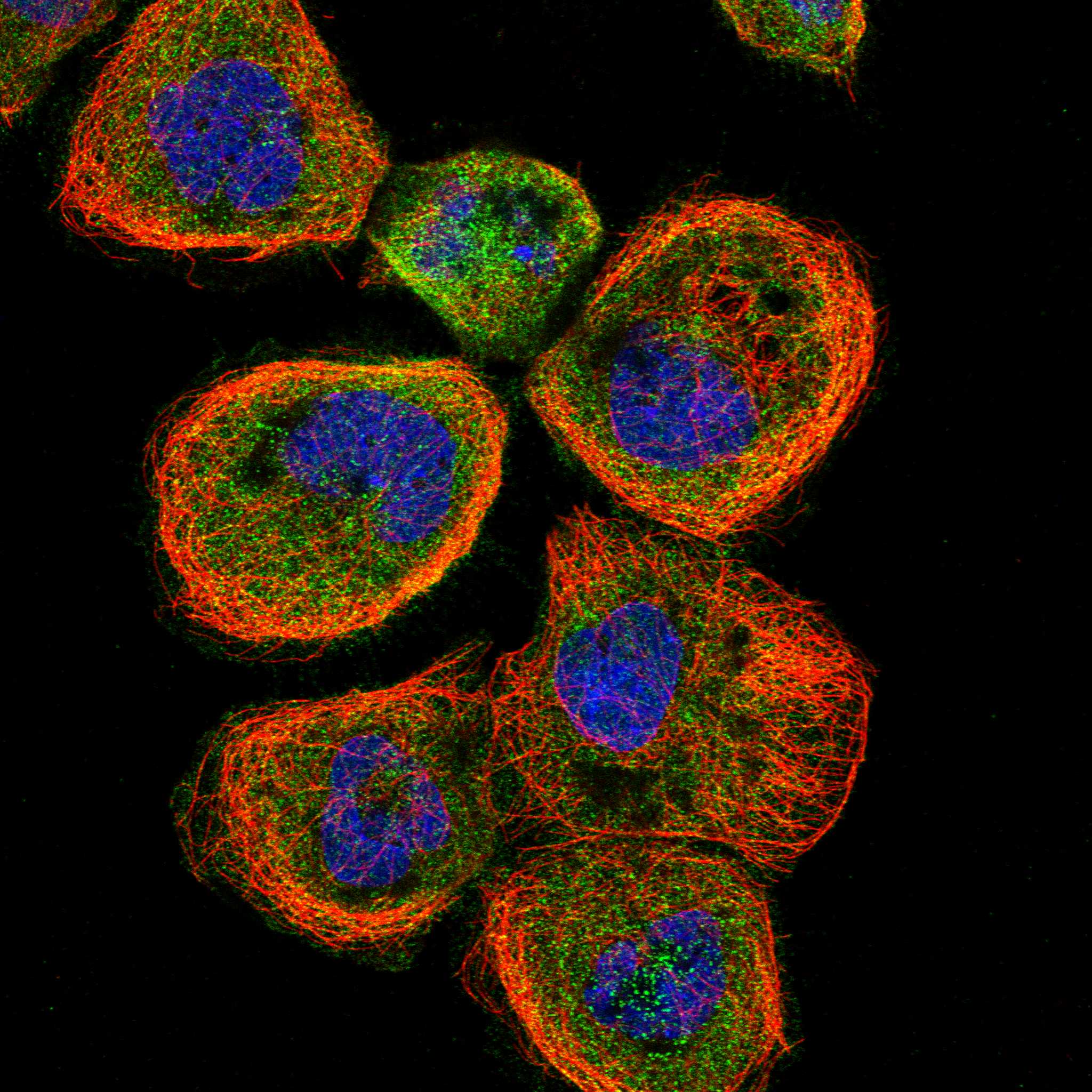
| IF/ICC Q: should a membrane or ciliary signal count? | A: HPA supports cytosol as the main ICC-IF location; plasma membrane and primary cilium are additional uncertain locations (HPA: subcellular). These IF observations do not establish the expected chromogenic paraffin-section IHC pattern (HPA: subcellular; HPA: tissue IHC). |
| Situation | Likely cause | Next action |
|---|---|---|
| HPA-listed positive cells show no staining. | The relevant cells may be absent from the section, or an IHC processing or detection step may have failed (standard IHC practice). | Confirm cell identity and assess a known-working tissue control; review retrieval, primary incubation and chromogen development (standard IHC practice). Treat HPA Medium staining as provisional because tissue IHC reliability is Uncertain (HPA: tissue IHC). |
| Nuclei dominate the signal. | This differs from the documented cytoplasmic location (UniProt Q9Y4K4: subcellular location; HPA: tissue IHC). | Check whether nuclear colour persists in a no-primary control, then reassess antibody conditions and compartment scoring (standard IHC practice). Do not score nuclear-only staining as the expected MAP4K5 pattern (UniProt Q9Y4K4: cytoplasm). |
| Cells reported as HPA-negative stain strongly. | Cross-reactivity or endogenous detection activity can produce cell-specific colour (standard IHC practice); HPA lists adipocytes and cardiomyocytes as Not detected (HPA: tissue IHC). | Compare a no-primary control, review detection blocking and check staining in a listed medium-staining cell population (standard IHC practice; HPA: tissue IHC). A single HPA-negative result cannot validate specificity because HPA reliability is Uncertain (HPA: tissue IHC). |
| Background colour obscures cell boundaries. | Nonspecific antibody binding or endogenous detection activity may contribute (standard IHC practice). | Inspect no-primary controls, revisit blocking and washing, and adjust primary antibody concentration using the antibody's IHC guidance if available (standard IHC practice). Score only where cytoplasmic signal is distinguishable from background (UniProt Q9Y4K4: cytoplasm; standard IHC practice). |
| Staining varies across a section or between runs. | Uneven reagent coverage, retrieval or development can alter chromogenic signal (standard IHC practice); no MAP4K5-specific fixation sensitivity is supplied (UniProt Q9Y4K4; HPA: tissue IHC). | Check section coverage and run controls, then compare like-for-like regions and cell populations (standard IHC practice). Avoid attributing the variation to a MAP4K5-specific fixation effect without direct evidence. |
| A bright membrane rim is the only positive feature. | HPA's additional plasma-membrane ICC-IF localization is Uncertain; the supported ICC-IF location is cytosol (HPA: subcellular). | Inspect cytoplasmic staining and controls before scoring the IHC result (UniProt Q9Y4K4: cytoplasm; standard IHC practice). Record a membrane-only result as discordant with the expected tissue IHC pattern (HPA: tissue IHC). |



Comprehensive Human Protein Atlas IHC scoring per tissue (reliability: Uncertain — Low consistency between antibody staining and RNA expression data. Pending external verification.). Rows are taken directly from the HPA tissue chart — click any row's HPA link to view the source.
| Tissue | Cell type | Level | Evidence | Source |
|---|---|---|---|---|
| Cerebellum | Purkinje cells | Medium | Protein (IHC) | HPA → |
| Colon | Endothelial cells | Medium | Protein (IHC) | HPA → |
| Duodenum | Glandular cells | Medium | Protein (IHC) | HPA → |
| Esophagus | Squamous epithelial cells | Medium | Protein (IHC) | HPA → |
| Fallopian tube | Glandular cells | Medium | Protein (IHC) | HPA → |
| Tissue | Cell type | Level | Evidence | Source |
|---|---|---|---|---|
| Adipose tissue | Adipocytes | Not detected | Protein (IHC) | HPA → |
| Adrenal gland | Glandular cells | Not detected | Protein (IHC) | HPA → |
| Bronchus | Respiratory epithelial cells | Not detected | Protein (IHC) | HPA → |
| Caudate | Glial cells | Not detected | Protein (IHC) | HPA → |
| Epididymis | Glandular cells | Not detected | Protein (IHC) | HPA → |
Troubleshoot MAP4K5 staining by checking retrieval, cytoplasmic localisation, cell type and controls before comparing chromogenic IHC results across sections.
The catalog antibody has IHC data from human paraffin-embedded ovarian cancer tissue and IF/ICC data from A431 cells (IHC and IF image captions).
A10186-1 was demonstrated on a human paraffin-embedded ovarian cancer section by chromogenic IHC (IHC image caption; catalog reactivity: Human). A10186-1 was also demonstrated by IF/ICC on A431 cells (IF image caption; catalog applications: IF, ICC).
Which to pick: Choose A10186-1 for paraffin-section IHC: its image caption documents heat retrieval in EDTA at pH 8.0 and DAB detection (IHC image caption). The same SKU is listed for IF/ICC and has an A431-cell IF image; its host is rabbit, while clonality is unreported. No cross-species choice is supported because reactivity is listed only for human, and the paraffin-section caption does not report the fixative (catalog reactivity: Human; IHC image caption).