MAPK11 / Mitogen-activated protein kinase 11 · IHC design guide

Design Immunohistochemistry for MAPK11

Plan MAPK11 chromogenic IHC on paraffin sections using the catalog antibody at 2–5 μg/mL (datasheet A03738-1). Kidney tubules and cerebellar granular-layer cells show high staining; assess the often granular cytoplasmic pattern with appropriate controls (HPA tissue IHC).

Evidence assembled Oct 2026 · For research use; verify linked source records and product datasheet before use
Immunohistochemistry protocol sheet for MAPK11 (IHC for MAPK11): expected localisation Tissue: granular cytoplasm; nucleus also annotated (HPA tissue IHC; UniProt), antibody A03738-1, validated IHC image, and IHC protocol steps
Printable MAPK11 IHC protocol sheet — expected localisation Tissue: granular cytoplasm; nucleus also annotated (HPA tissue IHC; UniProt), antibody A03738-1, controls and protocol steps. Open the full MAPK11 IHC guide →

MAPK11 Immunohistochemistry Experimental Design Guide

Expected localisation, validated protocols, controls and antibodies — the at-a-glance facts below, then the full design guide.

Must know before staining
Expected localisation Tissue: granular cytoplasm; nucleus also annotated (HPA tissue IHC; UniProt)
Staining pattern Cytoplasm, often granular, across many cell types (HPA tissue IHC)
Antigen retrieval EDTA pH 8.0 HIER, heat-mediated (datasheet A03738-1)
Positive control ⓘ Cerebellum+4 more · see all
Negative control ⓘ Ovary
Important caveats
Reasons your staining may differ from the expected pattern.
Fixation Keep formalin fixation consistent across paraffin sections. (standard IHC practice; not target-specific)
Caveat Mouse and rat IHC captions name MAPK1; verify specificity (datasheet A03738-1)
Regulation Brain-enhanced RNA expression (HPA RNA)
Isoform / epitope 2 isoforms; no transmembrane segment; epitope differences unknown (UniProt)
Section 1

Recommended MAPK11 IHC & IF Protocols

The catalog antibody has an IHC-P protocol (datasheet); the two published options below describe MAPK11 staining in liver and brain sections (PMC10693303; PMC5717400).

Recommended immunohistochemistry (IHC-P) protocol parameters
SampleParaffin-embedded human rectal cancer tissue; fixative not specified (datasheet A03738-1)
FixationImage fixative and duration unreported (datasheet A03738-1); verify before use.
Sectioning4–5 µm sections on charged slides (standard)
DeparaffinisationXylene, graded ethanol series to water (standard)
Antigen retrievalHeat retrieval: EDTA pH 8.0 (datasheet A03738-1); 20 min, 95–100 °C (standard)
Peroxidase block3% H2O2, 10 min, room temperature (standard)
Blocking10% goat serum (datasheet A03738-1)
Primary antibodyRabbit anti-MAPK11, 2-5μg/ml (datasheet A03738-1)
Primary incubationOvernight at 4 °C (datasheet A03738-1)
DetectionStreptavidin-biotin complex (SABC), DAB chromogen (datasheet A03738-1)
CounterstainHematoxylin, blue, dehydrate and mount (standard)
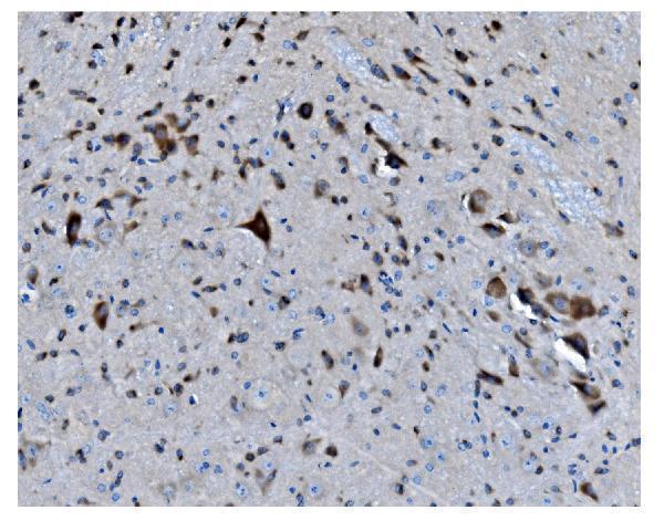
Expected resultMAPK11-positive staining in cells in granular layer of cerebellum (HPA tissue IHC: High). HPA tissue profile: Ubiquitous cytoplasmic expression often in a granular pattern. No signal in the no-primary control.
💡Decision noteStart with the catalog antibody’s heat-mediated EDTA retrieval at pH 8.0 (datasheet: A03738-1); the published protocols used citrate at pH 6.0 (PMC10693303; PMC5717400).
Section 2

What Is the Expected MAPK11 Staining Pattern?

MAPK11 staining should be predominantly cytoplasmic, often granular, in tissue sections (HPA: Approved IHC; medium consistency with RNA data; ubiquitous cytoplasmic profile). UniProt also lists the nucleus as a possible location and identifies no transmembrane segment (UniProt Q15759). Look for strong staining in cerebellar granular-layer cells, kidney tubule cells and selected glandular cells (HPA: High in those cells). Interpret intensity by cell type rather than treating an entire tissue as uniformly positive.

What am I looking at on my slide?
Granular cytoplasmic signal in cerebellar granular-layer cells or kidney tubule cells, with discernible cell boundaries and limited background.This matches the predominant compartment and two strong cell-level examples (HPA: ubiquitous, often granular cytoplasmic profile; High in these cells). Compare the stained cells with their neighbors and the counterstain before assigning intensity (standard IHC practice).
A crisp plasma-membrane rim or extracellular-only deposit dominates the section.That distribution conflicts with the reported cellular pattern and the absence of a transmembrane segment (HPA: cytoplasmic profile; UniProt Q15759 topology). Treat it as a likely staining artefact and check the detection controls. Nuclear signal alone needs more caution because UniProt also lists the nucleus (UniProt Q15759).
Ovarian stromal cells stain strongly, especially when the signal appears in no-primary controls.HPA reports ovarian stromal cells as not detected (HPA: ovary IHC). Strong staining there raises cross-reactivity or endogenous detection activity as possibilities; staining in a no-primary control supports the latter (standard IHC practice). It does not by itself identify which reagent caused the signal.
Diffuse color covers stroma, lumens or most of the section without a recognizable cellular pattern.That appearance is hard to score as MAPK11 because the reference pattern is cellular and predominantly cytoplasmic (HPA: tissue IHC profile). Nonspecific antibody binding or detection background are plausible causes (standard IHC practice); compare with controls before calling widespread expression.
Cerebellar granular-layer cells or kidney tubule cells show no detectable signal.Both are useful positive comparators because HPA reports High staining in those cell populations (HPA: cerebellum and kidney IHC). A blank result prompts a check of controls and the IHC workflow before concluding that MAPK11 is absent from the specimen (standard IHC practice).
💡Expected MAPK11 appearanceCall a section positive when defined cells show predominantly granular cytoplasmic staining, potentially strong in cerebellar granular-layer or kidney tubule cells (HPA: cytoplasmic profile; High in these cells); a membrane rim, extracellular deposit or diffuse tissue-wide haze is discordant with that pattern (HPA: tissue IHC profile; UniProt Q15759 topology).
How each factor affects the staining
Tissue and cell selectionHPA calls MAPK11 protein staining ubiquitous but assigns different levels to specific cells: High in duodenal, gallbladder and small-intestinal glandular cells, Medium in stomach glandular cells and adipocytes, and Not detected in ovarian stromal cells (HPA: tissue IHC). Score the named cells, not a whole-organ average.
RNA and protein evidenceBrain-enhanced RNA expression and the reported highest brain and heart levels can guide sample choice (HPA: tissue RNA; UniProt Q15759 tissue specificity). They do not replace the cell-level IHC calls; HPA rates the tissue staining Approved with only medium consistency against RNA data (HPA: tissue IHC reliability).
Compartment and protein formThe 1–364 MAPK11 chain has no signal peptide, propeptide or transmembrane segment (UniProt Q15759 processing and topology). Interpret cellular cytoplasmic staining in that context; the record supplies no basis for expecting a secreted or membrane-anchored staining pattern.
Total MAPK11 versus activationUniProt lists phosphorylation at Thr180, Tyr182 and Tyr323 (UniProt Q15759 modified residues). A total-MAPK11 stain cannot establish phosphorylation or pathway activation without evidence that the antibody recognizes a specified phospho-epitope (standard IHC interpretation).
Antibody validation and isoformsHPA lists two antibodies with Approved IHC status, HPA045069 and CAB012961, and two MAPK11 isoforms are recorded (HPA: antibody records; UniProt Q15759 isoforms). The supplied records give no epitope or isoform coverage, so do not infer that either stain distinguishes isoforms.
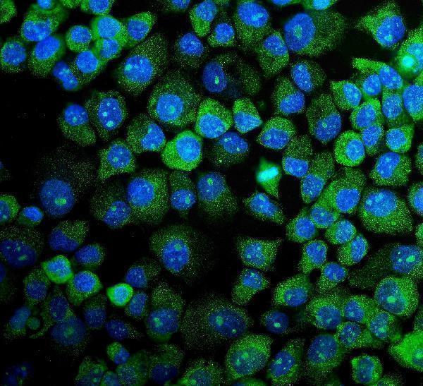
IF/ICC Q&AWhat localisation should IF/ICC show? Predominantly cytosolic signal is the approved HPA ICC-IF observation, with images listed for MCF-7, SH-SY5Y and U2OS (HPA: subcellular ICC-IF). This supports a localisation comparison only; the listed ICC-approved antibody, HPA073133, has no IHC status in the supplied record (HPA: antibody records).
Why is my staining missing, weak or wrong?
SituationLikely causeNext action
The known-positive cell population is blank.An IHC workflow or detection failure is possible; HPA reports High staining in cerebellar granular-layer cells and kidney tubules (HPA: tissue IHC; standard IHC practice).Check that the counterstain identifies the intended cells, then review the positive control, primary-antibody application and detection reagents (standard IHC practice).
Signal is weak across a positive control.Primary-antibody concentration, antigen-retrieval conditions or detection performance may need optimization (standard IHC practice). No MAPK11-specific fixation sensitivity is supplied.Optimize retrieval and antibody dilution on matched sections using the same control tissue, and retain a no-primary control to assess background (standard IHC practice).
A membrane rim or extracellular precipitate is prominent.The pattern conflicts with cytoplasmic tissue staining and MAPK11 topology (HPA: tissue IHC profile; UniProt Q15759 topology); deposited chromogen is one possible artefact (standard IHC practice).Inspect the no-primary control and reagent deposits, then repeat staining with clean detection reagents and compare the cellular pattern (standard IHC practice).
Ovarian stromal cells stain unexpectedly.HPA reports them as Not detected (HPA: ovary IHC); cross-reactivity or endogenous detection activity can produce apparent staining (standard IHC practice).Run a no-primary control and appropriate detection-blocking controls; reassess specificity against a known-positive cell population (standard IHC practice).
The section has diffuse background.Nonspecific binding or endogenous detection activity may obscure the expected cellular pattern (HPA: cytoplasmic profile; standard IHC practice).Compare no-primary and positive controls, then adjust blocking, washes or antibody dilution while preserving visible positive-cell staining (standard IHC practice).
A nucleus-only result is being scored as MAPK11 activation.UniProt permits nuclear localisation, but the HPA tissue profile is predominantly cytoplasmic; localisation alone does not establish phosphorylation (UniProt Q15759 subcellular location and modified residues; HPA: tissue IHC profile).Record nuclear and cytoplasmic signal separately, verify the antibody's stated epitope, and reserve activation claims for a validated phosphorylation-specific assay (standard IHC interpretation).

Sample controls for MAPK11 IHC & IF

🧪Run cerebellum first: cells in the granular layer should stain (HPA: High in cerebellar granular-layer cells). Run ovary as the negative tissue: ovarian stroma cells should show background staining (HPA: Not detected in ovarian stroma cells). On the cerebellum slide, treat unstained cells outside the granular layer as internal background references only after confirming their identity; HPA does not designate a specific internal negative cell type (HPA: High in granular-layer cells).
Positive control tissue: Cerebellum (Cells in granular layer, HPA High)
Negative control tissue: Ovary (HPA Not detected)
ICC-IF cell lines (HPA subcellular resource): HPA ICC-IF images show MAPK11 in MCF-7, SH-SY5Y, U2OS, with annotated localisation: Cytosol (approved) (HPA subcellular).
Technical controls: Include no-primary (secondary-only), host-species-matched rabbit IgG isotype, and MAPK11-knockout material as controls (hero caption: rabbit anti-MAPK11; standard IHC practice). Quench endogenous peroxidase and block endogenous biotin when using the caption’s biotin-based chromogenic detection in cerebellum (hero caption: biotinylated secondary, streptavidin-biotin complex and DAB; standard IHC practice).
⚠️Feasibility: A target-specific fixation window or fixation effect is unreported, and the selected A03738-1 paraffin-section caption does not state the fixative (hero caption: fixative not stated). The reported paraffin-section result used heat-mediated EDTA retrieval at pH 8.0, but the evidence does not establish whether retrieval is required for all MAPK11 antibodies or specimens (hero caption: EDTA retrieval, pH 8.0). The supplied evidence does not establish that frozen sections or IF are easier; for cerebellum, assess background outside the granular layer before scoring the expected signal (HPA: High in granular-layer cells; standard IHC practice).

HPA tissue IHC evidence for MAPK11

Comprehensive Human Protein Atlas IHC scoring per tissue (reliability: Approved — Medium consistency between antibody staining and RNA expression data.). Rows are taken directly from the HPA tissue chart — click any row's HPA link to view the source.

Positive expression · recommended positive controls

TissueCell typeLevelEvidenceSource
Cerebellum Cells in granular layer High Protein (IHC) HPA →
Cerebral cortex Neuropil High Protein (IHC) HPA →
Duodenum Glandular cells High Protein (IHC) HPA →
Gallbladder Glandular cells High Protein (IHC) HPA →
Kidney Cells in tubules High Protein (IHC) HPA →

Undetected expression · recommended negative controls

TissueCell typeLevelEvidenceSource
Ovary Ovarian stroma cells Not detected Protein (IHC) HPA →
Section 3

Advanced MAPK11 IHC Tips

Troubleshoot MAPK11 staining in paraffin sections by checking retrieval, controls, cell compartment, and scoring before interpreting chromogenic signal.

What should I change when MAPK11 staining is weak after antigen retrieval?
Start with heat mediated retrieval in EDTA at pH 8.0 for paraffin sections (datasheet A03738-1). The selected tissue image used that retrieval before incubation with 2 µg/ml primary antibody overnight at 4°C (datasheet A03738-1). If staining remains weak, compare a cautiously adjusted heating period on adjacent sections while keeping the buffer, antibody concentration, and detection system constant (standard IHC practice). Check tissue integrity and background alongside signal: stronger heating can damage morphology or expose nonspecific binding (standard IHC practice). The caption reports paraffin embedding but does not state the fixative, so it cannot establish performance for a particular fixation method (datasheet A03738-1).
How can I troubleshoot inconsistent MAPK11 staining across paraffin blocks?
MAPK11 specific sensitivity to fixation is unknown from the supplied evidence; the selected paraffin tissue caption does not report its fixative (datasheet A03738-1). Record each block's fixative, fixation duration, processing history, and section age before comparing stain intensity (standard IHC practice). Run adjacent sections from the same block together, using EDTA at pH 8.0 and the documented 2 µg/ml primary concentration as the starting conditions (datasheet A03738-1). If blocks differ, compare morphology and staining in matched tissue regions with a no primary control before attributing variation to MAPK11 abundance (standard IHC practice). Neither tissue staining patterns nor MAPK11 topology establishes a fixation effect (HPA: tissue IHC profile; UniProt Q15759 topology).
How should I evaluate nuclear MAPK11 staining when cytoplasmic staining dominates?
Assess cytoplasmic and nuclear signal separately because MAPK11 is annotated in both compartments, while the approved cell imaging location is cytosol (UniProt Q15759 subcellular; HPA: cytosol approved). In tissue IHC, the reported pattern is ubiquitous cytoplasmic staining, often granular, so nuclear staining needs particular scrutiny (HPA: tissue IHC profile). Compare nuclear signal with adjacent cytoplasm in intact cells and check the no primary control for nuclear background (standard IHC practice). Record the fraction of cells with convincing nuclear staining and retain the tissue region and cell type in the result (standard IHC practice). Localisation alone does not establish kinase activation (UniProt Q15759 modified residues).
Can this stain distinguish MAPK11 isoforms or activated MAPK11?
Treat the chromogenic signal as MAPK11 immunoreactivity unless the antibody's mapped epitope and specificity data support a narrower claim (standard IHC practice). MAPK11 has 2 listed isoforms and a kinase domain spanning residues 24–308; the supplied caption gives no epitope map (UniProt Q15759 isoforms and domains; datasheet A03738-1). Activation related modifications include phosphorylation at Thr180 and Tyr182, but total MAPK11 staining does not identify their phosphorylation state (UniProt Q15759 modified residues; standard IHC practice). Check the antibody's documented epitope against isoform sequences before making isoform claims, and use a separately validated phospho specific assay for activation claims (standard IHC practice). Report antibody identity and retrieval conditions with those conclusions (datasheet A03738-1).
How can I use IF to clarify ambiguous MAPK11 staining in tissue?
Use IF as a separate validation experiment and pair MAPK11 with a marker that identifies the cell population being evaluated (standard IF practice). Choose spectrally separated fluorophores and place the weaker signal in a channel with low measured tissue autofluorescence; include single stain and unstained controls (standard IF practice). MAPK11 lacks a transmembrane segment and is annotated in cytoplasm and nucleus, so select permeabilisation that gives antibody access to intracellular epitopes while preserving morphology (UniProt Q15759 topology and subcellular; standard IF practice). Compare cytosolic and nuclear distributions with the chromogenic result, remembering that approved cell imaging localises MAPK11 to cytosol (HPA: cytosol approved). The supplied paraffin caption establishes an IHC workflow, not IF performance (datasheet A03738-1).
What should I check when MAPK11 DAB staining looks diffuse or granular everywhere?
First compare the slide with a no primary control and inspect pigment, tissue edges, and damaged regions for staining unrelated to antibody binding (standard IHC practice). MAPK11 tissue IHC has a reported ubiquitous, often granular cytoplasmic pattern, so diffuse cytoplasmic signal alone is insufficient evidence of nonspecific staining (HPA: tissue IHC profile). The selected workflow used 10% goat serum, 2 µg/ml primary overnight at 4°C, and a biotin based DAB detection system (datasheet A03738-1). If background persists, assess endogenous peroxidase and endogenous biotin contributions with appropriate controls, then adjust blocking or antibody concentration one variable at a time (standard IHC practice). Judge changes against preserved cellular localisation and morphology (UniProt Q15759 subcellular; standard IHC practice).
How should I score MAPK11 staining across regions with different cellular makeup? ⚠ ANSWER MARKED FOR VERIFICATION
Define the cell population and tissue region before scoring, then use a consistent intensity scale with an H-score or report the percentage of positive cells (standard IHC practice). For spatial comparisons, positive cell density per mm² can supplement intensity scoring when cell abundance differs across regions (standard IHC practice). Normalise positive counts to eligible nucleated cells and density to the measured viable tissue area, excluding folds, edges, and necrosis (standard IHC practice). Score cytoplasmic and nuclear compartments separately because both are annotated, while tissue IHC chiefly reports cytoplasmic staining (UniProt Q15759 subcellular; HPA: tissue IHC profile). Use the same retrieval and detection settings across compared slides (datasheet A03738-1; standard IHC practice).
When should I distrust an apparent MAPK11 positive cell or region?
Look for intracellular signal in intact cells: MAPK11 is annotated in cytoplasm and nucleus and has no transmembrane segment (UniProt Q15759 subcellular and topology). Treat exclusively membranous staining, strong tissue edge signal, and staining confined to necrosis as suspect until controls and morphology support them (UniProt Q15759 topology; standard IHC practice). Match the apparent positive cells to tissue anatomy; HPA reports high staining in kidney tubular cells and low staining in bone marrow hematopoietic cells, for example (HPA: kidney and bone marrow tissue IHC). Check no primary and endogenous enzyme controls when DAB signal appears in unexpected compartments or cells (standard IHC practice). An IHC positive result indicates immunoreactivity, not MAPK11 phosphorylation or pathway activity (UniProt Q15759 modified residues; standard IHC practice).
Boster reagents

Best MAPK11 / Mitogen-activated protein kinase 11 IHC Antibodies

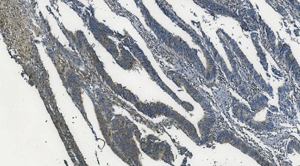
A03738-1 has MAPK11 IHC data from paraffin-embedded human rectal cancer tissue and IF/ICC data from A431 cells (catalog image captions). Listed reactivity covers human, mouse, and rat (catalog).

Real IHC data IHC analysis of MAPK11 using anti-MAPK11 antibody (A03738-1). MAPK11 was detected in paraffin-embedded section of human rectal cancer tissue. Heat mediated antigen retrieval was performed in EDTA buffer (pH8.0, epitope retrieval solution). The tissue section was blocked with 10% goat serum. The tissue section was then incubated with 2μg/ml rabbit anti-MAPK11 Antibody (A03738-1) overnight at 4°C. Biotinylated goat anti-rabbit IgG was used as secondary antibody and incubated for 30 minutes at 37°C. The tissue section was developed using Strepavidin-Biotin-Complex (SABC) (Catalog # SA1022) with DAB as the chromogen.
Anti-MAPK11 Antibody ®
Cat # A03738-1

A03738-1 is listed for IHC and IF/ICC, with a MAPK11 IHC image from paraffin-embedded human rectal cancer tissue (catalog; IHC image caption). Its MAPK11 IF image uses A431 cells, and its listed reactivity is human, mouse, and rat (IF image caption; catalog).

Which to pick: Choose A03738-1 for paraffin-section IHC: its MAPK11 caption documents EDTA retrieval at pH 8.0 and 2 μg/ml primary antibody; the fixative is unreported (IHC image caption). The same SKU supports IF/ICC in A431 cells at 5 μg/ml; its clone is unspecified (IF image caption; catalog). For mouse or rat work, A03738-1 lists both species as reactive, but its additional IHC captions identify MAPK1, so those images do not establish MAPK11 staining in either species (catalog; IHC image captions).

Each figure is that product's own IHC / IF validation image from its datasheet.

References

  1. UniProt Consortium. UniProt entry Q15759 (MK11_HUMAN, Mitogen-activated protein kinase 11).
  2. Human Protein Atlas. MAPK11 tissue IHC expression (reliability: Approved).
  3. Human Protein Atlas. MAPK11 subcellular location (ICC-IF): Localized to the cytosol..
  4. Human Protein Atlas. MAPK11 antibody validation summary (3 antibodies).
  5. Transcription factor ETV4 promotes the development of hepatocellular carcinoma by driving hepatic TNF-α signaling. Cancer communications (London, England) 2023 — PMC10693303.
  6. 18ï ¢-Glycyrrhetinic acid suppresses glioblastoma by regulating p38 signaling pathway: an integrative approach combining network analysis, transcriptomics, and experimental assessment. Frontiers in pharmacology 2026 — PMC12953517.
  7. Suppression of MAPK11 or HIPK3 reduces mutant Huntingtin levels in Huntington's disease models. Cell research 2017 — PMC5717400.
  8. PubMed PMID:8663524 — UniProt-cited evidence.
  9. PubMed PMID:9207191 — UniProt-cited evidence.
  10. PubMed PMID:9430721 — UniProt-cited evidence.