MAPK9 / Mitogen-activated protein kinase 9 · IHC design guide

Design Immunohistochemistry for MAPK9

Plan chromogenic MAPK9 IHC in paraffin sections using cytoplasmic and nuclear staining as the tissue pattern, with high staining in colon glandular cells (HPA tissue IHC). Score the compartments separately and interpret intensity cautiously because HPA reports medium agreement between staining and RNA expression (HPA tissue IHC).

Evidence assembled Oct 2026 · For research use; verify linked source records and product datasheet before use
Immunohistochemistry protocol sheet for MAPK9 (IHC for MAPK9): expected localisation Cytoplasmic and nuclear staining (HPA tissue IHC), antibody M02706, validated IHC image, and IHC protocol steps
Printable MAPK9 IHC protocol sheet — expected localisation Cytoplasmic and nuclear staining (HPA tissue IHC), antibody M02706, controls and protocol steps. Open the full MAPK9 IHC guide →

MAPK9 Immunohistochemistry Experimental Design Guide

Expected localisation, validated protocols, controls and antibodies — the at-a-glance facts below, then the full design guide.

Must know before staining
Expected localisation Cytoplasmic and nuclear staining (HPA tissue IHC)
Staining pattern General cytoplasmic and nuclear staining (HPA tissue IHC)
Antigen retrieval Tris-EDTA pH 9.0 HIER, 95–98 °C, 20 min (rule: nuclear antigen)
Positive control ⓘ Adrenal gland+4 more · see all
Negative control ⓘ Adipose tissue+4 more · see all
Important caveats
Reasons your staining may differ from the expected pattern.
Fixation Matched tissue-IHC evidence does not establish the fixation claim. Validate the specimen-specific method before use. (selected-SKU IHC image M02706)
Caveat Staining has medium agreement with RNA data (HPA tissue IHC)
Regulation Stress activates MAPK9 via MAP2K4/7 (UniProt)
Isoform / epitope 5 isoforms; epitope coverage unreported (UniProt)
Section 1

Recommended MAPK9 IHC & IF Protocols

Compare the catalog antibody’s IHC-P protocol with a published MAPK9/JNK2 chromogenic coverslip protocol (PMC5429546).

Recommended immunohistochemistry (IHC-P) protocol parameters
SampleParaffin-embedded Rat intestine tissue; fixative not specified (datasheet M02706)
FixationImage fixative and duration unreported (datasheet M02706); verify before use.
Sectioning4–5 µm sections on charged slides (standard)
DeparaffinisationXylene, graded ethanol series to water (standard)
Antigen retrievalHeat-induced epitope retrieval in Tris-EDTA buffer, pH 9.0, 20 min at 95–98 °C (standard rule: nuclear antigen)
Peroxidase block3% H2O2, 10 min, room temperature (standard)
Blocking10% normal serum of the secondary host, 30 min, room temperature (standard)
Primary antibodyRabbit monoclonal (clone EIF-13) anti-MAPK9, 1:50 recommended; image 1:100 (datasheet M02706)
Primary incubationOvernight at 4 °C (standard)
DetectionHRP-polymer secondary, DAB chromogen 5–10 min (standard)
CounterstainHematoxylin, blue, dehydrate and mount (standard)
Expected resultMAPK9-positive staining in glandular cells of adrenal gland (HPA tissue IHC: High). HPA tissue profile: General cytoplasmic and nuclear expression. No signal in the no-primary control.
💡Decision noteFor paraffin sections, start with Tris-EDTA HIER at pH 9.0, 95–98 °C for 20 min (page retrieval rule: nuclear antigen).
Section 2

What Is the Expected MAPK9 Staining Pattern?

In paraffin-section IHC, expect MAPK9 staining in the cytoplasm and nuclei of selected cells, including glandular cells in adrenal gland, breast, colon and duodenum, and respiratory epithelial cells in bronchus (HPA: general cytoplasmic and nuclear expression; High in these cell types). MAPK9 has no transmembrane segment (UniProt P45984 topology). The tissue IHC profile is rated Enhanced, with medium agreement between staining and RNA data and external verification pending (HPA: reliability).

What am I looking at on my slide?
Cytoplasmic and nuclear staining in colon glandular cells or bronchial respiratory epithelial cells.This fits the reported compartment pattern and High staining in those cell types (HPA: tissue IHC). Score the identified cell type and compartment; the HPA profile does not require identical intensity in every cell (HPA: tissue IHC profile).
Dominant extracellular staining, or staining confined to a crisp cell border with little intracellular signal.Question the localisation: tissue IHC is generally cytoplasmic and nuclear, and MAPK9 lacks a transmembrane segment (HPA: tissue IHC profile; UniProt P45984 topology). Plasma membrane localisation in ICC-IF is marked uncertain, so a border signal alone is inconclusive (HPA: subcellular).
Strong staining in adipocytes or cardiomyocytes while the expected positive cells are unstained.Those cell types were Not detected in the HPA tissue IHC examples (HPA: adipocytes; cardiomyocytes). Consider antibody cross-reactivity or detection background before assigning the signal to MAPK9; these examples do not establish universal absence.
Widespread brown haze across cells and surrounding section, with poor cell boundaries.Treat this as background until controls resolve it. General IHC causes include nonspecific antibody binding and endogenous detection activity (standard IHC practice). The reported MAPK9 pattern is intracellular in identifiable cells (HPA: tissue IHC profile).
No staining in glandular cells of a colon section used as a positive reference.Colon glandular cells were High in the HPA examples (HPA: tissue IHC). Check that the reference cells are present, then inspect staining controls and processing. A failed reference section cannot distinguish absent target from a failed IHC run (standard IHC practice).
💡Expected MAPK9 appearanceA credible positive is discernible cytoplasmic and/or nuclear signal in an identified HPA High cell type, such as colon glandular cells; diffuse haze or extracellular deposits are suspect (HPA: tissue IHC profile; standard IHC interpretation).
How each factor affects the staining
Tissue and cell choiceHPA reports High staining in adrenal, breast, colon and duodenal glandular cells, bronchial respiratory epithelium, cerebellar molecular-layer cells and cortical neurons; endometrial glandular cells are Medium (HPA: tissue IHC). Compare like cell types when judging intensity.
Reference-low and undetected cellsHPA lists Low staining in esophageal squamous cells, cholangiocytes, smooth muscle cells and skeletal myocytes, and Not detected in adipocytes and cardiomyocytes (HPA: tissue IHC). These are interpretation references, not proof that every specimen will be negative.
Antibody evidenceThe HPA tissue profile is Enhanced but has medium staining–RNA consistency and awaits external verification (HPA: reliability). CAB008910 has Enhanced IHC validation; HPA072462 has Supported ICC validation (HPA: antibody validation). Do not transfer an ICC validation label to IHC.
Isoforms and activation stateUniProt lists five MAPK9 isoforms and phosphorylation at Thr183 and Tyr185 (UniProt P45984: isoforms; modified residues). Without an antibody epitope or phospho-specificity record, staining cannot identify an isoform or establish kinase activation.
IF/ICC Q: Should its image match IHC?A: Expect supported cytosol and nuclear-speckle localisation in ICC-IF; plasma membrane localisation is uncertain (HPA: subcellular). Tissue IHC instead reports general cytoplasmic and nuclear expression (HPA: tissue IHC). Apply each assay's own validated interpretation.
Why is my staining missing, weak or wrong?
SituationLikely causeNext action
Positive-reference cells are unstained.The run may have failed, or the expected cell type may be missing from the section (standard IHC practice).Verify the HPA High reference cell type is present, then review reagent, chromogen and staining controls and the IHC-validated antibody's documented procedure (HPA: tissue IHC; standard IHC practice).
Signal is weak in an HPA High cell type.Low apparent signal can reflect assay conditions or section quality; HPA intensity is a reported observation, not a guaranteed result for every specimen (HPA: tissue IHC; standard IHC practice).Compare a reference section stained in the same run, inspect tissue preservation and follow the validated retrieval and antibody-use instructions; no MAPK9-specific fixation sensitivity is reported here.
Brown signal appears throughout the section.Nonspecific binding or endogenous detection activity can obscure cell-specific staining (standard chromogenic IHC practice).Check a reagent-omission control and the assay's blocking and detection controls; only score signal that resolves to identifiable cells and compartments (standard IHC practice; HPA: tissue IHC profile).
Adipocytes or cardiomyocytes stain strongly.These cells were Not detected in the HPA examples, so cross-reactivity or detection background is plausible (HPA: tissue IHC).Check controls and compare with a High reference cell type in the same run before calling the unexpected staining MAPK9 (HPA: tissue IHC; standard IHC practice).
Staining is mainly extracellular or restricted to cell borders.This conflicts with the general tissue IHC profile; ICC-IF plasma membrane localisation remains uncertain (HPA: tissue IHC; subcellular).Inspect morphology, counterstain and background controls, then reassess intracellular signal. Do not use uncertain ICC-IF membrane localisation to validate an isolated IHC border pattern (HPA: subcellular; standard IHC practice).

Sample controls for MAPK9 IHC & IF

🧪Run breast first: glandular cells should stain (HPA: High in breast glandular cells). Use adipose tissue as the negative comparator (HPA: Not detected in adipocytes); on the breast slide, cells established as internal negatives should show counterstain without specific chromogen, while adjacent stromal cells should not be assumed negative from the HPA glandular-cell result alone.
Positive control tissue: Adrenal gland (Glandular cells, HPA High)
Negative control tissue: Adipose tissue (HPA Not detected)
ICC-IF cell lines (HPA subcellular resource): HPA ICC-IF images show MAPK9 in EFO-21, GAMG, U2OS, hTERT-RPE1 (serum starved), Sperm, with annotated localisation: Plasma membrane (uncertain), Cytosol (supported) (HPA subcellular).
Technical controls: Include a no-primary or secondary-only slide, an isotype control matched to the primary antibody’s host species and class, and a MAPK9 knockout specimen as a biological negative (standard IHC practice). Check endogenous peroxidase in breast sections for chromogenic detection, and assess tissue autofluorescence if using IF (standard IHC/IF practice).
⚠️Feasibility: A target-specific fixation window and retrieval dependence are unreported in the supplied evidence; optimize antigen retrieval for paraffin IHC (standard IHC practice). The selected M02706 caption reports rat intestine paraffin-section staining at 1:100, but its fixative is unreported (selected SKU caption). These data do not establish whether frozen sections or IF would be easier; breast-section endogenous peroxidase can complicate chromogenic interpretation (standard IHC practice).

HPA tissue IHC evidence for MAPK9

Comprehensive Human Protein Atlas IHC scoring per tissue (reliability: Enhanced — Medium consistency between antibody staining and RNA expression data. Pending external verification.). Rows are taken directly from the HPA tissue chart — click any row's HPA link to view the source.

Positive expression · recommended positive controls

TissueCell typeLevelEvidenceSource
Adrenal gland Glandular cells High Protein (IHC) HPA →
Breast Glandular cells High Protein (IHC) HPA →
Bronchus Respiratory epithelial cells High Protein (IHC) HPA →
Cerebellum Cells in molecular layer High Protein (IHC) HPA →
Cerebral cortex Neuronal cells High Protein (IHC) HPA →

Undetected expression · recommended negative controls

TissueCell typeLevelEvidenceSource
Adipose tissue Adipocytes Not detected Protein (IHC) HPA →
Bone marrow Hematopoietic cells Not detected Protein (IHC) HPA →
Caudate Glial cells Not detected Protein (IHC) HPA →
Cervix Glandular cells Not detected Protein (IHC) HPA →
Heart muscle Cardiomyocytes Not detected Protein (IHC) HPA →
Section 3

Advanced MAPK9 IHC Tips

Troubleshoot MAPK9 chromogenic IHC in paraffin sections using the page retrieval setting, cell type controls, and compartment aware scoring.

How should I retrieve MAPK9 when staining is weak in paraffin sections?
Use Tris-EDTA pH 9.0 heat-induced epitope retrieval at 95–98 °C for 20 min (page retrieval setting). Allow slides to cool in retrieval buffer before washing, and keep heating and cooling consistent across comparison sections (standard IHC practice). Check whether stronger signal comes with tissue lifting, damaged nuclei, or diffuse background before increasing retrieval intensity (standard IHC practice). Include a positive cell population, such as colon glandular cells, and assess signal in its cellular context (HPA: High in colon glandular cells). The rat intestine image used 1:100 antibody dilution, but its caption does not establish retrieval performance across tissues (M02706 caption).
Could fixation explain absent or patchy MAPK9 staining?
MAPK9-specific fixation sensitivity is unknown: the paraffin-embedded rat intestine image reports 1:100 dilution but no fixative (M02706 caption). Record fixative, fixation duration, specimen thickness, and processing history before comparing staining between blocks (standard IHC practice). For a new series, process matched specimens consistently and compare adjacent sections under the same retrieval and detection conditions (standard IHC practice; page retrieval setting). Review morphology alongside staining, since poor preservation can make weak or patchy chromogen difficult to interpret (standard IHC practice). Do not assign a fixation effect from MAPK9 localisation or phosphorylation annotations alone (UniProt P45984: cytoplasm, nucleus, and modified residues).
Should MAPK9 stain nuclei, cytoplasm, or cell membranes?
Evaluate cytoplasmic and nuclear staining separately because both compartments are annotated for MAPK9 (UniProt P45984: cytoplasm and nucleus; HPA tissue IHC: general cytoplasmic and nuclear expression). HPA cell imaging supports cytosol and nuclear speckles, while its plasma membrane assignment is uncertain (HPA subcellular localisation). A membrane-only chromogenic pattern therefore needs stronger validation than a pattern that includes the expected compartments (HPA subcellular localisation; standard IHC practice). Compare glandular cells in colon or duodenum with adjacent structures on the same section (HPA: High in colon and duodenum glandular cells). Check nuclear counterstain and tissue morphology before calling focal nuclear chromogen positive (standard IHC practice).
Can this stain distinguish MAPK9 isoforms or activation states?
The record lists 5 MAPK9 isoforms, but the supplied antibody evidence does not identify a binding epitope or establish which isoforms it detects (UniProt P45984: isoforms; M02706 caption). MAPK9 contains a protein kinase domain spanning residues 26–321 (UniProt P45984: domain annotation). Phosphorylation is annotated at Thr183 and Tyr185, but a total MAPK9 stain cannot be interpreted as an activation-specific readout without antibody specificity evidence (UniProt P45984: modified residues; standard IHC practice). If isoform or phosphorylation status matters, document the antibody epitope and validate that claim with an appropriate orthogonal assay (standard IHC practice). Score the present chromogenic result as MAPK9 immunoreactivity, subject to antibody validation (standard IHC practice).
How should I adapt the MAPK9 localisation check to multiplex immunofluorescence?
On the separate IF/ICC workflow, pair MAPK9 with a glandular epithelial marker when examining colon glandular cells (HPA: High in colon glandular cells; standard IF practice). Choose a spectrally distinct, preferably far-red fluorophore when tissue autofluorescence overlaps shorter wavelength channels, and inspect an unstained section (standard IF practice). MAPK9 has no transmembrane segment, so use permeabilisation appropriate for a cytosolic or nuclear epitope, while recognising that this antibody’s epitope is unspecified (UniProt P45984: topology and localisation; M02706 caption; standard IF practice). Include single-stain and secondary-only controls to assess bleed-through and nonspecific fluorescence (standard IF practice). Compare cytosolic and nuclear signal separately; HPA also reports supported nuclear speckles in cell imaging (HPA subcellular localisation).
How can I separate MAPK9 signal from chromogenic background?
Run a section without primary antibody to assess detection-system staining, and inspect negative areas for precipitate or pigment (standard IHC practice). In a peroxidase-based workflow, use a peroxidase block before DAB development and keep development time consistent across sections (standard IHC practice). Reduce nonspecific staining by checking blocking, antibody concentration, washing, and the clarity of the cell boundaries around chromogen (standard IHC practice). The catalog image documents 1:100 in paraffin-embedded rat intestine; treat that dilution as its image condition, not a universal optimum (M02706 caption). Compare suspicious staining with adipocytes, where HPA reports MAPK9 as not detected, while respecting tissue and assay differences (HPA: adipocytes Not detected).
What is a defensible way to quantify MAPK9 IHC across samples? ⚠ ANSWER MARKED FOR VERIFICATION
Define the cell population and score cytoplasmic and nuclear compartments separately before reviewing group labels (UniProt P45984: cytoplasm and nucleus; standard IHC practice). For each compartment, report the percentage of positive viable cells and an intensity category, or calculate an H-score on the 0–300 scale (standard IHC practice). If comparing infiltrating or scattered cells, report positive cell density per mm² of viable tissue as well (standard IHC practice). Normalise comparisons to the same annotated cell type and evaluable tissue area, excluding folds and necrosis (standard IHC practice). Keep retrieval, antibody dilution, DAB development, imaging, and thresholds consistent across the series (page retrieval setting; standard IHC practice).
Which MAPK9 staining patterns warrant skepticism despite strong colour?
A convincing result should follow preserved cell boundaries and show plausible cytoplasmic or nuclear localisation in the scored cells (UniProt P45984: localisation; standard IHC practice). Colon or duodenum glandular cells offer reported positive contexts, whereas adipocytes are reported as not detected (HPA tissue IHC). Treat isolated membrane-only staining cautiously because the HPA membrane assignment is uncertain (HPA subcellular localisation). Discount staining concentrated at section edges, folds, necrotic regions, or sites that also stain without primary antibody (standard IHC practice). Finally, chromogenic MAPK9 intensity alone does not establish kinase activation or distinguish its 5 isoforms (UniProt P45984: modified residues and isoforms; standard IHC practice).
Boster reagents

Best MAPK9 / Mitogen-activated protein kinase 9 IHC Antibodies

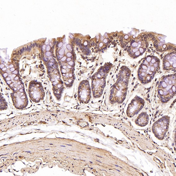
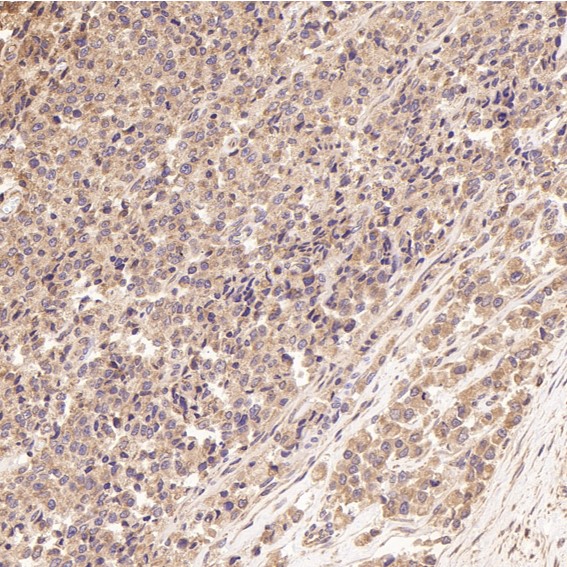
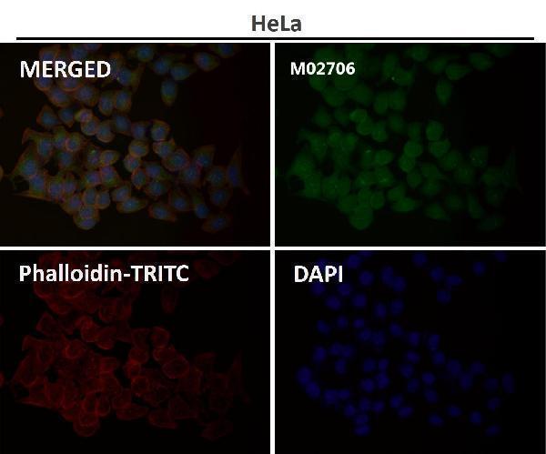
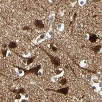
Catalog anti-MAPK9 antibodies cover IHC images from paraffin-embedded rat intestine, human prostate cancer and astrocytoma, and mouse skin (M02706 IHC captions), plus an IF image with sample type unreported (M02706 IF caption).

Real IHC data Immunohistochemical analysis of paraffin-embedded Rat intestine, using the Antibody at 1:100 dilution.
Anti-JNK2 MAPK9 Rabbit Monoclonal Antibody
Cat # M02706

M02706 lists IHC and IF/ICC for human, monkey, mouse, and rat; its IHC captions show paraffin-embedded rat intestine, human prostate cancer and astrocytoma, and mouse skin (M02706 applications, reactivity, IHC captions). RP1077 lists IF/ICC for human, mouse, and rat, with no supplied IHC or IF image captions (RP1077 applications, reactivity, image captions).

Which to pick: Choose M02706 for tissue IHC: it is a rabbit monoclonal with paraffin-section IHC images, including rat intestine at 1:100; the fixative is unreported (M02706 catalog, IHC captions). For IF/ICC, M02706 has a supplied IF image caption at 1:50, while RP1077 lists IF/ICC without a supplied image caption (M02706 IF caption; RP1077 applications, image captions). M02706 offers the broader listed species reactivity, including monkey; RP1077 lists human, mouse, and rat (M02706 and RP1077 reactivity).

Each figure is that product's own IHC / IF validation image from its datasheet.

References

  1. UniProt Consortium. UniProt entry P45984 (MK09_HUMAN, Mitogen-activated protein kinase 9).
  2. Human Protein Atlas. MAPK9 tissue IHC expression (reliability: Enhanced).
  3. Human Protein Atlas. MAPK9 subcellular location (ICC-IF): Mainly localized to the plasma membrane and cytosol. In addition localized to the nuclear speckles..
  4. Human Protein Atlas. MAPK9 antibody validation summary (2 antibodies).
  5. A predictive model of immune infiltration and prognosis of head and neck squamous cell carcinoma based on cell adhesion-related genes: including molecular biological validation. Frontiers in immunology 2023 — PMC10484396.
  6. Genome-wide DNA methylation analysis in lung fibroblasts co-cultured with silica-exposed alveolar macrophages. Respiratory research 2017 — PMC5429546.
  7. Insulin-like growth factor 1 receptor (IGF1R) expression and survival in operable squamous-cell laryngeal cancer. PloS one 2013 — PMC3554755.
  8. PubMed PMID:7969172 — UniProt-cited evidence.
  9. PubMed PMID:8001819 — UniProt-cited evidence.
  10. PubMed PMID:8654373 — UniProt-cited evidence.