This website uses cookies to ensure you get the best experience on our website.


- Table of Contents
Plan MARCKS chromogenic IHC in paraffin sections using appendix glandular cells or kidney tubules as high-staining reference tissues (HPA tissue IHC). Interpret cytoplasmic and membranous staining with the IHC-validated antibody, while allowing for PKC-dependent redistribution between those compartments (HPA tissue IHC; UniProt).

Expected localisation, validated protocols, controls and antibodies — the at-a-glance facts below, then the full design guide.
| Expected localisation | Cytoplasmic and membranous staining (HPA tissue IHC) | |
| Staining pattern | Glandular and respiratory epithelial cells: cytoplasmic/membranous (HPA tissue IHC) | |
| Antigen retrieval | EDTA pH 8.0 HIER, heat-mediated (datasheet A04133-4) | |
| Positive control | Appendix+4 more · see all | |
| Negative control | Adipose tissue+4 more · see all |
| Fixation | Keep paraffin-section fixation consistent (standard IHC practice; not target-specific); Matched tissue-IHC evidence does not establish the fixation claim. Validate the specimen-specific method before use. (selected-SKU IHC image A04133-4) | |
| Caveat | Tissue staining has medium RNA concordance (HPA tissue IHC) | |
| Regulation | PKC phosphorylation shifts membrane signal (UniProt) | |
| Isoform / epitope | No isoforms annotated; epitope map unspecified (UniProt) |
Compare the catalog antibody’s IHC-P protocol (datasheet A04133-4) with two published MARCKS IHC protocols (PMC5351628; PMC5795991).
| Sample | Paraffin-embedded mouse brain tissue; fixative not specified (datasheet A04133-4) |
| Fixation | Image fixative and duration unreported (datasheet A04133-4); verify before use. |
| Sectioning | 4–5 µm sections on charged slides (standard) |
| Deparaffinisation | Xylene, graded ethanol series to water (standard) |
| Antigen retrieval | Heat retrieval: EDTA pH 8.0 (datasheet A04133-4); 20 min, 95–100 °C (standard) |
| Peroxidase block | 3% H2O2, 10 min, room temperature (standard) |
| Blocking | 10% goat serum (datasheet A04133-4) |
| Primary antibody | Rabbit anti-MARCKS, 2-5 μg/ml (datasheet A04133-4) |
| Primary incubation | Overnight at 4 °C (datasheet A04133-4) |
| Detection | HRP-conjugated secondary, DAB chromogen (datasheet A04133-4) |
| Counterstain | Hematoxylin, blue, dehydrate and mount (standard) |
| Expected result | MARCKS-positive staining in glandular cells of appendix (HPA tissue IHC: High). HPA tissue profile: Cytoplasmic and membranous expression in most tissues. No signal in the no-primary control. |
In paraffin-section IHC, expect cytoplasmic and membranous MARCKS staining in many tissues, including glandular and respiratory epithelial cells, kidney tubule cells, and cerebellar neuropil (HPA tissue IHC: high in these cells or regions). MARCKS has no transmembrane segment and can move between membrane and cytoplasm (UniProt P29966: topology and subcellular location). HPA rates the tissue pattern Enhanced, with medium consistency between staining and RNA data pending external verification (HPA tissue IHC: reliability).
| Membranous and cytoplasmic staining marks glandular cells in colon or appendix, respiratory epithelium in bronchus, or kidney tubule cells; the cerebellar molecular-layer neuropil also stains (HPA tissue IHC: High). | These are supported positive patterns for a total-MARCKS stain. Score the specified cells or region and record membrane and cytoplasmic signal separately when distinguishable (HPA tissue IHC: cell-level pattern; UniProt P29966: subcellular location). HPA reports staining levels, not an expected staining percentage or a numerical scoring cutoff (HPA tissue IHC: High). |
| The dominant signal is nuclear with little membrane or cytoplasmic staining in an HPA-positive tissue (HPA tissue IHC: cytoplasmic and membranous profile). | Treat a nuclear-only IHC result as discordant and review the antibody, controls, and detection conditions (HPA tissue IHC: profile; standard IHC practice). HPA lists nucleoplasmic localisation as uncertain in ICC-IF, so that observation alone does not validate dominant nuclear staining in paraffin sections (HPA subcellular: nucleoplasm uncertain). |
| Cardiomyocytes, skeletal myocytes, or adipocytes stain strongly while the expected positive cells are weak (HPA tissue IHC: Not detected in those cell types; High in listed positive cells). | This distribution raises concern for nonspecific binding, cross-reactivity, or endogenous chromogenic activity; it does not identify which cause applies (standard IHC practice). Compare tissue architecture and matched controls. HPA's Not detected calls apply to the named cell types, not every cell in their tissues (HPA tissue IHC: cell-level negatives). |
| Brown signal spreads evenly through the section or appears outside recognizable cell boundaries (standard chromogenic IHC observation). | Diffuse deposition obscures the membrane and cytoplasmic pattern used to interpret MARCKS (HPA tissue IHC: profile). Check background on a no-primary control, then assess blocking, antibody concentration, washes, and chromogen development as general IHC variables (standard IHC practice). |
| A run shows no convincing signal in colon glandular cells, bronchial respiratory epithelium, or kidney tubule cells (HPA tissue IHC: High in these cells). | The result is inconclusive until a known-positive control works in the same run (standard IHC practice). Check the catalog antibody's IHC-P instructions, retrieval conditions, and detection reagents before calling the test tissue negative (standard IHC practice). The supplied sources do not establish MARCKS-specific fixation sensitivity. |
| Compartment and phosphorylation state (UniProt P29966: subcellular location) | MARCKS associates with the cell membrane and cytoplasm; PKC-dependent phosphorylation displaces it from the membrane, and dephosphorylation accompanies reassociation (UniProt P29966: subcellular location). Therefore, relative membrane and cytoplasmic prominence can vary without changing the expected set of compartments. A total-MARCKS IHC image alone cannot establish phosphorylation state (UniProt P29966: phosphorylation-linked movement; standard interpretation). |
| Cell-level tissue reference (HPA tissue IHC: profile and examples) | HPA reports High staining in selected glandular cells, respiratory epithelial cells, kidney tubule cells, and cerebellar neuropil, alongside Not detected calls for specified adipocytes, cardiomyocytes, and myocytes (HPA tissue IHC: levels). Use the named cells as reference points; low tissue RNA specificity does not mean every cell should stain alike (HPA tissue IHC: RNA specificity and cell-level staining). |
| Strength of reference evidence (HPA tissue IHC: reliability; HPA antibodies: IHC validation) | The tissue profile is rated Enhanced, although its staining-to-RNA consistency is medium and external verification is pending (HPA tissue IHC: reliability description). HPA lists three antibodies with Enhanced IHC status (HPA antibodies: HPA067595, HPA069443, CAB022062). These observations support comparison with the pattern; they do not validate an unlisted catalog antibody or define a universal staining cutoff (HPA tissue IHC; standard interpretation). |
| Retrieval and epitope information (supplied UniProt P29966 and HPA records) | No epitope location or MARCKS-specific retrieval response is supplied. Follow the catalog antibody's IHC-P procedure and evaluate its stated retrieval condition with a positive control (standard IHC practice). The lack of a transmembrane segment does not identify an optimal retrieval method or predict fixation effects (UniProt P29966: topology). |
| Detection system background (standard chromogenic IHC practice) | Endogenous enzyme activity and nonspecific reagent binding can produce chromogenic signal unrelated to antibody binding (standard IHC practice). A no-primary control helps reveal detection-system background in the tested tissue, while a positive control checks that the run can reveal the expected cellular pattern (standard IHC practice; HPA tissue IHC: positive cell examples). |
| IF/ICC Q&A: where should fluorescence appear? (HPA subcellular: ICC-IF) | Mainly at the plasma membrane, which HPA marks supported; HPA also lists cytosol, nucleoplasm, and primary cilium as uncertain additional locations (HPA subcellular: locations). This ICC-IF observation helps interpret fluorescence images. It is separate from the paraffin-section IHC pattern and supplies no IF/ICC protocol option (HPA subcellular; HPA tissue IHC). |
| Situation | Likely cause | Next action |
|---|---|---|
| No signal in a section expected to contain colon glandular cells, bronchial respiratory epithelium, or kidney tubules (HPA tissue IHC: High in these cells). | The run may have failed at antibody incubation, antigen retrieval, or detection; the absent signal alone cannot identify the failed step (standard IHC practice). | Run a known-positive section alongside the sample, confirm tissue preservation and the catalog antibody's IHC-P instructions, and check detection reagents (standard IHC practice). Interpret a negative sample only after the positive control stains in the expected cells (HPA tissue IHC: High examples). |
| The entire section develops diffuse brown colour, including areas without clear cellular staining (standard chromogenic IHC observation). | Possible causes include detection background, insufficient blocking or washing, excessive primary-antibody concentration, or prolonged chromogen development (standard IHC practice). | Review a no-primary control and reagent controls; then adjust blocking, washes, antibody concentration, or development time one variable at a time (standard IHC practice). Reassess whether membrane and cytoplasmic boundaries are readable (HPA tissue IHC: expected profile). |
| A strong nuclear-only pattern replaces membrane and cytoplasmic staining in otherwise positive cells (HPA tissue IHC: expected profile). | The pattern is discordant with tissue IHC; HPA's additional nucleoplasmic ICC-IF location is uncertain and does not explain a nuclear-only paraffin result by itself (HPA subcellular: nucleoplasm uncertain; HPA tissue IHC: profile). | Compare with a no-primary control, verify antibody and detection setup, and examine an HPA-positive tissue before assigning biological meaning (standard IHC practice; HPA tissue IHC: High examples). |
| Adipocytes, cardiomyocytes, or skeletal myocytes appear strongly positive (HPA tissue IHC: Not detected in these named cell types). | Nonspecific staining or endogenous chromogenic activity is possible; HPA's cell-level negatives cannot determine the cause from one slide (standard IHC practice; HPA tissue IHC: negatives). | Confirm which cells carry the colour, inspect a no-primary control, and compare a named positive cell population in the same run (standard IHC practice; HPA tissue IHC: positives and negatives). |
| Membrane staining is weaker than cytoplasmic staining, but the expected cells retain a readable signal (HPA tissue IHC: cytoplasmic and membranous profile). | MARCKS can shift between membrane and cytoplasm with phosphorylation and dephosphorylation; relative intensity does not establish that mechanism in this specimen (UniProt P29966: phosphorylation-linked movement). | Record both compartments and score the named cells against controls using the same readout criteria (standard IHC practice; HPA tissue IHC: cell-level pattern). Do not infer phosphorylation status from total-MARCKS chromogenic staining alone (UniProt P29966: subcellular movement). |
| An IF/ICC image shows fluorescence outside the plasma membrane (HPA subcellular: ICC-IF locations). | HPA calls plasma membrane localisation supported but labels cytosol, nucleoplasm, and primary cilium uncertain; an additional location is therefore not independently confirmed by that listing (HPA subcellular: location confidence). | Interpret the image within the separate IF/ICC guide using its own controls, and avoid transferring an uncertain ICC-IF location into the paraffin-section IHC scoring rule (HPA subcellular: ICC-IF; HPA tissue IHC: profile; standard interpretation). |
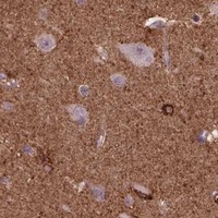
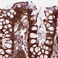


Comprehensive Human Protein Atlas IHC scoring per tissue (reliability: Enhanced — Medium consistency between antibody staining and RNA expression data. Pending external verification.). Rows are taken directly from the HPA tissue chart — click any row's HPA link to view the source.
| Tissue | Cell type | Level | Evidence | Source |
|---|---|---|---|---|
| Appendix | Glandular cells | High | Protein (IHC) | HPA → |
| Bronchus | Respiratory epithelial cells | High | Protein (IHC) | HPA → |
| Cerebellum | Molecular layer - neuropil | High | Protein (IHC) | HPA → |
| Colon | Glandular cells | High | Protein (IHC) | HPA → |
| Endometrium | Glandular cells | High | Protein (IHC) | HPA → |
| Tissue | Cell type | Level | Evidence | Source |
|---|---|---|---|---|
| Adipose tissue | Adipocytes | Not detected | Protein (IHC) | HPA → |
| Adrenal gland | Glandular cells | Not detected | Protein (IHC) | HPA → |
| Heart muscle | Cardiomyocytes | Not detected | Protein (IHC) | HPA → |
| Liver | Cholangiocytes | Not detected | Protein (IHC) | HPA → |
| Parathyroid gland | Glandular cells | Not detected | Protein (IHC) | HPA → |
Use the catalog antibody’s paraffin section protocol as the starting point, then assess MARCKS staining by cell type, compartment and controls.
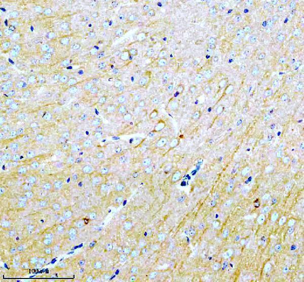
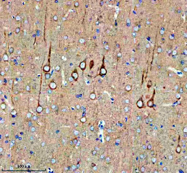
The catalog antibody has real paraffin-section IHC images from mouse and rat brain and human stomach cancer tissue (A04133-4 image captions); no IF image is supplied (catalog payload).
A04133-4 is listed for human, mouse and rat IHC, with paraffin-section images from human stomach cancer tissue and mouse and rat brain (catalog applications/reactivity; A04133-4 image captions). A04133S163 is listed for human, mouse and rat IHC targeting phospho-S163, but has no IHC image in the payload (catalog applications/reactivity; A04133S163 catalog entry).
Which to pick: Choose A04133-4 for tissue IHC and cross-species comparisons: its own captions document paraffin sections from all three listed species, while the fixative and clonality are unreported (A04133-4 image captions; catalog reactivity/clone field). Choose A04133S163 when phospho-S163 is the target, allowing for its lack of an IHC image; its listed IHC dilution is 1:50–1:200 and clonality is unreported (A04133S163 catalog entry). Neither SKU lists IF/ICC validation or supplies an IF image, so there is no catalog-supported IF/ICC pick here (catalog applications; IF image fields).