This website uses cookies to ensure you get the best experience on our website.


- Table of Contents
Plan chromogenic IHC of paraffin sections using the catalog antibody’s documented conditions (datasheet A00216-1). Assess the general cytoplasmic tissue pattern (HPA tissue IHC), allowing for proliferation-dependent nucleolar localization (UniProt).

Expected localisation, validated protocols, controls and antibodies — the at-a-glance facts below, then the full design guide.
| Expected localisation | General cytoplasmic staining (HPA tissue IHC) | |
| Staining pattern | Cytoplasmic staining in glandular cells (HPA tissue IHC) | |
| Antigen retrieval | EDTA pH 8.0 HIER, heat-mediated (datasheet A00216-1) | |
| Positive control | Breast+4 more · see all | |
| Negative control | None in HPA (detected in all 45 tissues); use no-primary + isotype controls |
| Fixation | Keep paraffin-section fixation consistent. (standard IHC practice; not target-specific) | |
| Caveat | Nucleolar signal can disappear in quiescent cells (UniProt) | |
| Regulation | Nucleolar localization in proliferative cells (UniProt) | |
| Isoform / epitope | 2 isoforms; epitope impact unknown; no TM segment (UniProt) |
Compare the catalog antibody’s IHC-P protocol (datasheet A00216-1) with four published MARS1 IHC protocols (PMC12293401; PMC10670752; PMC12676747; PMC12624232).
| Sample | Paraffin-embedded human prostate cancer tissue; fixative not specified (datasheet A00216-1) |
| Fixation | Image fixative and duration unreported (datasheet A00216-1); verify before use. |
| Sectioning | 4–5 µm sections on charged slides (standard) |
| Deparaffinisation | Xylene, graded ethanol series to water (standard) |
| Antigen retrieval | Heat retrieval: EDTA pH 8.0 (datasheet A00216-1); 20 min, 95–100 °C (standard) |
| Peroxidase block | 3% H2O2, 10 min, room temperature (standard) |
| Blocking | 10% goat serum (datasheet A00216-1) |
| Primary antibody | Rabbit anti-MARS1, 2-5 μg/ml (datasheet A00216-1) |
| Primary incubation | Overnight at 4 °C (datasheet A00216-1) |
| Detection | HRP-conjugated secondary, DAB chromogen (datasheet A00216-1) |
| Counterstain | Hematoxylin, blue, dehydrate and mount (standard) |
| Expected result | MARS1-positive staining in glandular cells of breast (HPA tissue IHC: High). HPA tissue profile: General cytoplasmic expression. No signal in the no-primary control. |
MARS1 should show mainly cytoplasmic staining in paraffin sections, including glandular, respiratory epithelial and neuronal cells reported as high by HPA (HPA: tissue IHC). Cytosolic localization is consistent with a protein lacking a transmembrane segment (UniProt P56192: localization and topology). HPA rates the tissue staining pattern Supported, with medium consistency between staining and RNA expression (HPA: tissue IHC reliability).
| Cytoplasmic staining in breast or colon glandular cells, or bronchial respiratory epithelial cells. | This fits HPA’s general cytoplasmic pattern and its High staining calls for those cells (HPA: tissue IHC). Compare signal with adjacent tissue structures; a positive call should identify stained cells and their compartment. |
| Nucleolar staining in some cells alongside cytoplasmic staining. | This can fit MARS1 in proliferative cells: UniProt reports nucleolar localization there and disappearance from the nucleolus in quiescent cells (UniProt P56192: subcellular location). Nucleolar staining alone needs careful review against the expected cytoplasmic pattern (HPA: tissue IHC). |
| Predominantly membranous or extracellular staining, with little cytoplasmic signal. | That is discordant with the reported cytoplasmic pattern and lack of a transmembrane segment (HPA: tissue IHC; UniProt P56192: topology). Check morphology and controls before interpreting it as MARS1; the pattern alone cannot identify the artefact. |
| Strong chondrocyte staining, especially if it resembles the signal in high-staining glandular cells. | HPA lists chondrocytes as Low, so unexpectedly strong staining warrants a check for cross-reactivity or detection background (HPA: tissue IHC; general IHC practice). Low is an observed level, not a verified negative control. |
| Diffuse stain across cells, matrix or the whole section, or no signal in a known high-staining cell population. | Diffuse stain obscures the reported cytoplasmic pattern; absent signal in HPA High cells fails to reproduce that reference observation (HPA: tissue IHC). Neither finding alone distinguishes a technical problem from a specimen difference. |
| Tissue and cell selection | HPA reports High staining in breast, colon, duodenum, endometrium and epididymis glandular cells, bronchial respiratory epithelium, cortical neurons and cerebellar synaptic glomeruli cores (HPA: tissue IHC). Choose a reference with recognizable anatomy; HPA lists no negative tissue. |
| Subcellular location and cell state | Cytosol is the principal location (UniProt P56192: subcellular location; HPA: subcellular ICC-IF). Nucleolar localization depends on proliferation and is reported to disappear in quiescent cells (UniProt P56192: subcellular location); its absence does not negate cytoplasmic staining. |
| Antibody evidence | Both listed antibodies have Supported IHC status; HPA004125 has Enhanced ICC status, while CAB017097 has Supported ICC status (HPA: antibody validation). These application ratings do not demonstrate identical staining in every tissue or establish a fixation effect. |
| Isoforms and epitope coverage | UniProt lists two MARS1 isoforms (UniProt P56192: isoforms). The supplied records give no antibody epitope or isoform coverage, so an isoform-specific staining difference cannot be assigned from this evidence. |
| IF/ICC Q: What localization should be expected? | A: Mainly cytosolic signal; HPA additionally reports sperm mid piece, principal piece and end piece localization (HPA: subcellular ICC-IF). These are IF observations, while this page’s primary readout is chromogenic IHC on paraffin sections. |
| Fixation sensitivity | Target-specific fixation sensitivity is unreported in the supplied UniProt and HPA records. Interpret changes across specimen preparations with matched controls (general IHC practice); do not attribute a change specifically to MARS1 epitope masking. |
| Situation | Likely cause | Next action |
|---|---|---|
| No stain in a reference section expected to contain high-staining glandular or respiratory epithelial cells. | The result does not reproduce HPA’s High calls; the cause is unresolved by those observations (HPA: tissue IHC). | Confirm the expected cells are present, then review antigen retrieval, antibody dilution and detection controls as general IHC workflow checks (general IHC practice). |
| Stain appears mainly on cell borders or in extracellular material. | The distribution conflicts with cytoplasmic MARS1 and its lack of a transmembrane segment (HPA: tissue IHC; UniProt P56192: topology). | Review cellular boundaries and compare with a known cytoplasmic reference pattern; check a no-primary control for detection background (general IHC practice). |
| Nucleolar signal is absent although cytoplasmic signal is clear. | UniProt reports nucleolar localization in proliferative cells and its disappearance in quiescent cells (UniProt P56192: subcellular location). | Score the cytoplasmic pattern separately; assess cell state before treating missing nucleolar signal as a failed stain (UniProt P56192: subcellular location). |
| Strong signal appears in chondrocytes. | This exceeds HPA’s Low call and could reflect cross-reactivity or detection background; the record cannot distinguish them (HPA: tissue IHC; general IHC practice). | Compare intensity and compartment with a high-staining reference, and inspect a no-primary control (HPA: tissue IHC; general IHC practice). |
| Brown chromogen covers tissue broadly and obscures cell boundaries. | Diffuse background prevents assessment of HPA’s general cytoplasmic pattern (HPA: tissue IHC). Endogenous enzyme activity is one possible cause with enzyme-based detection (general IHC practice). | Check a no-primary control and the detection-only background; review blocking, washes and endogenous enzyme blocking appropriate to the detection system (general IHC practice). |
| Two IHC antibodies give different staining patterns. | Both have Supported IHC status, but that rating does not establish agreement in this specimen; epitope and isoform coverage are unspecified (HPA: antibody validation; UniProt P56192: isoforms). | Compare the same cell types and compartments on matched sections, then retain the interpretation supported by tissue morphology and controls (general IHC practice). |


Comprehensive Human Protein Atlas IHC scoring per tissue (reliability: Supported — Medium consistency between antibody staining and RNA expression data.). Rows are taken directly from the HPA tissue chart — click any row's HPA link to view the source.
| Tissue | Cell type | Level | Evidence | Source |
|---|---|---|---|---|
| Breast | Glandular cells | High | Protein (IHC) | HPA → |
| Bronchus | Respiratory epithelial cells | High | Protein (IHC) | HPA → |
| Cerebellum | Synaptic glomeruli - core | High | Protein (IHC) | HPA → |
| Cerebral cortex | Neuronal cells | High | Protein (IHC) | HPA → |
| Colon | Glandular cells | High | Protein (IHC) | HPA → |
| Tissue | Cell type | Level | Evidence | Source |
|---|---|---|---|---|
| None in HPA: MARS1 is detected in all 45 scored tissues. Use a no-primary (secondary-only) and an isotype control instead. | ||||
Troubleshoot MARS1 staining in paraffin sections by checking retrieval, staining conditions and cellular distribution against the reported IHC evidence.
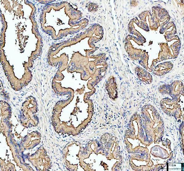
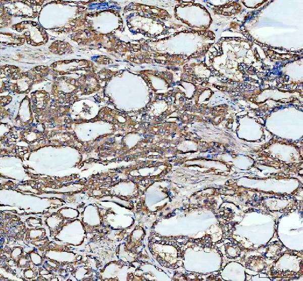
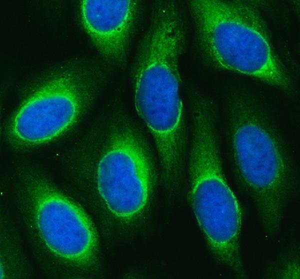
A00216-1 has IHC data from human paraffin-embedded prostate and thyroid cancer sections and IF data from U2OS cells and human prostate cancer sections (catalog image captions).
A00216-1 is listed for human IHC and IF/ICC (catalog: applications and reactivity). Its captions show IHC in paraffin-embedded human prostate and thyroid cancer sections, plus IF in U2OS cells and a paraffin-embedded human prostate cancer section (catalog image captions).
Which to pick: Choose A00216-1 for human paraffin-section IHC: its own captions document both tissue examples, and the listed IHC concentration is 2–5 μg/ml (catalog IHC captions; datasheet: IHC dilution). For IF/ICC, A00216-1 has documented U2OS cell IF and prostate-section IF at 5 μg/ml (catalog IF captions; datasheet: IF dilution). No cross-species option is listed because A00216-1 specifies human reactivity; its tissue captions do not report the fixative (catalog: reactivity; catalog IHC/IF captions).