This website uses cookies to ensure you get the best experience on our website.


- Table of Contents
Plan MAT2A chromogenic IHC in paraffin sections using the catalog antibody's 2–5 μg/ml range (datasheet A04557-1). Assess nuclear and cytoplasmic staining, and validate the pattern because antibody staining and RNA show low consistency (HPA tissue IHC).

Expected localisation, validated protocols, controls and antibodies — the at-a-glance facts below, then the full design guide.
| Expected localisation | Nuclear and cytoplasmic in several tissues (HPA tissue IHC) | |
| Staining pattern | Several cell types show nuclear and cytoplasmic staining (HPA tissue IHC) | |
| Antigen retrieval | EDTA pH 8.0 HIER, heat-mediated (datasheet A04557-1) | |
| Positive control | Bone marrow+4 more · see all | |
| Negative control | Adipose tissue+4 more · see all |
| Fixation | Keep fixation consistent across sections. (standard IHC practice; not target-specific) | |
| Caveat | Staining and RNA show low consistency; validate the pattern (HPA tissue IHC) | |
| Regulation | Expression regulation unreported (UniProt) | |
| Isoform / epitope | 2 isoforms; epitope coverage needs validation (UniProt) |
The catalog antibody protocol uses EDTA retrieval (datasheet A04557-1). Two published MAT2A IHC protocols provide additional conditions for kidney and liver tissue (PMC4003826; PMC3089560).
| Sample | Paraffin-embedded human rectal cancer tissue; fixative not specified (datasheet A04557-1) |
| Fixation | Image fixative and duration unreported (datasheet A04557-1); verify before use. |
| Sectioning | 4–5 µm sections on charged slides (standard) |
| Deparaffinisation | Xylene, graded ethanol series to water (standard) |
| Antigen retrieval | Heat retrieval: EDTA pH 8.0 (datasheet A04557-1); 20 min, 95–100 °C (standard) |
| Peroxidase block | 3% H2O2, 10 min, room temperature (standard) |
| Blocking | 10% goat serum (datasheet A04557-1) |
| Primary antibody | Rabbit anti-MAT2A, 2-5 μg/ml (datasheet A04557-1) |
| Primary incubation | Overnight at 4 °C (datasheet A04557-1) |
| Detection | HRP-conjugated secondary, DAB chromogen (datasheet A04557-1) |
| Counterstain | Hematoxylin, blue, dehydrate and mount (standard) |
| Expected result | MAT2A-positive staining in hematopoietic cells of bone marrow (HPA tissue IHC: Medium). HPA tissue profile: Nuclear and cytoplasmic expression in several tissues. No signal in the no-primary control. |
MAT2A staining in paraffin sections is reported in nuclei and cytoplasm across several tissues (HPA tissue IHC). Expect cell-specific staining, including medium staining in bone marrow hematopoietic cells and bronchial respiratory epithelial cells (HPA tissue IHC). MAT2A has no annotated transmembrane segment (UniProt P31153 topology). Treat the tissue pattern as provisional: HPA rates it Approved but reports low consistency with RNA data and pending external verification (HPA tissue IHC).
| Nuclear and cytoplasmic signal appears in hematopoietic cells in bone marrow or respiratory epithelial cells in bronchus (HPA tissue IHC). | This fits the reported compartments and medium staining in those specific cells (HPA tissue IHC). Judge the named cells, since a whole-tissue label does not establish uniform staining across every cell. |
| Signal is restricted to cell outlines, extracellular material, or a sharply membrane-bound pattern. | That distribution conflicts with the reported nuclear and cytoplasmic IHC profile (HPA tissue IHC) and the absence of a transmembrane segment (UniProt P31153 topology). Investigate an artefact before calling it MAT2A. |
| Strong staining appears in adipocytes or cardiomyocytes, while the intended positive cells are absent. | HPA reports MAT2A as not detected in those cell types (HPA tissue IHC). Consider antibody cross-reactivity or endogenous chromogenic activity; check omission controls and the cell-specific reference pattern (general IHC practice). |
| Color spreads across tissue and blank spaces, obscuring cell boundaries. | Diffuse signal cannot establish the reported nuclear and cytoplasmic distribution (HPA tissue IHC). Check blocking, wash stringency, antibody concentration, and detection-only controls as general IHC practice. |
| No signal appears in bone marrow hematopoietic cells or bronchial respiratory epithelial cells. | Both are reported at medium intensity (HPA tissue IHC), so inspect tissue preservation, retrieval, primary antibody application, and detection controls (general IHC practice). HPA’s low RNA–staining consistency limits certainty about any single specimen (HPA tissue IHC). |
| Cell and tissue selection | Use HPA’s cell-level calls: bone marrow hematopoietic cells and bronchial respiratory epithelial cells are Medium, whereas adipocytes and cardiomyocytes are Not detected (HPA tissue IHC). These are comparison patterns, not universal guarantees. |
| Compartment and topology | HPA reports nuclear and cytoplasmic tissue staining (HPA tissue IHC); UniProt annotates no transmembrane segment (UniProt P31153 topology). Prioritize intracellular, cell-resolved signal when interpreting chromogenic sections. |
| IF/ICC expectation? | The separate IF/ICC evidence places MAT2A mainly in nucleoplasm, with additional cytosol localization (HPA subcellular ICC-IF). This supports a compartment comparison; the reported tissue IHC profile remains the reference for this guide (HPA tissue IHC). |
| Isoforms and epitope coverage | UniProt lists 2 isoforms (UniProt P31153). The supplied records do not map antibody epitopes to either isoform, so staining cannot identify an isoform without additional validation. |
| Evidence strength | HPA lists HPA043028 and CAB009968 as IHC Approved, yet reports low consistency with RNA expression and pending external verification (HPA antibodies; HPA tissue IHC). Approval alone does not establish the stronger Enhanced validation designation. |
| Situation | Likely cause | Next action |
|---|---|---|
| Positive-control cells are blank despite a clean counterstain. | Retrieval, primary antibody application, or chromogenic detection may have failed (general IHC practice); HPA reports medium staining in bone marrow hematopoietic cells and bronchial respiratory epithelial cells (HPA tissue IHC). | Check the control cells on the slide, confirm each incubation and reagent, then review retrieval and detection against the antibody’s validated IHC conditions (general IHC practice). |
| Nuclei stain, but cytoplasm does not. | The balance of compartments can vary across a section; HPA reports both nuclear and cytoplasmic tissue expression without requiring equal intensity in every cell (HPA tissue IHC). | Score nuclear and cytoplasmic compartments separately in identifiable cells. Compare the result with a suitable positive control before treating absent cytoplasmic color as assay failure (general IHC practice). |
| Only membranes or extracellular deposits are prominent. | This disagrees with the reported intracellular IHC pattern (HPA tissue IHC) and the lack of a transmembrane segment (UniProt P31153 topology); precipitated chromogen or nonspecific staining is possible (general IHC practice). | Inspect morphology and a detection-only control, then reassess washing and chromogen development (general IHC practice). Do not score deposits as MAT2A-positive cells. |
| Adipocytes or cardiomyocytes stain strongly. | Those specific cell types are listed as Not detected (HPA tissue IHC). Antibody cross-reactivity or endogenous detection activity may explain unexpected color (general IHC practice). | Run a primary-omission control and inspect the detection chemistry; compare staining with a reported positive cell population (general IHC practice; HPA tissue IHC). |
| Nearly every cell and open area develops faint color. | Background from incomplete blocking, excessive antibody or detection reagent, or insufficient washing can hide cell-specific localization (general IHC practice). | Check omission controls and adjust blocking, washing, or reagent concentration within the validated IHC workflow (general IHC practice). Interpret only clearly bounded cellular signal. |
| A reported positive population varies between specimens. | HPA’s tissue calls are observational, and its IHC summary reports low consistency with RNA data and pending external verification (HPA tissue IHC). A single discrepancy does not establish biological absence. | Compare the same named cell type across sections, document compartment and intensity, and repeat with an appropriate positive control or independent validation when the conclusion depends on the discrepancy (general IHC practice). |
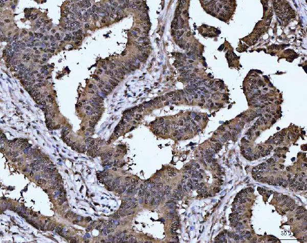
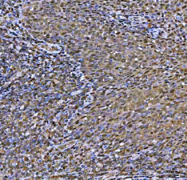


Comprehensive Human Protein Atlas IHC scoring per tissue (reliability: Approved — Low consistency between antibody staining and RNA expression data. Pending external verification.). Rows are taken directly from the HPA tissue chart — click any row's HPA link to view the source.
| Tissue | Cell type | Level | Evidence | Source |
|---|---|---|---|---|
| Bone marrow | Hematopoietic cells | Medium | Protein (IHC) | HPA → |
| Breast | Glandular cells | Medium | Protein (IHC) | HPA → |
| Bronchus | Respiratory epithelial cells | Medium | Protein (IHC) | HPA → |
| Cerebellum | Purkinje cells | Medium | Protein (IHC) | HPA → |
| Cervix | Squamous epithelial cells | Medium | Protein (IHC) | HPA → |
| Tissue | Cell type | Level | Evidence | Source |
|---|---|---|---|---|
| Adipose tissue | Adipocytes | Not detected | Protein (IHC) | HPA → |
| Appendix | Glandular cells | Not detected | Protein (IHC) | HPA → |
| Caudate | Glial cells | Not detected | Protein (IHC) | HPA → |
| Duodenum | Glandular cells | Not detected | Protein (IHC) | HPA → |
| Heart muscle | Cardiomyocytes | Not detected | Protein (IHC) | HPA → |
Troubleshoot MAT2A chromogenic IHC in paraffin sections by checking retrieval, controls, compartment, and cell type before interpreting staining intensity.
A04557-1 has IHC images from human paraffin sections (catalog image captions); M04557 is listed for ICC/IF in human, mouse and rat, without an IF image (catalog applications, reactivity and image alts).
A04557-1 is listed for IHC and has images from human paraffin sections, including rectal cancer and tonsil; its listed reactivity is human, mouse and rat (catalog applications, image captions and reactivity). M04557 is listed for ICC/IF in human, mouse and rat, but has no IF image in the payload (catalog applications, reactivity and image alts).
Which to pick: Choose A04557-1 for tissue IHC: its own images document human paraffin sections, with 2 μg/ml primary antibody in the captions; the fixative is unreported (A04557-1 image captions). Choose rabbit monoclonal M04557 for ICC/IF based on its listed application and clone 25M89, while treating IF performance as undocumented by an image here (M04557 catalog applications, clone and image alts). Both list human, mouse and rat reactivity, but the supplied tissue IHC images demonstrate human samples only (catalog reactivity; A04557-1 image captions).