This website uses cookies to ensure you get the best experience on our website.


- Table of Contents
Plan paraffin IHC for MATR3 using its nuclear tissue pattern (HPA tissue IHC) and the catalog antibody’s 1–2 μg/ml starting range (datasheet A02931-2). Compare nuclear staining across consistently fixed sections, keeping HPA’s cross-gene staining caution in mind (HPA tissue IHC).

Expected localisation, validated protocols, controls and antibodies — the at-a-glance facts below, then the full design guide.
| Expected localisation | Nuclear staining in tissue (HPA tissue IHC) | |
| Staining pattern | Nuclei across many tissue cell types (HPA tissue IHC) | |
| Antigen retrieval | EDTA pH 8.0 HIER, heat-mediated (datasheet A02931-2) | |
| Positive control | Adipose tissue+4 more · see all | |
| Negative control | None in HPA (detected in all 45 tissues); use no-primary + isotype controls |
| Fixation | Keep fixation conditions consistent across sections. (standard IHC practice; not target-specific) | |
| Caveat | Staining may reflect proteins from other genes (HPA tissue IHC) | |
| Regulation | Low tissue specificity (HPA tissue RNA) | |
| Isoform / epitope | Two isoforms; epitope differences are unspecified (UniProt) |
The catalog antibody’s IHC-P protocol is accompanied by published MATR3 IHC protocols for mouse spinal cord and muscle, renal tissue, and human entorhinal cortex (PMC6299607; PMC9952985; PMC7603783).
| Sample | Paraffin-embedded human breast cancer tissue; fixative not specified (datasheet A02931-2) |
| Fixation | Image fixative and duration unreported (datasheet A02931-2); verify before use. |
| Sectioning | 4–5 µm sections on charged slides (standard) |
| Deparaffinisation | Xylene, graded ethanol series to water (standard) |
| Antigen retrieval | Heat retrieval: EDTA pH 8.0 (datasheet A02931-2); 20 min, 95–100 °C (standard) |
| Peroxidase block | 3% H2O2, 10 min, room temperature (standard) |
| Blocking | 10% goat serum (datasheet A02931-2) |
| Primary antibody | Rabbit anti-MATR3, 1-2 μg/ml (datasheet A02931-2) |
| Primary incubation | Overnight at 4 °C (datasheet A02931-2) |
| Detection | HRP-conjugated secondary, DAB chromogen (datasheet A02931-2) |
| Counterstain | Hematoxylin, blue, dehydrate and mount (standard) |
| Expected result | MATR3-positive staining in adipocytes of adipose tissue (HPA tissue IHC: High). HPA tissue profile: Ubiquitous nuclear expression. No signal in the no-primary control. |
MATR3 is a nuclear matrix protein with no transmembrane segment (UniProt P43243: subcellular location and topology). Expect nuclear staining across many cell types, including adipocytes, glandular cells, hematopoietic cells, and neurons (HPA tissue IHC: ubiquitous nuclear expression; High in the listed cells). Interpret this expectation cautiously: HPA rates the tissue pattern Supported, reports medium agreement with RNA data, and warns that the staining targets proteins from more than one gene (HPA tissue IHC).
| Distinct nuclear chromogen in tissue cells, with limited staining outside nuclei. | This matches the reported ubiquitous nuclear pattern (HPA tissue IHC) and nuclear matrix location (UniProt P43243). Nuclear staining in listed High examples—adipocytes, adrenal glandular cells, bone marrow hematopoietic cells, or caudate neurons—is a useful positive check (HPA tissue IHC). The pattern supports interpretation but does not, by itself, establish antibody specificity (HPA tissue IHC: multiple-gene caution). |
| Predominantly cytoplasmic, membranous, or extracellular staining while nuclei remain faint. | The dominant compartment conflicts with the expected nuclear location (UniProt P43243; HPA tissue IHC). Review morphology and detection controls before calling it MATR3. A stained section alone cannot distinguish antibody binding to another protein from nonspecific chromogen deposition; the HPA multiple-gene warning makes that distinction especially relevant (HPA tissue IHC). |
| Strong signal confined to an unexpected cell population while nearby expected nuclei are unstained. | Check whether the apparent cell identity and nuclear boundaries are correct. HPA describes broad nuclear expression and lists several High cell populations (HPA tissue IHC), but supplies no negative tissue or cell population. An isolated unexpected pattern raises cross-reactivity or endogenous detection activity as possibilities; it is not proof of either. Compare a negative detection control and another validated antibody when available (general IHC practice). |
| Haze or granular chromogen covers nuclei and surrounding tissue alike. | Diffuse deposit obscures the compartment needed to interpret MATR3 (UniProt P43243: nuclear matrix; HPA tissue IHC: nuclear expression). Excess detection background, inadequate washing, or endogenous enzyme activity are general chromogenic IHC possibilities. Assess the no-primary control and preserve visible nuclear counterstain before scoring cells (general IHC practice). |
| No convincing nuclear signal in a listed High tissue and no widespread background. | A technical false negative is possible, particularly if internal tissue morphology or the control section is poor (general IHC practice). HPA lists High staining in adipocytes, adrenal glandular cells, bone marrow hematopoietic cells, and caudate neurons (HPA tissue IHC). Confirm that the expected cell type is present, then review the antibody's IHC-P instructions and positive control; HPA levels do not establish a target-specific fixation effect. |
| Compartment and tissue distribution | MATR3 is assigned to the nuclear matrix (UniProt P43243); HPA reports ubiquitous nuclear tissue staining and low tissue RNA specificity (HPA tissue IHC). Use nuclear location and the identity of cells in the section together; broad tissue positivity alone is a weak specificity check. |
| Strength of tissue evidence | The tissue profile is Supported, with medium agreement between antibody staining and RNA expression, and a warning that staining targets proteins from more than one gene (HPA tissue IHC). Treat an otherwise plausible nuclear pattern as supported, not uniquely attributable to MATR3 without further validation. |
| Antibody validation | Three listed antibodies have Supported IHC status: HPA036564, HPA036565, and CAB033552 (HPA antibodies). No listed antibody has an IHC Enhanced designation in this payload (HPA antibodies). Confirm which antibody produced a reference image before comparing its pattern with the catalog antibody. |
| IF/ICC Q&A: what location should be expected? | The HPA ICC-IF location is approved nucleoplasm, with images from A-431, U-251MG, and U2OS; HPA also warns that the antibodies target proteins from multiple genes (HPA subcellular). Nuclear fluorescence is compatible with that record. This IF/ICC observation does not define an IHC-P staining protocol. |
| Situation | Likely cause | Next action |
|---|---|---|
| Expected nuclei are blank in an HPA-listed High tissue. | Target-specific fixation effects are not established by the supplied assay evidence. Verify with a matched IHC source before attributing a result to fixation. | Verify tissue morphology and the listed High cells, then check the positive control and catalog antibody's IHC-P instructions. Review retrieval and detection conditions as general IHC workflow variables. |
| Chromogen is mainly cytoplasmic or membranous. | That distribution conflicts with nuclear matrix localization and the nuclear tissue profile (UniProt P43243; HPA tissue IHC). Misread cell boundaries or nonspecific staining may contribute (general IHC practice). | Inspect nuclear counterstain and a no-primary control. Score only a reproducible nuclear pattern; if the discrepancy persists, compare another IHC-supported antibody (HPA antibodies). |
| Only an unusual cell population stains strongly. | HPA reports ubiquitous nuclear expression but provides no negative cell population here; cross-reactivity or endogenous detection activity remains possible (HPA tissue IHC; general IHC practice). | Identify the stained cells morphologically, check the no-primary control, and compare an independent IHC-supported antibody if available (HPA antibodies). Avoid declaring the population MATR3-specific from this pattern alone. |
| The whole section has diffuse brown background. | Nonspecific reagent binding, insufficient washing, or endogenous peroxidase are general chromogenic IHC causes; diffuse deposit cannot be read as nuclear MATR3 (general IHC practice; UniProt P43243). | Check a no-primary control, review blocking and washes, and verify the detection system's endogenous-activity control (general IHC practice). Reassess nuclear localization after background is controlled. |
| Nuclear staining is too strong to distinguish individual cells. | Excess chromogen or high antibody concentration can obscure nuclear boundaries in chromogenic IHC (general IHC practice). Intensity alone cannot overcome HPA's multiple-gene caution (HPA tissue IHC). | Follow the catalog antibody's IHC-P dilution guidance and optimize detection development with a counterstain that leaves nuclei readable (general IHC practice). Score localization only where cell boundaries remain clear. |
| A nuclear pattern differs between antibodies. | The supplied antibodies are IHC Supported, while HPA flags possible staining of proteins from multiple genes (HPA antibodies; HPA tissue IHC). Different patterns therefore require scrutiny rather than averaging. | Compare matched tissue regions and controls, document each antibody separately, and restrict conclusions to the reproducible nuclear pattern (general IHC practice; UniProt P43243). |
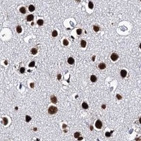


Comprehensive Human Protein Atlas IHC scoring per tissue (reliability: Supported — Medium consistency between antibody staining and RNA expression data. Caution, targets protein from more than one gene.). Rows are taken directly from the HPA tissue chart — click any row's HPA link to view the source.
| Tissue | Cell type | Level | Evidence | Source |
|---|---|---|---|---|
| Adipose tissue | Adipocytes | High | Protein (IHC) | HPA → |
| Adrenal gland | Glandular cells | High | Protein (IHC) | HPA → |
| Appendix | Glandular cells | High | Protein (IHC) | HPA → |
| Bone marrow | Hematopoietic cells | High | Protein (IHC) | HPA → |
| Breast | Adipocytes | High | Protein (IHC) | HPA → |
| Tissue | Cell type | Level | Evidence | Source |
|---|---|---|---|---|
| None in HPA: MATR3 is detected in all 45 scored tissues. Use a no-primary (secondary-only) and an isotype control instead. | ||||
Troubleshoot chromogenic MATR3 staining in paraffin sections by checking nuclear localisation, retrieval conditions and assay controls before comparing staining scores.
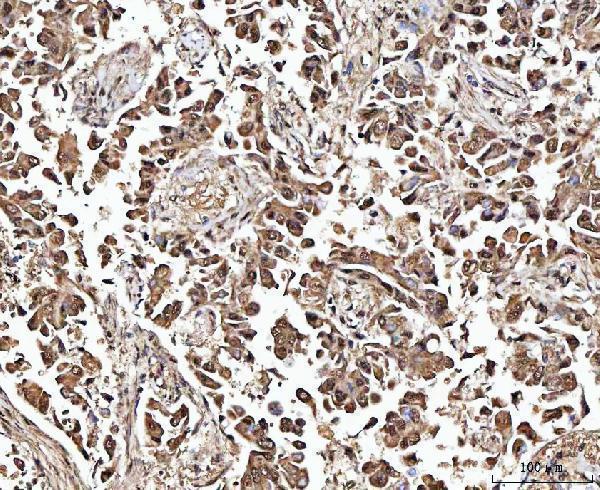
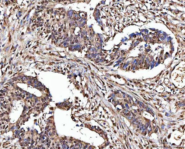
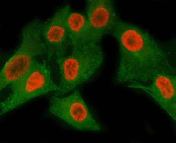
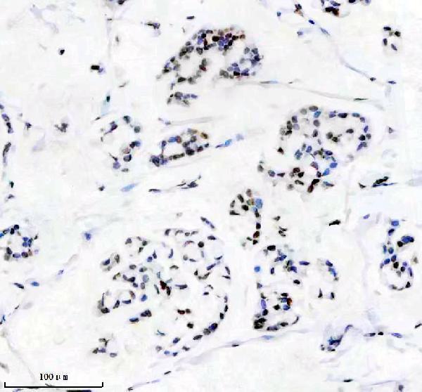
Both antibodies have paraffin-section IHC images from human breast cancer tissue; A02931-2 also has an IF image from A549 cells (catalog image captions). Human, Mouse and Rat are listed as reactive species (catalog reactivity).
A02931-2 has IHC images from human breast cancer, rectum adenocarcinoma, placenta and testicular germ cell tumor sections, plus an IF image from A549 cells (A02931-2 image captions). M02931-1 has an IHC image from human breast cancer tissue; IF/ICC is listed as an application, but no IF image is supplied (M02931-1 catalog applications and image captions).
Which to pick: For tissue IHC, A02931-2 has images across more sample types; both SKUs have paraffin-section images using EDTA pH 8.0 retrieval, and neither caption reports the fixative (catalog IHC image captions). For IF/ICC, A02931-2 has an A549 IF image, while M02931-1 is a rabbit monoclonal with IF/ICC listed but no IF image (catalog IF image captions; M02931-1 catalog clone and applications). Both list Human, Mouse and Rat reactivity, though the supplied IHC images show human tissue only (catalog reactivity and IHC image captions).