MBD2 / Methyl-CpG-binding domain protein 2 · IHC design guide

Design Immunohistochemistry for MBD2

Plan chromogenic MBD2 IHC in paraffin sections around the nuclear staining reported in most tissues (HPA tissue IHC). Use high-staining squamous epithelium as a reference and interpret cytoplasmic signal cautiously (HPA tissue IHC).

Evidence assembled Oct 2026 · For research use; verify linked source records and product datasheet before use
Immunohistochemistry protocol sheet for MBD2 (IHC for MBD2): expected localisation Nuclear in most tissues (HPA tissue IHC), antibody M01746-1, validated IHC image, and IHC protocol steps
Printable MBD2 IHC protocol sheet — expected localisation Nuclear in most tissues (HPA tissue IHC), antibody M01746-1, controls and protocol steps. Open the full MBD2 IHC guide →

MBD2 Immunohistochemistry Experimental Design Guide

Expected localisation, validated protocols, controls and antibodies — the at-a-glance facts below, then the full design guide.

Must know before staining
Expected localisation Nuclear in most tissues (HPA tissue IHC)
Staining pattern Nuclear staining across most tissue cell types (HPA tissue IHC)
Antigen retrieval Citrate pH 6 HIER, heat-mediated (datasheet M01746-1)
Positive control ⓘ Cerebellum+4 more · see all
Negative control ⓘ Hippocampus+1 more · see all
Important caveats
Reasons your staining may differ from the expected pattern.
Fixation Formaldehyde-fixed paraffin sections (selected-SKU IHC image M01746-1); keep fixation consistent (standard IHC practice; not target-specific)
Caveat Cytoplasmic staining was disregarded (HPA tissue IHC)
Regulation Expression regulation not established (UniProt)
Isoform / epitope 2 isoforms (1 and 3); epitope coverage unknown (UniProt)
Section 1

Recommended MBD2 IHC & IF Protocols

The catalog antibody protocol uses citrate pH 6 heat retrieval (datasheet M01746-1). The published MBD2 IHC methods below provide tissue-specific starting points (PMC12478297; PMC4940051; PMC7400232; PMC8933641).

Recommended immunohistochemistry (IHC-P) protocol parameters
SampleFormaldehyde-fixed, paraffin-embedded human colon tissue (datasheet M01746-1)
FixationImage formalin-fixed; duration unreported (datasheet M01746-1); verify before use.
Sectioning4–5 µm sections on charged slides (standard)
DeparaffinisationXylene, graded ethanol series to water (standard)
Antigen retrievalHeat retrieval: Citrate pH 6 (datasheet M01746-1); 20 min, 95–100 °C (standard)
Peroxidase block3% H2O2, 10 min, room temperature (standard)
Blocking10% normal serum of the secondary host, 30 min, room temperature (standard)
Primary antibodyMouse monoclonal (clone 1812CT625.9.45) anti-MBD2, 1:25 (datasheet M01746-1)
Primary incubation1 hours at 37°C (datasheet M01746-1)
DetectionHRP-polymer secondary, DAB chromogen 5–10 min (standard)
CounterstainHematoxylin, blue, dehydrate and mount (standard)
Expected resultMBD2-positive staining in purkinje cells of cerebellum (HPA tissue IHC: High). HPA tissue profile: Nuclear expression in most tissues. No signal in the no-primary control.
💡Decision noteStart with citrate pH 6 heat retrieval for the catalog antibody (datasheet M01746-1); adjust retrieval only after checking nuclear staining and controls (UniProt Q9UBB5 localization).
Section 2

What Is the Expected MBD2 Staining Pattern?

MBD2 should stain nuclei in most tissues, including Purkinje cells, squamous epithelial cells, keratinocytes and urothelial cells at HPA high-staining sites (HPA tissue IHC). Nuclear localisation fits a chromatin-associated protein with no transmembrane segment (UniProt Q9UBB5). Interpret the tissue pattern cautiously: HPA rates its IHC evidence Approved, reports low agreement with RNA data, and disregards observed cytoplasmic staining (HPA tissue IHC).

What am I looking at on my slide?
Distinct nuclear stain in Purkinje cells or in squamous epithelial cells of cervix, esophagus, oral mucosa or tonsil.This matches HPA's High staining in those cell types and its broader nuclear profile (HPA tissue IHC). Score the named cells and their nuclei; staining elsewhere in the section does not substitute for that reference pattern.
Prominent cytoplasmic colour with weak or absent nuclear staining.Treat this as a suspect pattern: HPA observed cytoplasmic staining but disregarded it, while UniProt places MBD2 in nuclei and on chromosomes (HPA tissue IHC; UniProt Q9UBB5). Review the negative reagent control and detection background before assigning target signal.
Strong stain in a cell population expected to be unstained, without the anticipated nuclear signal in reference cells.Consider cross-reactivity or endogenous detection activity as general IHC possibilities. HPA reports glial cells in hippocampus and ovarian stroma cells as Not detected; that finding applies to those cells, not every cell in either tissue (HPA tissue IHC).
Diffuse colour over nuclei, cytoplasm and surrounding section.This is difficult to score as MBD2 because the reference pattern is predominantly nuclear (HPA tissue IHC; UniProt Q9UBB5). In general IHC practice, compare a negative reagent control and review blocking, washing and chromogen development for background.
No nuclear signal in a named high-staining reference population.A missing signal in, for example, skin keratinocytes or rectal glandular cells conflicts with their HPA High staining (HPA tissue IHC). Check section quality and the IHC detection run before interpreting an experimental sample as negative; HPA's Approved rating still carries a consistency caveat.
💡Expected MBD2 appearanceCall a result positive when the expected cells show clear, predominantly nuclear chromogenic stain—for example, High nuclear staining in skin keratinocytes—with cytoplasmic-only or diffuse stain treated as suspect (HPA tissue IHC; UniProt Q9UBB5).
How each factor affects the staining
Tissue and cell selectionHPA reports nuclear expression in most tissues, but staining differs by cell type: skin keratinocytes are High, liver hepatocytes Low, and ovarian stroma cells Not detected (HPA tissue IHC). Identify the cell population before comparing sections.
Evidence strengthThe HPA tissue pattern is Approved, with low agreement between antibody staining and RNA data; HPA disregarded cytoplasmic staining (HPA tissue IHC). Use the reported nuclear pattern as a reference, while treating a discrepant sample as a result to investigate.
Antibody validation by applicationCAB037282 has an Approved IHC rating; HPA067582 and HPA067615 have Enhanced ICC ratings, with no IHC rating listed for either (HPA antibodies). Keep these application-specific ratings separate when evaluating a paraffin-section result.
Protein form and localisationUniProt lists two MBD2 isoforms, one chain spanning residues 1–411, no signal peptide, no propeptide and no transmembrane segment (UniProt Q9UBB5). These facts support a nuclear interpretation but do not establish which isoform an antibody detects or where its epitope lies.
IF/ICC appearance?In ICC-IF, HPA places MBD2 mainly in the nucleoplasm with supported localisation and lists images from A-431, HaCaT and U2OS cells (HPA subcellular). UniProt also reports discrete nuclear foci, including late-S-phase replication foci (UniProt Q9UBB5); a foci pattern is not required for an IHC positive call.
Why is my staining missing, weak or wrong?
SituationLikely causeNext action
Positive reference tissue shows no nuclear colour.A detection-run problem is possible in general IHC practice; the expected cells may also have been missed during review.Recheck the cell population and compare a run control. Use an HPA High reference such as skin keratinocytes or rectal glandular cells (HPA tissue IHC); then review the standard IHC detection steps.
Only cytoplasmic staining is visible.Its assignment to MBD2 is unsupported because HPA disregarded cytoplasmic staining (HPA tissue IHC).Inspect a negative reagent control and background in adjacent tissue, then judge positivity from nuclear staining in expected cells (HPA tissue IHC; UniProt Q9UBB5).
A low-staining reference appears faint.The chosen population may have limited reported signal: HPA rates cardiomyocytes, hepatocytes and lung macrophages Low (HPA tissue IHC).Compare a named High cell population in the same run before changing general IHC conditions (HPA tissue IHC). Record the cell type rather than calling the whole tissue negative.
Colour spreads through the section and obscures nuclear boundaries.Nonspecific background or excess chromogen development is possible under general IHC practice; the diffuse result cannot be mapped confidently to HPA's nuclear profile (HPA tissue IHC).Compare the negative reagent control; review blocking, washing and development, then rescore identifiable nuclei.
Unexpected cells stain while reference cells do not.Cross-reactivity or endogenous detection activity is possible under general IHC practice. HPA's Not detected calls concern hippocampal glial cells and ovarian stroma cells specifically (HPA tissue IHC).Confirm cell identity and compartment, inspect the negative reagent control, and compare a named HPA High population before attributing the signal to MBD2.
ICC-IF looks nucleoplasmic, but the paraffin-section IHC result differs.The validation records cover different applications: two antibodies have Enhanced ICC ratings, while CAB037282 has an Approved IHC rating (HPA antibodies).Evaluate each result against its own application rating and localisation evidence; for IHC, prioritize the reported nuclear tissue pattern and its consistency caveat (HPA tissue IHC).

Sample controls for MBD2 IHC & IF

🧪Run rectum first: glandular cells should show nuclear staining (HPA: High in rectal glandular cells; UniProt Q9UBB5: nucleus). Use ovarian stroma as the negative tissue (HPA: Not detected in ovarian stroma cells); on the rectal slide, non-nuclear areas should remain unstained, but the supplied evidence does not identify a negative cell type within that tissue.
Positive control tissue: Cerebellum (Purkinje cells, HPA High)
Negative control tissue: Hippocampus (HPA Not detected)
ICC-IF cell lines (HPA subcellular resource): HPA ICC-IF images show MBD2 in A-431, HaCaT, U2OS, with annotated localisation: Nucleoplasm (supported) (HPA subcellular).
Technical controls: Include a no-primary, secondary-only control; an isotype control matched to the primary antibody’s host species, clonality and concentration; and MBD2 knockout tissue or cognate-peptide competition where available (standard IHC practice). For chromogenic rectal sections, block endogenous peroxidase and, if using biotin detection, endogenous biotin (standard IHC practice; selected M01746-1 caption: biotinylated secondary antibody).
⚠️Feasibility: A target-specific fixation window or fixation effect is unreported, and the selected M01746-1 caption’s fixative field is unreported; its citrate buffer at pH 6 with heat retrieval documents one IHC-P condition, not a retrieval requirement (selected M01746-1 caption). The supplied evidence does not establish whether frozen sections are easier; IF/ICC is feasible in A-431, HaCaT and U2OS, with mainly nucleoplasmic signal (HPA: ICC-IF images and supported nucleoplasm location). In rectal sections, assess glandular nuclei against mucus and chromogenic background (HPA: rectal glandular cells High; standard IHC scoring practice).

HPA tissue IHC evidence for MBD2

Comprehensive Human Protein Atlas IHC scoring per tissue (reliability: Approved — Low consistency between antibody staining and RNA expression data. Cytoplasmic staining observed and disregarded.). Rows are taken directly from the HPA tissue chart — click any row's HPA link to view the source.

Positive expression · recommended positive controls

TissueCell typeLevelEvidenceSource
Cerebellum Purkinje cells High Protein (IHC) HPA →
Cervix Squamous epithelial cells High Protein (IHC) HPA →
Esophagus Squamous epithelial cells High Protein (IHC) HPA →
Oral mucosa Squamous epithelial cells High Protein (IHC) HPA →
Rectum Glandular cells High Protein (IHC) HPA →

Undetected expression · recommended negative controls

TissueCell typeLevelEvidenceSource
Hippocampus Glial cells Not detected Protein (IHC) HPA →
Ovary Ovarian stroma cells Not detected Protein (IHC) HPA →
Section 3

Advanced MBD2 IHC Tips

Troubleshoot MBD2 staining by checking nuclear localisation, section processing, controls and cell specific scoring (UniProt Q9UBB5; HPA tissue IHC).

How should I adjust retrieval when MBD2 nuclear staining is weak?
Start with heat mediated retrieval in citrate buffer at pH 6 (datasheet M01746-1). Keep heating time and cooling conditions consistent across control and test sections, because variable retrieval can change chromogenic staining intensity (standard IHC practice). The selected colon section image used citrate retrieval, a 1:25 primary dilution and 1 hour incubation at 37°C; those conditions are an example, not proof of optimal sensitivity elsewhere (caption M01746-1). If nuclear signal remains weak, compare a modestly longer citrate retrieval with an untreated control section and watch for tissue damage or rising background (standard IHC practice; UniProt Q9UBB5 localisation).
Could fixation explain weak MBD2 staining in my paraffin sections?
Target specific fixation sensitivity for MBD2 is unknown from the supplied evidence (record: fixation evidence). The selected paraffin section image reports formaldehyde fixation, but that single example does not establish how different fixation durations affect MBD2 detection (caption M01746-1). Record fixative type and duration for each block, then compare sections processed with the same pH 6 citrate retrieval and detection conditions (datasheet M01746-1; standard IHC practice). If morphology is preserved but nuclear staining varies, test a retrieval series and a known positive section before attributing the difference to fixation (HPA tissue IHC; standard IHC practice).
Should cytoplasmic MBD2 staining count as a positive result?
Score nuclear chromogen as the expected MBD2 pattern: UniProt places MBD2 in nuclei and chromatin, while HPA reports nuclear expression in most tissues (UniProt Q9UBB5 localisation; HPA tissue IHC). HPA specifically disregarded cytoplasmic staining and reported low consistency between antibody staining and RNA expression (HPA tissue IHC). Inspect intact nuclei at higher magnification and compare adjacent cells with a matched negative control before counting diffuse cytoplasmic colour (standard IHC practice). Discrete nuclear foci can be biologically plausible, including at late S phase replication sites, but morphology alone cannot establish their identity (UniProt Q9UBB5 localisation; standard IHC practice).
How do MBD2 isoforms and epitope location affect IHC interpretation?
MBD2 has 2 recorded isoforms, 1 and 3, and its methyl CpG binding domain spans residues 145–213 (UniProt Q9UBB5 isoforms and domains). The supplied evidence does not map the catalog antibody epitope, so do not assume it detects both isoforms equally (record: antibody epitope unspecified). Nuclear staining also cannot identify a particular isoform or prove methylated DNA binding in that cell (UniProt Q9UBB5 function and localisation; standard IHC interpretation). If isoform specificity matters, obtain the immunogen sequence and validate it against isoform sequences with an independent assay before assigning an IHC pattern (standard antibody validation practice).
How can I check MBD2 localisation by IF alongside a cell marker?
On the separate IF/ICC workflow, pair MBD2 with a marker for the cell population being examined and use the marker to identify cells before judging nuclear overlap (standard IF practice; UniProt Q9UBB5 localisation). Choose fluorophores after checking unstained tissue for autofluorescence, and reserve a channel with low background for the weaker signal (standard IF practice). MBD2 is nuclear and has no transmembrane segment, so permeabilisation must allow antibody access through the plasma and nuclear envelopes; the antibody epitope itself is unspecified (UniProt Q9UBB5 topology and localisation; record: antibody epitope unspecified). Include single stain and secondary only controls to assess bleed through and nonspecific fluorescence (standard IF practice).
What should I check when MBD2 IHC has widespread brown background?
Compare a no primary control with the stained section to assess detection reagent background, and check whether colour is nuclear or diffuse (standard chromogenic IHC practice; UniProt Q9UBB5 localisation). For peroxidase detection, block endogenous peroxidase and inspect blood rich or damaged areas before interpreting brown DAB deposits (standard chromogenic IHC practice). The selected image used 3% BSA for 0.5 hour at room temperature and a 1:25 primary dilution; these are reported conditions, not universal settings (caption M01746-1). If controls are clean but background persists, titrate primary concentration and washing while retaining a positive nuclear reference section (standard IHC practice; HPA tissue IHC).
How should I quantify MBD2 staining across sections? ⚠ ANSWER MARKED FOR VERIFICATION
Define cell populations and tissue regions before scoring, then record the percentage of intact nuclei with chromogen above a threshold set from negative controls (standard IHC quantification practice; UniProt Q9UBB5 localisation). An H score can combine percentage at each intensity category, yielding a range of 0–300 when intensities are scored 0–3 (standard IHC quantification practice). Alternatively report positive nuclei per mm², normalised to evaluable tissue area, or positive nuclei as a fraction of all evaluable nuclei in the same compartment (standard IHC quantification practice). Keep retrieval, imaging and thresholds constant, and exclude folds and necrosis from the denominator (standard IHC practice).
How can I distinguish true MBD2 staining from section artefacts?
A credible MBD2 pattern is nuclear in intact cells, consistent with its chromatin association and HPA's predominantly nuclear tissue profile (UniProt Q9UBB5 localisation; HPA tissue IHC). Use an expected positive population, such as rectal glandular cells, alongside a no primary control; HPA reports high staining in that population (HPA tissue IHC; standard IHC practice). Discount colour restricted to cut edges, folds or necrotic regions, and investigate brown deposits that persist without primary antibody as possible endogenous enzyme or detection background (standard chromogenic IHC practice). Because HPA disregarded cytoplasmic staining and reported low staining versus RNA consistency, avoid inferring MBD2 activity or methylation status from chromogen intensity alone (HPA tissue IHC; UniProt Q9UBB5 function).
Boster reagents

Best MBD2 / Methyl-CpG-binding domain protein 2 IHC Antibodies

MBD2 has a human colon paraffin-section IHC figure (M01746-1 image caption). IF/ICC is listed for human, mouse, and rat samples, but no IF figure is supplied (M01746 catalog applications/reactivity; image alts).

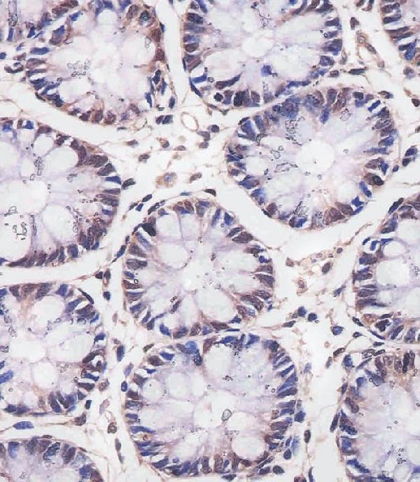
Real IHC data M01746-1 staining MBD2 in human colon tissue sections by Immunohistochemistry (IHC-P -paraformaldehyde-fixed, paraffin-embedded sections). Tissue was fixed with formaldehyde and blocked with 3% BSA for 0. 5 hour at room temperature; antigen retrieval was by heat mediation with a citrate buffer (pH6). Samples were incubated with primary antibody (1/25) for 1 hours at 37°C. A undiluted biotinylated goat polyvalent antibody was used as the secondary antibody.
Anti-MBD2 Antibody
Cat # M01746-1

The rendered card is M01746-1, a human-reactive IHC-P antibody shown staining human colon paraffin sections (catalog applications/reactivity; M01746-1 image caption). Its figure documents citrate pH 6 retrieval and a 1:25 primary dilution (M01746-1 image caption).

Which to pick: For human tissue IHC-P, choose mouse monoclonal M01746-1: its own figure supports paraffin sections, although the caption gives conflicting fixative terms (M01746-1 catalog host/clone; image caption). For human IF/ICC, rabbit monoclonal M01746-2 lists both applications; no IF figure or fixative is reported (M01746-2 catalog host/clone, applications/reactivity, image alts). For cross-species work, rabbit monoclonal M01746 lists human, mouse, and rat reactivity alongside IHC and IF/ICC applications; no figure or fixative is reported (M01746 catalog host/clone, applications/reactivity, image alts).

Each figure is that product's own IHC / IF validation image from its datasheet.

References

  1. UniProt Consortium. UniProt entry Q9UBB5 (MBD2_HUMAN, Methyl-CpG-binding domain protein 2).
  2. Human Protein Atlas. MBD2 tissue IHC expression (reliability: Approved).
  3. Human Protein Atlas. MBD2 subcellular location (ICC-IF): Mainly localized to the nucleoplasm..
  4. Human Protein Atlas. MBD2 antibody validation summary (3 antibodies).
  5. Loss of MBD2 is associated with tumor growth in small intestinal neuroendocrine tumors. Endocrine connections 2025 — PMC12478297.
  6. MBD2 as a novel marker associated with poor survival of patients with hepatocellular carcinoma after hepatic resection. Molecular medicine reports 2016 — PMC4940051.
  7. Expression and clinical significance of methyl-CpG binding domain protein 2 in high-grade serous ovarian cancer. Oncology letters 2020 — PMC7400232.
  8. Genetic or siRNA inhibition of MBD2 attenuates the UUO- and I/R-induced renal fibrosis via downregulation of EGR1. Molecular therapy. Nucleic acids 2022 — PMC8933641.
  9. PubMed PMID:9774669 — UniProt-cited evidence.
  10. PubMed PMID:10441743 — UniProt-cited evidence.
  11. PubMed PMID:15489334 — UniProt-cited evidence.