This website uses cookies to ensure you get the best experience on our website.


- Table of Contents
Plan MCM3 paraffin IHC around nuclear staining in proliferating cells (HPA tissue IHC). Bone marrow hematopoietic cells offer a high-expression reference, while adipocytes are reported as not detected (HPA tissue IHC).

Expected localisation, validated protocols, controls and antibodies — the at-a-glance facts below, then the full design guide.
| Expected localisation | Nuclei of proliferating cells (HPA tissue IHC) | |
| Staining pattern | High nuclear signal in marrow hematopoietic cells (HPA tissue IHC) | |
| Antigen retrieval | EDTA pH 8.0 HIER, heat-mediated (datasheet PA1651) | |
| Positive control | Bone marrow+4 more · see all | |
| Negative control | Adipose tissue+4 more · see all |
| Fixation | Keep fixation consistent across sections (standard IHC practice; not target-specific) | |
| Caveat | Proliferation varies within tissue, changing the positive-cell fraction (HPA tissue IHC) | |
| Regulation | Enriched in proliferating cells (HPA tissue IHC) | |
| Isoform / epitope | 2 isoforms; epitope differences are unspecified (UniProt) |
Compare the catalog antibody’s IHC-P protocol (datasheet: PA1651) with published breast biopsy, melanoma, and ovarian carcinoma IHC methods (PMC12534050; PMC3433373; PMC9035053).
| Sample | Paraffin-embedded human cervical cancer tissue; fixative not specified (datasheet PA1651) |
| Fixation | Image fixative and duration unreported (datasheet PA1651); verify before use. |
| Sectioning | 4–5 µm sections on charged slides (standard) |
| Deparaffinisation | Xylene, graded ethanol series to water (standard) |
| Antigen retrieval | Heat retrieval: EDTA pH 8.0 (datasheet PA1651); 20 min, 95–100 °C (standard) |
| Peroxidase block | 3% H2O2, 10 min, room temperature (standard) |
| Blocking | 10% goat serum (datasheet PA1651) |
| Primary antibody | Rabbit anti-MCM3, 2-5μg/ml (datasheet PA1651) |
| Primary incubation | Overnight at 4 °C (datasheet PA1651) |
| Detection | HRP-conjugated secondary, DAB chromogen (datasheet PA1651) |
| Counterstain | Hematoxylin, blue, dehydrate and mount (standard) |
| Expected result | MCM3-positive staining in hematopoietic cells of bone marrow (HPA tissue IHC: High). HPA tissue profile: Nuclear expression in proliferating cells. No signal in the no-primary control. |
MCM3 should stain nuclei, especially in proliferating cells (HPA tissue IHC: nuclear expression in proliferating cells; UniProt P25205: nucleus and chromosome). High staining is reported in bone marrow hematopoietic cells and placental cytotrophoblasts (HPA tissue IHC). The tissue pattern has Supported reliability, with medium consistency between staining and RNA data (HPA tissue IHC). MCM3 has no transmembrane segment (UniProt P25205 topology).
| Distinct nuclear staining in bone marrow hematopoietic cells or placental cytotrophoblasts. | This fits the reported high staining in both cell populations and the expected nuclear location (HPA tissue IHC; UniProt P25205). Judge the named cells, rather than calling every cell in the section positive. Staining strength elsewhere may differ: skin basal-layer cells and lymph-node germinal-center cells are reported at medium levels (HPA tissue IHC). |
| Predominantly cytoplasmic staining, with little convincing nuclear staining. | Treat this as a possible assay artefact because the reported tissue pattern is nuclear and the approved ICC-IF location is nucleoplasmic (HPA tissue IHC; HPA subcellular). Review a known-positive section and a control omitting the primary antibody. Chromatin association changes during replication, so a single cell’s appearance alone is insufficient to define a new compartment pattern (UniProt P25205). |
| Strong staining in adipocytes or adrenal glandular cells. | These particular cell populations were reported as not detected (HPA tissue IHC); staining there merits a cross-reactivity or endogenous-detection check. The HPA designation applies to those named cells, not every cell in adipose tissue or adrenal gland. Compare nuclear morphology and a control omitting the primary antibody before interpreting unexpected color as MCM3. |
| Diffuse color covers tissue, including spaces between cells. | Diffuse, noncellular color does not match nuclear expression in proliferating cells (HPA tissue IHC). As general IHC practice, assess blocking, antibody concentration, detection background and wash steps, then compare with a control omitting the primary antibody. This observation alone cannot identify which reagent or step caused the background. |
| No nuclear signal in bone marrow hematopoietic cells or placental cytotrophoblasts. | Absence conflicts with the reported high staining in those cell populations (HPA tissue IHC), but does not by itself prove that MCM3 is absent. First verify that the expected cells are present and assess the IHC-validated antibody and detection controls. The supplied sources do not establish a target-specific fixation sensitivity. |
| Cell population and scoring region | MCM3 shows nuclear expression in proliferating cells (HPA tissue IHC). HPA reports high staining in bone marrow hematopoietic cells and placental cytotrophoblasts, medium staining in skin basal-layer cells, and no detection in adipocytes (HPA tissue IHC). Score the specified cells and their nuclear signal; these entries do not assign one intensity to an entire tissue. |
| Replication-stage localization | MCM3 associates with chromatin before nuclei form and detaches as DNA replication progresses (UniProt P25205). Its reported locations are nucleus and chromosome (UniProt P25205), while HPA reports an approved nucleoplasmic ICC-IF location (HPA subcellular). Interpret unusual single-cell appearances in the context of the overall nuclear tissue pattern. |
| Antibody evidence and molecular variation | HPA lists three antibodies with Supported IHC status; CAB002162 also has Approved ICC status (HPA antibodies). Supported tissue reliability means medium consistency with RNA data, not proof that every stained cell is specific (HPA tissue IHC). UniProt lists two isoforms (UniProt P25205); no supplied epitope information establishes whether the catalog antibody detects both. |
| IF/ICC Q: Where should signal appear? | A: The approved ICC-IF main location is the nucleoplasm, with no additional location listed (HPA subcellular). HPA lists ICC-IF images for A-431, U-251MG and U2OS (HPA subcellular). This localization answer does not supply an IF/ICC protocol or establish that an IHC-P antibody dilution transfers to IF/ICC. |
| Situation | Likely cause | Next action |
|---|---|---|
| High background obscures nuclear boundaries. | As general IHC practice, nonspecific antibody binding or detection-system background can produce diffuse color; the HPA pattern is nuclear (HPA tissue IHC). | As general IHC practice, compare with a control omitting the primary antibody, then review blocking, antibody concentration and washes. Keep assessment focused on resolved nuclei rather than total brown area. |
| Signal is mainly cytoplasmic. | This differs from the nucleus/chromosome annotation and the nuclear tissue pattern (UniProt P25205; HPA tissue IHC); the image alone cannot identify the cause. | As general IHC practice, inspect morphology and compare a known-positive section with a control omitting the primary antibody. Reassess the pattern before treating cytoplasmic color as MCM3. |
| No staining appears in a known-positive cell population. | Bone marrow hematopoietic cells and placental cytotrophoblasts are reported high (HPA tissue IHC); absence may reflect cell selection or an assay problem. | Confirm the named cells are present, then check antibody and detection controls using general IHC practice. Do not attribute failure specifically to fixation or antigen retrieval: target-specific effects are unreported in the supplied sources. |
| Unexpected cells stain strongly. | For adipocytes and adrenal glandular cells, HPA reports no detection (HPA tissue IHC); cross-reactivity or endogenous detection activity is possible, not established. | Compare the staining with nuclear morphology and a control omitting the primary antibody; as general IHC practice, assess endogenous detection activity if the control is colored. Interpret HPA's negative calls at the named-cell level. |
| Only a subset of nuclei stains. | HPA describes nuclear expression in proliferating cells, while UniProt describes a role in cell-cycle DNA replication (HPA tissue IHC; UniProt P25205). A mixed field need not stain uniformly. | Identify the cell populations and score their nuclear staining separately. Compare expected high and medium HPA populations before increasing assay sensitivity merely to make all nuclei positive (HPA tissue IHC). |
| IF/ICC and IHC images appear different. | HPA's approved ICC-IF location is nucleoplasmic, while tissue IHC reports nuclear expression in proliferating cells (HPA subcellular; HPA tissue IHC). The supplied records do not compare protocol performance. | Evaluate nuclear localization within each application. Use the separate IF/ICC guide for its workflow; do not transfer an IHC-P procedure or interpretation threshold to IF/ICC solely from these localization records. |
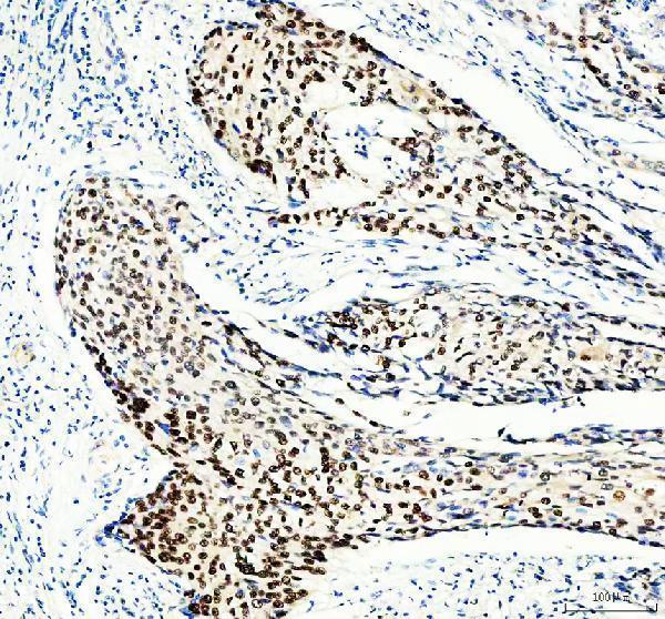
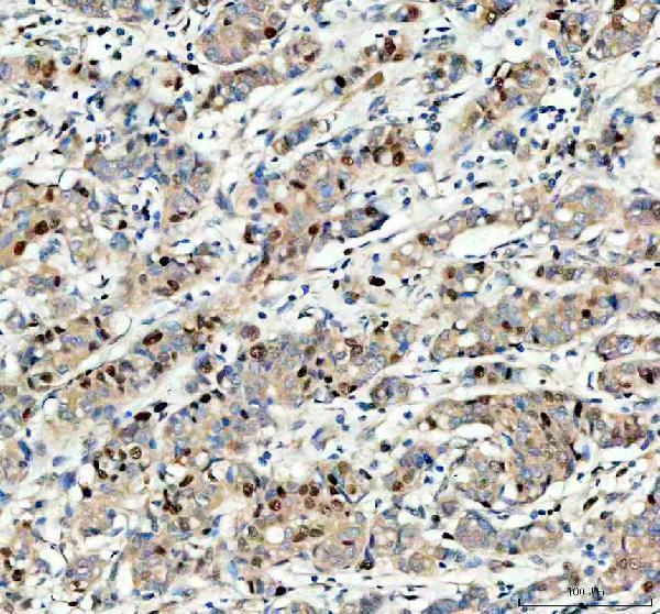
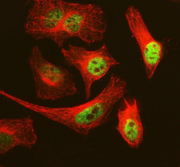
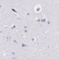


Comprehensive Human Protein Atlas IHC scoring per tissue (reliability: Supported — Medium consistency between antibody staining and RNA expression data.). Rows are taken directly from the HPA tissue chart — click any row's HPA link to view the source.
| Tissue | Cell type | Level | Evidence | Source |
|---|---|---|---|---|
| Bone marrow | Hematopoietic cells | High | Protein (IHC) | HPA → |
| Placenta | Cytotrophoblasts | High | Protein (IHC) | HPA → |
| Duodenum | Enterocytes | Medium | Protein (IHC) | HPA → |
| Lymph node | Germinal center cells | Medium | Protein (IHC) | HPA → |
| Nasopharynx | Ciliated cells (cell body) | Medium | Protein (IHC) | HPA → |
| Tissue | Cell type | Level | Evidence | Source |
|---|---|---|---|---|
| Adipose tissue | Adipocytes | Not detected | Protein (IHC) | HPA → |
| Adrenal gland | Glandular cells | Not detected | Protein (IHC) | HPA → |
| Appendix | Endocrine cells | Not detected | Protein (IHC) | HPA → |
| Bronchus | Basal cells | Not detected | Protein (IHC) | HPA → |
| Caudate | Glial cells | Not detected | Protein (IHC) | HPA → |
Troubleshoot MCM3 staining by checking nuclear localisation, tissue controls and detection background before interpreting the fraction of positive cells.
Catalog-listed anti-MCM3 antibodies cover IHC and IF/ICC in human, mouse, and rat (catalog: applications and reactivity). PA1651 has IHC images from human and rat tissues and IF/ICC data from HeLa cells (PA1651 captions).
PA1651 has IHC images from paraffin sections of human cervical cancer, human breast cancer, and rat spleen, plus an IF/ICC image from HeLa cells (PA1651 image captions). M02580 is listed for IHC and IF/ICC in human, mouse, and rat, but has no supplied specimen images (catalog: M02580 applications, reactivity, image alts).
Which to pick: Choose PA1651 for paraffin-section tissue IHC: its own captions document human and rat sections; the fixative is unreported (PA1651 IHC captions). For IF/ICC, PA1651 has a HeLa-cell image, while M02580 lists IF/ICC without a supplied image (PA1651 IF caption; catalog: M02580 applications and image alts). For cross-species planning, both list human, mouse, and rat reactivity; PA1651 shows human and rat IHC, while M02580 is a rabbit monoclonal without supplied IHC images (catalog: reactivity and M02580 clone; PA1651 IHC captions).