This website uses cookies to ensure you get the best experience on our website.


- Table of Contents
Plan chromogenic MCM4 IHC in paraffin sections around variable nuclear staining (HPA tissue IHC). Start with the catalog antibody’s 2–5 μg/ml IHC range and compare high-staining with low-staining cell populations (datasheet A02301-1; HPA tissue IHC).

Expected localisation, validated protocols, controls and antibodies — the at-a-glance facts below, then the full design guide.
| Expected localisation | Variable nuclear staining across tissues (HPA tissue IHC) | |
| Staining pattern | Nuclei stain at variable levels across tissue cell types (HPA tissue IHC) | |
| Antigen retrieval | EDTA pH 8.0 HIER, heat-mediated (datasheet A02301-1) | |
| Positive control | Appendix+4 more · see all | |
| Negative control | None in HPA (detected in all 45 tissues); use no-primary + isotype controls |
| Fixation | Keep paraffin-section fixation consistent across samples (standard IHC practice; not target-specific) | |
| Caveat | Chromatin association changes as replication proceeds (UniProt) | |
| Regulation | Nuclear expression varies across tissues (HPA tissue IHC) | |
| Isoform / epitope | No annotated isoforms; mature chain spans residues 2–863 (UniProt) |
The catalog antibody uses EDTA pH 8.0 retrieval (datasheet A02301-1). Published MCM4 IHC methods below cover urothelial, pancreatic, and esophageal samples (PMC10515259; PMC12967608; PMC5250507).
| Sample | Paraffin-embedded mouse colon tissue; fixative not specified (datasheet A02301-1) |
| Fixation | Image fixative and duration unreported (datasheet A02301-1); verify before use. |
| Sectioning | 4–5 µm sections on charged slides (standard) |
| Deparaffinisation | Xylene, graded ethanol series to water (standard) |
| Antigen retrieval | Heat retrieval: EDTA pH 8.0 (datasheet A02301-1); 20 min, 95–100 °C (standard) |
| Peroxidase block | 3% H2O2, 10 min, room temperature (standard) |
| Blocking | 10% goat serum (datasheet A02301-1) |
| Primary antibody | Rabbit anti-MCM4, 2-5μg/ml (datasheet A02301-1) |
| Primary incubation | Overnight at 4 °C (datasheet A02301-1) |
| Detection | Streptavidin-biotin complex (SABC), DAB chromogen (datasheet A02301-1) |
| Counterstain | Hematoxylin, blue, dehydrate and mount (standard) |
| Expected result | MCM4-positive staining in germinal center cells of appendix (HPA tissue IHC: High). HPA tissue profile: Nuclear expression at variable levels in all tissues. No signal in the no-primary control. |
MCM4 should stain nuclei, including germinal center cells in appendix and hematopoietic cells in bone marrow (HPA tissue IHC: High). Nuclear staining varies across tissues (HPA tissue IHC: Enhanced reliability; medium staining–RNA consistency). Its nuclear and chromosomal localization, with no transmembrane segment, supports a nuclear interpretation (UniProt P33991: subcellular location and topology).
| Clear nuclear chromogen in appendix germinal center cells or bone marrow hematopoietic cells. | This matches reported high staining in those cells (HPA tissue IHC: High). Score the proportion and intensity of stained nuclei within the identified cell population; compare runs against the same positive tissue (general IHC practice). |
| Predominantly cytoplasmic or membrane staining, with nuclei unstained. | That compartment conflicts with the nuclear tissue pattern (HPA tissue IHC: nuclear expression) and nuclear/chromosomal localization (UniProt P33991). Check morphology, counterstain and detection controls before interpreting it as MCM4 (general IHC practice). |
| Strong signal in cells expected to stain weakly, such as smooth muscle cells or liver cholangiocytes. | These cells have low reported staining (HPA tissue IHC: Low). Unexpected intensity may reflect cross-reactivity or endogenous detection activity; it does not establish either cause without controls (general IHC practice). |
| Diffuse chromogen obscures nuclei and tissue boundaries. | The distribution cannot be scored reliably against the expected nuclear pattern (HPA tissue IHC: nuclear expression). Review no-primary control, blocking, washes and antibody concentration to locate nonspecific background (general IHC practice). |
| No nuclear signal in appendix germinal center cells or bone marrow hematopoietic cells. | Those cells are reported High (HPA tissue IHC). First verify tissue identity, section integrity, positive control and detection reagents; absence alone cannot distinguish a technical failure from a sample-specific result (general IHC practice). |
| Compartment and cell cycle | MCM4 is nuclear and chromosomal and participates in the MCM2–7 replication helicase (UniProt P33991). It associates with chromatin before nuclei form and detaches as replication progresses (UniProt P33991); interpret variation with morphology and controls, without assigning a cell-cycle phase from stain intensity alone. |
| Tissue range and control choice | Nuclear expression varies across all tissues; the supplied HPA profile lists high and low examples but no negative tissue (HPA tissue IHC). Appendix germinal center cells or bone marrow hematopoietic cells provide documented high-staining reference populations (HPA tissue IHC: High). |
| Strength of pattern evidence | The tissue profile has Enhanced reliability with medium agreement between staining and RNA (HPA tissue IHC). Two listed antibodies have Enhanced IHC validation, HPA004873 and CAB004497 (HPA antibodies); this supports the pattern without validating every reagent or specimen. |
| Processing and epitope claims | No signal peptide or propeptide is annotated; the reported chain spans residues 2–863 (UniProt P33991). Phosphorylation and an MCM ATPase domain are annotated (UniProt P33991), but the payload gives no antibody epitope or target-specific retrieval or fixation effect; optimize retrieval empirically (general IHC practice). |
| What should IF/ICC show? | Nucleoplasmic localization is the reported IF pattern (HPA subcellular: Nucleoplasm, enhanced), consistent with nuclear localization (UniProt P33991). IF/ICC images and antibody validation are separate from this paraffin-section chromogenic IHC interpretation (HPA subcellular; HPA antibodies). |
| Situation | Likely cause | Next action |
|---|---|---|
| Known high-staining cells have no nuclear signal. | Tissue selection, retrieval, antibody incubation or detection may have failed (general IHC practice); high staining is reported for appendix germinal center and bone marrow hematopoietic cells (HPA tissue IHC). | Confirm the reference cells on the counterstain, run a known-positive section, then check retrieval conditions and each detection step (general IHC practice). |
| Cytoplasm or membranes stain more strongly than nuclei. | This disagrees with nuclear localization (UniProt P33991; HPA tissue IHC). Nonspecific binding or misread chromogen is possible (general IHC practice). | Inspect nuclear boundaries, compare a no-primary control and repeat with an IHC-validated antibody if available (general IHC practice; HPA antibodies: Enhanced IHC validation). |
| Low-staining reference cells show intense chromogen. | Smooth muscle cells and liver cholangiocytes are reported Low (HPA tissue IHC). Cross-reactivity or endogenous detection activity is possible (general IHC practice). | Check a no-primary control and any detection-only control; review enzyme blocking and whether signal stays within nuclei (general IHC practice). |
| Background covers the section or pools at edges. | A diffuse deposit prevents comparison with the reported nuclear pattern (HPA tissue IHC); excess reagent or inadequate washing is possible (general IHC practice). | Review antibody concentration, blocking, wash steps and section handling; retain the same positive tissue while changing one condition at a time (general IHC practice). |
| Nuclear staining varies markedly between tissue regions. | Variable nuclear expression is reported (HPA tissue IHC). Uneven processing or different cell populations can also alter the apparent pattern (general IHC practice). | Score identified cell types separately and compare intact regions with the same run's positive control before attributing the variation to MCM4 (general IHC practice). |
| An unstained cell population is being used as a negative control. | The supplied profile reports nuclear expression at variable levels in all tissues and lists no negative tissue (HPA tissue IHC). | Use no-primary and detection controls to assess background; treat HPA Low populations as relative comparators, not proven MCM4-negative cells (general IHC practice; HPA tissue IHC: Low). |
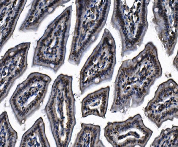
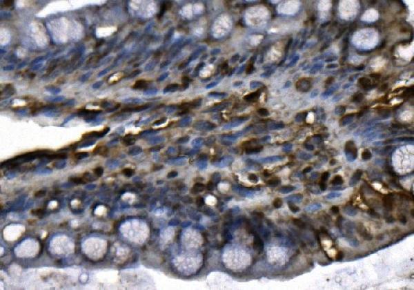
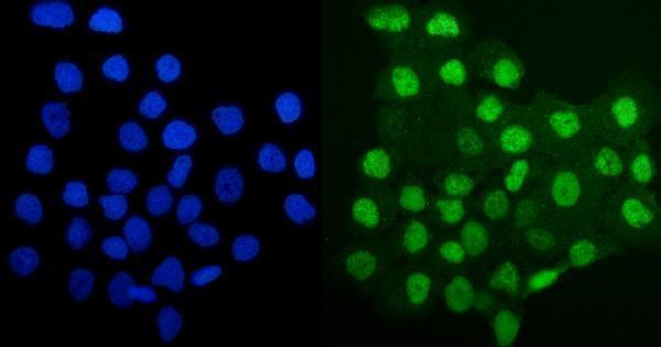
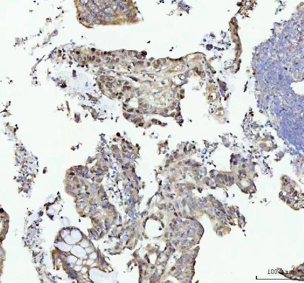
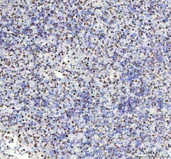
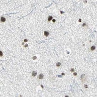


Comprehensive Human Protein Atlas IHC scoring per tissue (reliability: Enhanced — Medium consistency between antibody staining and RNA expression data.). Rows are taken directly from the HPA tissue chart — click any row's HPA link to view the source.
| Tissue | Cell type | Level | Evidence | Source |
|---|---|---|---|---|
| Appendix | Germinal center cells | High | Protein (IHC) | HPA → |
| Bone marrow | Hematopoietic cells | High | Protein (IHC) | HPA → |
| Breast | Glandular cells | High | Protein (IHC) | HPA → |
| Bronchus | Basal cells | High | Protein (IHC) | HPA → |
| Caudate | Glial cells | High | Protein (IHC) | HPA → |
| Tissue | Cell type | Level | Evidence | Source |
|---|---|---|---|---|
| None in HPA: MCM4 is detected in all 45 scored tissues. Use a no-primary (secondary-only) and an isotype control instead. | ||||
Troubleshoot MCM4 staining by checking nuclear localisation, retrieval, controls and scoring against the documented paraffin-section workflow.
Anti-MCM4 IHC images cover human, mouse and rat paraffin sections; IF/ICC imaging covers A431 cells (catalog image captions).
A02301-1 has IHC images from mouse and rat colon and human breast cancer and gallbladder adenocarcinoma, plus an IF/ICC image from A431 cells (A02301-1 image captions). M02301 has IHC images from human colorectal adenocarcinoma and spleen (M02301 image captions).
Which to pick: For paraffin-section IHC, either antibody has its own image: A02301-1 used EDTA retrieval at pH 8.0 and 2 μg/ml primary antibody in mouse colon, while monoclonal M02301 used EDTA retrieval at pH 8.0 and 1:50 in human colorectal adenocarcinoma (respective IHC image captions). Choose A02301-1 for IF/ICC because its application list includes both and its A431 image uses 5 μg/ml; M02301 lists neither IF nor ICC (catalog applications; A02301-1 IF image caption). A02301-1 also lists human, mouse and rat reactivity, versus human and mouse for M02301; the fixative is unreported in their paraffin-section captions (catalog reactivity; respective IHC image captions).