This website uses cookies to ensure you get the best experience on our website.


- Table of Contents
Plan MCM7 chromogenic IHC around nuclear staining in highly proliferative cells (HPA tissue IHC). This guide covers paraffin-section controls, consistent fixation, antibody dilution, detection, counterstaining and nuclear scoring (standard IHC practice).

Expected localisation, validated protocols, controls and antibodies — the at-a-glance facts below, then the full design guide.
| Expected localisation | Nuclear staining observed in proliferative cells (HPA tissue IHC) | |
| Staining pattern | Nuclear staining in highly proliferative cells (HPA tissue IHC) | |
| Antigen retrieval | EDTA pH 8.0 HIER, heat-mediated (datasheet PB9261) | |
| Positive control | Appendix+4 more · see all | |
| Negative control | Adipose tissue+4 more · see all |
| Fixation | Keep fixation consistent across sections (standard IHC practice; not target-specific) | |
| Caveat | Proliferation differences can change the stained-cell fraction (HPA tissue IHC) | |
| Regulation | Enriched in highly proliferative cells (HPA tissue IHC) | |
| Isoform / epitope | Three isoforms; epitope coverage is unspecified (UniProt) |
The catalog antibody uses EDTA pH 8.0 heat retrieval (datasheet: PB9261). Four published MCM7 IHC protocols provide additional conditions (PMC3964599; PMC3125391; PMC4156209; PMC5250507).
| Sample | Paraffin-embedded Mouse Testis tissue; fixative not specified (datasheet PB9261) |
| Fixation | Image fixative and duration unreported (datasheet PB9261); verify before use. |
| Sectioning | 4–5 µm sections on charged slides (standard) |
| Deparaffinisation | Xylene, graded ethanol series to water (standard) |
| Antigen retrieval | Heat retrieval: EDTA pH 8.0 (datasheet PB9261); 20 min, 95–100 °C (standard) |
| Peroxidase block | 3% H2O2, 10 min, room temperature (standard) |
| Blocking | 10% goat serum (datasheet PB9261) |
| Primary antibody | Rabbit anti-MCM7, 0.5-1μg/ml (datasheet PB9261) |
| Primary incubation | Overnight at 4 °C (datasheet PB9261) |
| Detection | Streptavidin-biotin complex (SABC), DAB chromogen (datasheet PB9261) |
| Counterstain | Hematoxylin, blue, dehydrate and mount (standard) |
| Expected result | MCM7-positive staining in endocrine cells of appendix (HPA tissue IHC: High). HPA tissue profile: Nuclear expression in highly proliferative cells. No signal in the no-primary control. |
MCM7 should stain nuclei, chiefly in highly proliferative cells (HPA tissue IHC: nuclear expression in highly proliferative cells; UniProt P33993: nucleus and chromosome). Expect strong staining in the specific cell populations HPA rates High, such as bone marrow hematopoietic cells and lymph node germinal center cells (HPA tissue IHC: High). HPA rates the tissue pattern Enhanced, reflecting high consistency between antibody staining and RNA expression data (HPA tissue IHC: Enhanced).
| Strong nuclear staining in bone marrow hematopoietic cells or lymph node germinal center cells. | This matches the expected compartment and two High-rated cell populations (UniProt P33993: nucleus; HPA tissue IHC: High in these cells). Score the proportion and intensity of stained nuclei within the relevant population; a positive field need not mean every cell is positive (standard IHC practice). |
| Predominantly cytoplasmic staining, with little nuclear signal, in an otherwise positive field. | Treat this as an unexpected IHC pattern and review specificity and detection controls (UniProt P33993: nucleus; HPA tissue IHC: nuclear expression). HPA also reports cytosol and basal body in ICC-IF; those approved additional locations do not establish a predominantly cytoplasmic chromogenic tissue pattern (HPA subcellular ICC-IF). |
| Strong staining in adipocytes or cardiomyocytes, especially without corresponding positive nuclei. | These cells are rated Not detected, so investigate cross-reactivity or nonspecific detection before interpreting the stain as MCM7 (HPA tissue IHC: Not detected in adipocytes and cardiomyocytes). Compare cell identity and compartment on the same section and with appropriate controls (standard IHC practice). |
| Diffuse color over nuclei, cytoplasm, and tissue spaces, obscuring cell boundaries. | This cannot be scored as the expected nuclear pattern (HPA tissue IHC: nuclear expression). Background from detection reagents, insufficient blocking, or inadequate washing is possible; use a no-primary control to help identify reagent-related staining (standard IHC practice). |
| No nuclear staining in bone marrow hematopoietic cells despite intact tissue morphology. | A High-rated reference population has failed to stain, so the result is inconclusive until the assay is checked (HPA tissue IHC: High in bone marrow hematopoietic cells). Examine the IHC-validated antibody, retrieval and detection workflow, and a positive control processed alongside the section (standard IHC practice). |
| Compartment and replication state | MCM7 is nuclear and chromosome associated; it associates with chromatin before nuclei form and detaches as DNA replication progresses (UniProt P33993). These facts support nuclear interpretation but do not define a slide-based cell-cycle cutoff or require every nucleus to stain (UniProt P33993; HPA tissue IHC: nuclear expression). |
| Cell population within a tissue | HPA rates bronchial basal cells, esophageal squamous epithelial cells, and appendix endocrine cells High, while adipocytes and cardiomyocytes are Not detected (HPA tissue IHC). Identify the cell population before calling a tissue positive or negative; an organ-wide label would obscure these distinctions (HPA tissue IHC). |
| Strength of IHC evidence | The tissue pattern is Enhanced, with high consistency between staining and RNA expression (HPA tissue IHC: Enhanced). HPA lists HPA003898, CAB002163, and CAB016312 as IHC Enhanced; that validation supports their reported patterns but does not validate an unlisted antibody or a particular staining run (HPA antibodies). |
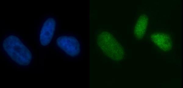
| ICC-IF localization boundary | HPA approves nucleoplasm as the main ICC-IF location and cytosol and basal body as additional locations (HPA subcellular ICC-IF). This is a useful answer when comparing modalities, but ICC-IF observations do not supply a chromogenic IHC protocol or override the tissue IHC nuclear profile (HPA subcellular ICC-IF; HPA tissue IHC). |
| Protein architecture and variants | UniProt lists no transmembrane segment or signal peptide, three isoforms, and an MCM C-terminal AAA(+) ATPase domain at residues 316–647 (UniProt P33993). These annotations do not identify the catalog antibody's epitope or predict its retrieval needs; confirm those assay details from the antibody's own documentation (standard IHC practice). |
| Target-specific fixation sensitivity | No target-specific fixation effect is established by the supplied UniProt localization or HPA staining data (UniProt P33993; HPA tissue IHC). If retrieval needs adjustment, treat it as assay optimization and judge the result against positive and negative control tissues, rather than assuming a documented MCM7 fixation response (standard IHC practice). |
| Situation | Likely cause | Next action |
|---|---|---|
| Known-positive tissue has no nuclear signal. | The staining run may have failed; bone marrow hematopoietic cells and lymph node germinal center cells are High-rated references (HPA tissue IHC). | Check that a matched positive control stained, then review the IHC-validated antibody, retrieval, dilution, and detection steps under the assay documentation (standard IHC practice). Do not score the test section as biologically negative until the control works. |
| Signal is weak in a population expected to be High. | Weak detection or an unsuitable assay setting is possible; HPA's High category describes the observed reference pattern, not a guaranteed intensity in every run (HPA tissue IHC: High). | Compare morphology and a concurrently processed positive control, then adjust one documented assay variable at a time and reassess nuclear contrast (standard IHC practice). |
| Color is mainly cytoplasmic. | The result conflicts with the tissue IHC nuclear profile; ICC-IF's additional cytosol location alone cannot establish this chromogenic pattern (HPA tissue IHC: nuclear expression; HPA subcellular ICC-IF: cytosol). | Review the no-primary control, cell boundaries, and antibody specificity; accept MCM7 IHC only when the expected nuclear pattern is demonstrable in a suitable positive population (standard IHC practice; HPA tissue IHC). |
| Adipocytes or cardiomyocytes stain strongly. | Those populations are Not detected in the HPA tissue profile; cross-reactivity or nonspecific chromogen is possible (HPA tissue IHC: Not detected). | Confirm cell identity and compare a no-primary control with a positive reference on the same run (standard IHC practice). If unexpected staining persists only with primary antibody, reassess specificity before reporting it as MCM7. |
| Diffuse background obscures positive nuclei. | Detection reagent background, insufficient blocking, or inadequate washing can reduce interpretability (standard IHC practice). | Inspect a no-primary control, then review blocking and wash steps and the detection system (standard IHC practice). Reassess whether discrete nuclear staining remains in High-rated cells (HPA tissue IHC: nuclear expression and High populations). |
| A section seems negative because most cells are unstained. | HPA assigns staining levels to named cell populations, not every cell in an organ; it also lists several populations as Not detected (HPA tissue IHC). | Evaluate the relevant cell type and nuclear compartment separately, using morphology and a working positive control (standard IHC practice; HPA tissue IHC: nuclear expression). Report the observed population and pattern rather than an organ-wide positive or negative call. |


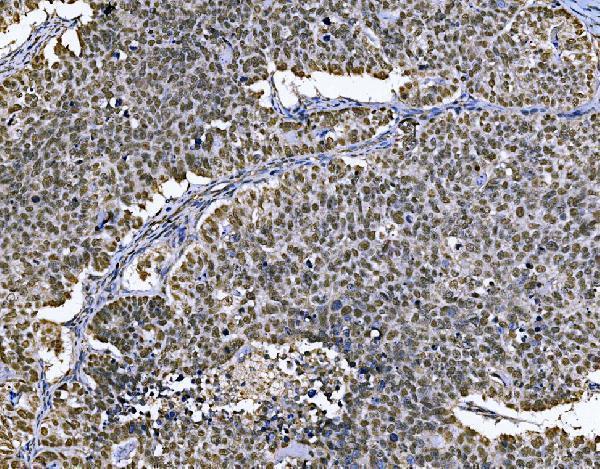
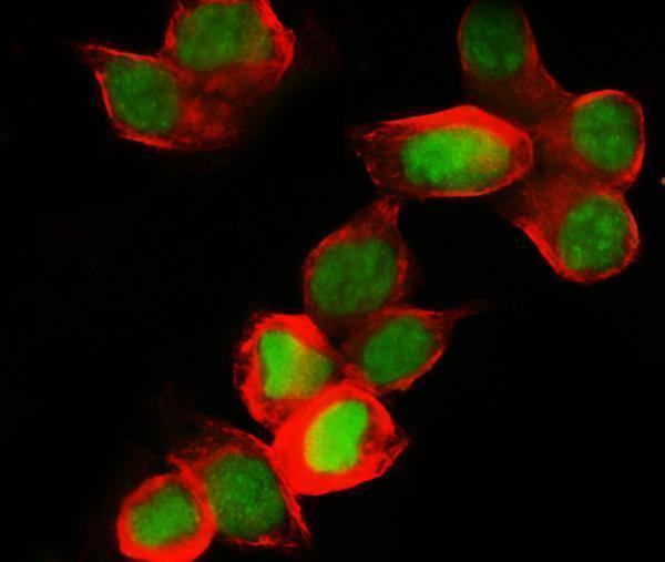


Comprehensive Human Protein Atlas IHC scoring per tissue (reliability: Enhanced — High consistency between antibody staining and RNA expression data.). Rows are taken directly from the HPA tissue chart — click any row's HPA link to view the source.
| Tissue | Cell type | Level | Evidence | Source |
|---|---|---|---|---|
| Appendix | Endocrine cells | High | Protein (IHC) | HPA → |
| Bone marrow | Hematopoietic cells | High | Protein (IHC) | HPA → |
| Bronchus | Basal cells | High | Protein (IHC) | HPA → |
| Colon | Endocrine cells | High | Protein (IHC) | HPA → |
| Duodenum | Endocrine cells | High | Protein (IHC) | HPA → |
| Tissue | Cell type | Level | Evidence | Source |
|---|---|---|---|---|
| Adipose tissue | Adipocytes | Not detected | Protein (IHC) | HPA → |
| Caudate | Glial cells | Not detected | Protein (IHC) | HPA → |
| Cerebellum | Cells in granular layer | Not detected | Protein (IHC) | HPA → |
| Cerebral cortex | Endothelial cells | Not detected | Protein (IHC) | HPA → |
| Heart muscle | Cardiomyocytes | Not detected | Protein (IHC) | HPA → |
Troubleshoot MCM7 staining in paraffin sections by checking retrieval, nuclear localisation, controls, and cell type before comparing scores (datasheet PB9261; UniProt P33993; HPA tissue IHC).
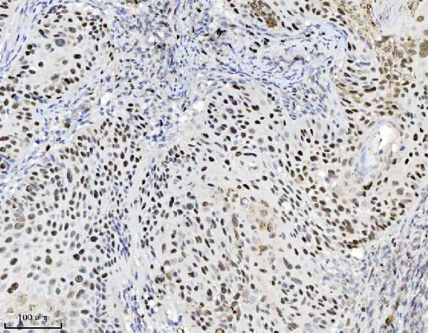
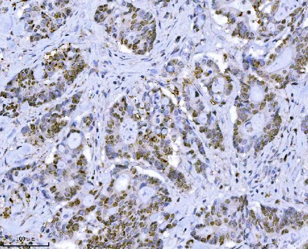
Anti-MCM7 IHC figures cover mouse testis and human laryngeal squamous cell carcinoma and bladder cancer (catalog IHC captions); IF figures cover human colon cancer, PC-3, and MCF-7 (catalog IF captions).
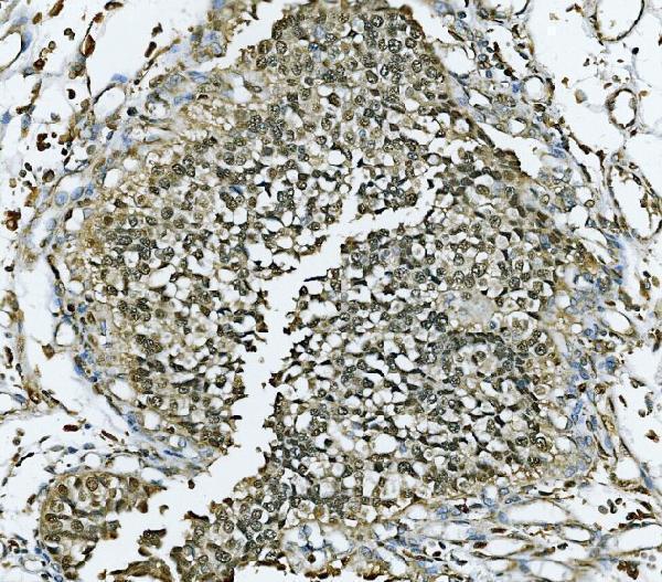
PB9261 has paraffin-section IHC images in mouse testis, rat intestine, and human lung cancer (PB9261 IHC captions), plus IF images in U20S cells and human colon cancer sections (PB9261 IF captions). M01649-3 has paraffin-section IHC images in human laryngeal squamous cell carcinoma, colon adenocarcinoma, and chronic tonsillitis (M01649-3 IHC captions), plus IF in PC-3 cells (M01649-3 IF caption); PA1792 has paraffin-section IHC images in human bladder cancer, ovarian serous adenocarcinoma, and rectal adenocarcinoma (PA1792 IHC captions), plus IF in MCF-7 cells (PA1792 IF caption).
Which to pick: For human paraffin-section IHC, M01649-3 is a mouse monoclonal antibody, clone 3H11 (catalog: host and clone), pictured in laryngeal squamous cell carcinoma after EDTA retrieval (M01649-3 IHC caption); PA1792 is pictured in bladder cancer after EDTA retrieval (PA1792 IHC caption). For IF/ICC in PC-3 cells, M01649-3 has an IF image and lists both applications (M01649-3 IF caption; catalog: applications). For work across human, mouse, and rat, PB9261 lists all three species and IHC/IF/ICC applications (PB9261 catalog), with paraffin-section IHC images in each species (PB9261 IHC captions); the fixative is unreported in those captions (PB9261 IHC captions).