MDH1 / Malate dehydrogenase, cytoplasmic · IHC design guide

Design Immunohistochemistry for MDH1

Plan chromogenic MDH1 IHC-P using the catalog antibody's 1:10–1:50 dilution (datasheet: A04262-1). Use cytoplasmic staining in stomach glandular cells as a reference, and interpret results cautiously because HPA rates its tissue staining reliability as uncertain (HPA tissue IHC).

Evidence assembled Oct 2026 · For research use; verify linked source records and product datasheet before use
Immunohistochemistry protocol sheet for MDH1 (IHC for MDH1): expected localisation Cytoplasmic tissue staining (HPA tissue IHC), antibody A04262-1, validated IHC image, and IHC protocol steps
Printable MDH1 IHC protocol sheet — expected localisation Cytoplasmic tissue staining (HPA tissue IHC), antibody A04262-1, controls and protocol steps. Open the full MDH1 IHC guide →

MDH1 Immunohistochemistry Experimental Design Guide

Expected localisation, validated protocols, controls and antibodies — the at-a-glance facts below, then the full design guide.

Must know before staining
Expected localisation Cytoplasmic tissue staining (HPA tissue IHC)
Staining pattern Cytoplasmic staining in stomach glandular cells (HPA tissue IHC)
Antigen retrieval Citrate pH 6.0 HIER, 95–98 °C, 20 min (rule: cytoplasmic / membrane antigen)
Positive control ⓘ Cerebral cortex+4 more · see all
Negative control ⓘ Adipose tissue+4 more · see all
Important caveats
Reasons your staining may differ from the expected pattern.
Fixation Keep formalin fixation consistent across sections (standard IHC practice; not target-specific)
Caveat Antibody staining and RNA show low consistency (HPA tissue IHC)
Regulation No specific expression regulator reported (UniProt)
Isoform / epitope 3 isoforms; C-terminal epitope coverage unknown (UniProt; datasheet A04262-1)
Section 1

Recommended MDH1 IHC & IF Protocols

The catalog antibody’s IHC-P protocol is accompanied by two published MDH1 IHC methods: gerbil hippocampal sections (PMC10079925) and primary NSCLC tissue microarrays (PMC5559971).

Recommended immunohistochemistry (IHC-P) protocol parameters
SampleFFPE human hepatocarcinoma tissue (datasheet A04262-1)
FixationImage formalin-fixed; duration unreported (datasheet A04262-1); verify before use.
Sectioning4–5 µm sections on charged slides (standard)
DeparaffinisationXylene, graded ethanol series to water (standard)
Antigen retrievalHeat-induced epitope retrieval in citrate buffer, pH 6.0, 20 min at 95–98 °C (standard rule: cytoplasmic / membrane antigen)
Peroxidase block3% H2O2, 10 min, room temperature (standard)
Blocking10% normal serum of the secondary host, 30 min, room temperature (standard)
Primary antibodyRabbit anti-MDH1, 1:10-1:50 (datasheet A04262-1)
Primary incubationOvernight at 4 °C (standard)
DetectionHRP-polymer secondary, DAB chromogen 5–10 min (standard)
CounterstainHematoxylin, blue, dehydrate and mount (standard)
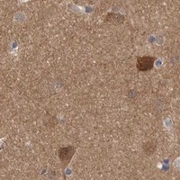
Expected resultMDH1-positive staining in neuropil of cerebral cortex (HPA tissue IHC: High). HPA tissue profile: Cytoplasmic expression in several different tissue types. No signal in the no-primary control.
💡Decision noteFor paraffin sections, start with citrate pH 6.0 HIER at 95–98 °C for 20 min (page antigen-retrieval rule).
Section 2

What Is the Expected MDH1 Staining Pattern?

MDH1 is a cytosolic protein without a transmembrane segment (UniProt P40925). In paraffin-section IHC, expect cytoplasmic staining in HPA-observed cells, including stomach glandular cells, heart cardiomyocytes and cerebral cortex neuropil (HPA: tissue IHC). Treat tissue intensity as a reference, not a definitive pass criterion: HPA rates the tissue pattern Uncertain because antibody staining has low consistency with RNA expression (HPA: tissue IHC).

What am I looking at on my slide?
Cytoplasmic signal in stomach glandular cells or heart cardiomyocytes, with tissue structure still visible.This matches the reported cytoplasmic tissue pattern (HPA: tissue IHC) and cytosolic location (UniProt P40925). HPA reports High staining in stomach glandular cells and Medium staining in cardiomyocytes; intensity can be judged against neighboring structures within the same section (HPA: tissue IHC; general IHC practice).
Predominantly nuclear or sharply plasma-membrane staining in cells expected to show cytoplasm.That distribution conflicts with MDH1's cytosolic location and lack of a transmembrane segment (UniProt P40925). Check whether the pattern persists with an independent antibody and appropriate controls before calling it MDH1; an off-target signal or staining artefact is possible (general IHC practice).
Strong signal in a cell population that HPA reports as Not detected, such as adipocytes or lymph-node germinal center cells.The result differs from the reported IHC observations for those cells (HPA: tissue IHC). Consider antibody cross-reactivity or endogenous chromogenic detection activity, and inspect control sections (general IHC practice). HPA's Uncertain reliability means its Not detected calls are reference observations, not proof that every such cell lacks MDH1 (HPA: tissue IHC).
Diffuse color across tissue and empty spaces, obscuring cell borders and tissue architecture.This is difficult to score as cytoplasmic MDH1 (UniProt P40925; general IHC practice). Broad background can arise from nonspecific antibody binding, residual detection activity or excess chromogen development (general IHC practice). Compare a no-primary control and review blocking, washing and development conditions (general IHC practice).
No visible signal in stomach glandular cells or cerebral cortex neuropil.Both are reported as High in HPA tissue IHC, so an absent result warrants a technical check (HPA: tissue IHC). Confirm that tissue morphology and counterstain are intact, then review the catalog antibody's IHC-P instructions and detection controls (general IHC practice). HPA's Uncertain rating limits how strongly one section can be treated as a biological positive control (HPA: tissue IHC).
💡Expected MDH1 appearanceA convincing positive is interpretable cytoplasmic staining in HPA-observed cells, potentially High in stomach glandular cells or cerebral cortex neuropil; dominant nuclear or membrane staining is suspect (HPA: tissue IHC; UniProt P40925).
How each factor affects the staining
Tissue and cell contextHPA reports High staining in stomach glandular cells and cerebral cortex neuropil, Medium in heart cardiomyocytes, and Low in lung macrophages (HPA: tissue IHC). Choose the cell population before scoring; a whole-section average can hide the reported pattern (general IHC practice).
Intracellular locationMDH1 is cytosolic and has no transmembrane segment or signal peptide (UniProt P40925). For IHC, score cellular cytoplasm against morphology rather than treating membrane or nuclear color as the expected distribution (UniProt P40925; general IHC practice).
Antibody and isoform interpretationHPA rates tissue reliability Uncertain and lists three antibodies with Uncertain IHC validation (HPA: tissue IHC; HPA: antibodies). UniProt lists three isoforms, but the supplied record gives no antibody epitope or isoform coverage; do not infer that a negative stain excludes all isoforms (UniProt P40925).
IF/ICC: where should signal appear?HPA reports cytosol as Supported and centrosome as Approved in ICC-IF images from A-431, U-251MG and U2OS (HPA: subcellular). This is an IF/ICC localization reference; the tissue IHC profile describes cytoplasmic expression and does not establish a visible centrosomal pattern in paraffin sections (HPA: tissue IHC).
Why is my staining missing, weak or wrong?
SituationLikely causeNext action
No staining in an HPA High reference cell population.The staining run may have failed, or the sampled section may differ from the HPA observation (general IHC practice; HPA: tissue IHC).Check tissue identity and cell morphology, review the catalog antibody's IHC-P instructions, and include a detection control (general IHC practice). Interpret a negative result cautiously because HPA rates tissue reliability Uncertain (HPA: tissue IHC).
Nuclear or membrane color dominates the slide.That compartment differs from the reported cytosolic location and lacks support from MDH1 topology (UniProt P40925).Compare no-primary and independent-antibody controls; score MDH1 only where the pattern is interpretable in cytoplasm (general IHC practice; UniProt P40925).
Cells reported as Not detected show strong color.Cross-reactivity or endogenous detection activity is possible (general IHC practice); the result differs from the HPA cell-specific observation (HPA: tissue IHC).Inspect a no-primary control, verify blocking and chromogenic detection controls, and repeat with an independent antibody if available (general IHC practice).
Uniform haze makes cytoplasm hard to distinguish.Nonspecific binding, inadequate washing or excess chromogen development can obscure cellular localization (general IHC practice).Compare background on no-primary sections, review blocking and wash steps, and adjust development within the detection system's instructions (general IHC practice).
Staining is weak in a tissue reported as Low.A weak result may match the HPA observation; lung macrophages and intestinal glandular cells are listed as Low (HPA: tissue IHC).Judge the specified cell population and compare it with an HPA High reference tissue in the same run before changing conditions (HPA: tissue IHC; general IHC practice).
A second antibody produces a different tissue pattern.HPA reports low consistency between antibody staining and RNA data and rates all three listed antibodies Uncertain for IHC (HPA: tissue IHC; HPA: antibodies).Document the cell types, compartments and controls for each stain; treat disagreement as unresolved specificity rather than selecting the stronger signal as correct (general IHC practice; HPA: tissue IHC).

Sample controls for MDH1 IHC & IF

🧪Run stomach first: glandular cells should stain (HPA: High in stomach glandular cells). Use appendix glandular cells as the negative tissue (HPA: Not detected in appendix glandular cells); on the stomach slide, assess nonglandular cells for background without assuming they are MDH1-negative.
Positive control tissue: Cerebral cortex (Neuropil, HPA High)
Negative control tissue: Adipose tissue (HPA Not detected)
ICC-IF cell lines (HPA subcellular resource): HPA ICC-IF images show MDH1 in A-431, U-251MG, U2OS, with annotated localisation: Centrosome (approved), Cytosol (supported) (HPA subcellular).
Technical controls: Include no-primary (secondary-only) and host-species-matched isotype controls, matching monoclonal isotype or polyclonal IgG class as appropriate, plus MDH1-knockout material as a biological negative. Quench endogenous peroxidase for chromogenic detection and check blood or inflammatory cells in the stomach section for residual signal (standard IHC practice).
⚠️Feasibility: The selected A04262-1 caption explicitly describes formalin-fixed, paraffin-embedded tissue, but no supplied source reports an MDH1-specific fixation window or fixation effect (selected tissue-IHC caption). Antigen-retrieval dependency and any advantage of frozen sections or IF over paraffin IHC are unreported. Cytosolic staining is expected (UniProt P40925: cytosol); in stomach glands, interpret staining alongside the peroxidase controls because blood or inflammatory cells can contribute background (standard IHC practice).

HPA tissue IHC evidence for MDH1

Comprehensive Human Protein Atlas IHC scoring per tissue (reliability: Uncertain — Low consistency between antibody staining and RNA expression data.). Rows are taken directly from the HPA tissue chart — click any row's HPA link to view the source.

Positive expression · recommended positive controls

TissueCell typeLevelEvidenceSource
Cerebral cortex Neuropil High Protein (IHC) HPA →
Stomach Glandular cells High Protein (IHC) HPA →
Adrenal gland Glandular cells Medium Protein (IHC) HPA →
Caudate Neuronal cells Medium Protein (IHC) HPA →
Cerebellum Purkinje cells Medium Protein (IHC) HPA →

Undetected expression · recommended negative controls

TissueCell typeLevelEvidenceSource
Adipose tissue Adipocytes Not detected Protein (IHC) HPA →
Appendix Glandular cells Not detected Protein (IHC) HPA →
Bone marrow Hematopoietic cells Not detected Protein (IHC) HPA →
Breast Adipocytes Not detected Protein (IHC) HPA →
Cervix Glandular cells Not detected Protein (IHC) HPA →
Section 3

Advanced MDH1 IHC Tips

Troubleshoot MDH1 staining in paraffin sections by checking cytosolic localisation, tissue controls, detection background and the limits of the available IHC evidence.

How should I adjust retrieval when cytoplasmic MDH1 staining is weak?
Start with citrate buffer at pH 6.0, heated to 95–98 °C for 20 minutes (page retrieval setting). If staining remains weak, compare a modestly longer heating time on adjacent sections while keeping the buffer and detection conditions constant (general IHC practice). Include a section processed without primary antibody to identify retrieval-associated background, and compare both conditions in an expected positive cell population (general IHC practice; HPA: high staining in stomach glandular cells). Score cytoplasmic signal separately from tissue damage, since MDH1 is cytosolic and has no annotated transmembrane segment (UniProt P40925 localisation and topology).
How can I distinguish fixation effects from genuinely low MDH1 expression?
Target-specific MDH1 sensitivity to fixation is unknown from the supplied evidence; assess it experimentally rather than assigning a particular effect to this antigen. For chromogenic IHC, compare sections with documented fixation histories using the same citrate pH 6.0 retrieval at 95–98 °C for 20 minutes (page retrieval setting; general IHC practice). Process an expected positive tissue alongside each batch, and inspect morphology and counterstaining before interpreting a weak reaction (general IHC practice; HPA: high staining in stomach glandular cells). Record fixation conditions with the staining result so differences between samples are not mistaken for biological variation (general IHC practice).
What pattern should count as MDH1 positive in a paraffin section?
Prioritise cytoplasmic staining within identifiable cells, since MDH1 is annotated in the cytosol and has no transmembrane segment (UniProt P40925 localisation and topology). HPA describes cytoplasmic expression across several tissue types, although its tissue IHC reliability is uncertain (HPA: tissue profile and reliability). Score convincing cytoplasmic signal against a matched negative control and the local background, using the same counterstain and illumination for comparisons (general IHC practice). Treat isolated nuclear staining or a crisp membrane outline as a reason to review specificity, while recognising that HPA also reports a centrosomal location from subcellular imaging (HPA: centrosome approved, cytosol supported).
Could epitope selection explain inconsistent MDH1 staining between antibodies?
Check each antibody’s stated immunogen and epitope before comparing section scores: MDH1 has 3 annotated isoforms, but the supplied record does not map their sequence differences (UniProt P40925 isoforms). The selected IHC image identifies a C-terminal MDH1 antibody without establishing whether its epitope distinguishes those isoforms (selected A04262-1 tissue-IHC caption). MDH1 also has annotated modified residues, including acetylation at lysines 118 and 121, so an epitope near a modification merits direct validation rather than an assumed staining effect (UniProt P40925 modified residues). Compare antibodies on adjacent sections with identical retrieval and detection, and resolve discordance using an independent specificity control (general IHC practice).
How should I investigate a discordant MDH1 immunofluorescence result?
Use this IF comparison to investigate the IHC result, pairing MDH1 with a validated marker for the expected cell type and comparing the same anatomical compartment across sections (general IF practice; UniProt P40925 cytosolic localisation). Choose a fluorophore in a channel with low measured tissue autofluorescence, and include single-channel and no-primary controls before interpreting overlap (general IF practice). Allow permeabilisation to expose the cytosolic epitope, since MDH1 has no annotated transmembrane segment; optimise that step for the IF specimen rather than assuming IHC retrieval conditions apply (UniProt P40925 topology and localisation; general IF practice). HPA reports cytosolic and centrosomal localisation in subcellular imaging, which can guide pattern review but does not validate a particular IF protocol here (HPA: subcellular summary).
What should I check when DAB staining obscures cytoplasmic MDH1?
Run a no-primary section through the full detection sequence to identify background from the secondary reagent or DAB workflow (general chromogenic IHC practice). Check that endogenous peroxidase was blocked before peroxidase-based detection, and examine pigment or precipitate under brightfield before assigning it to MDH1 (general chromogenic IHC practice). Compare background in an HPA-reported negative population, such as adipocytes in adipose tissue, with an HPA-reported high population, such as stomach glandular cells (HPA: not detected in adipose adipocytes; high in stomach glandular cells). If diffuse staining persists, adjust blocking, washing or primary-antibody concentration one variable at a time and retain the same exposure to DAB across comparison sections (general IHC practice).
How should I quantify MDH1 staining across paraffin sections? ⚠ ANSWER MARKED FOR VERIFICATION
For cell-rich regions, report the percentage of positive cells and an H-score based on the proportions at each staining intensity; define the positivity threshold using matched controls (general IHC quantification practice). Normalise counts to the number of evaluable cells in the same cell type, or report positive-cell density per mm² when cell counting is impractical (general IHC quantification practice). Record cytoplasmic staining separately from other compartments, consistent with the annotated cytosolic location of MDH1 (UniProt P40925 localisation). Apply one segmentation rule, counterstain setting and DAB threshold across batches, and stratify by tissue compartment because HPA reports differing levels among cell populations (general IHC quantification practice; HPA: tissue IHC profile).
When is apparent MDH1 positivity more likely to be artefact?
Expect a reproducible cytoplasmic pattern in identifiable cells, consistent with MDH1’s cytosolic annotation and the broad cytoplasmic HPA profile (UniProt P40925 localisation; HPA: tissue profile). Investigate staining confined to nuclei, section edges, folds or necrotic regions before calling it positive (general IHC interpretation practice; UniProt P40925 localisation). Check a no-primary control for endogenous peroxidase activity or nonspecific detection, then compare the same cell population in independently processed sections (general chromogenic IHC practice). Interpret apparent tissue specificity cautiously: HPA labels its tissue IHC reliability uncertain because antibody staining and RNA expression have low consistency, and the selected hepatocarcinoma IHC caption does not establish clinical relevance (HPA: reliability; selected A04262-1 tissue-IHC caption).
Boster reagents

Best MDH1 / Malate dehydrogenase, cytoplasmic IHC Antibodies

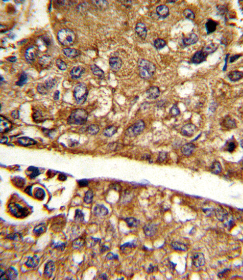
An anti-MDH1 IHC antibody has a DAB image from formalin-fixed, paraffin-embedded human hepatocarcinoma (catalog image caption); an IF/ICC-listed option covers human, mouse, and rat (catalog: M04262).

Real IHC data Formalin-fixed and paraffin-embedded human hepatocarcinoma reacted with MDH1 Antibody (C-term), which was peroxidase-conjugated to the secondary antibody, followed by DAB staining. This data demonstrates the use of this antibody for immunohistochemistry; clinical relevance has not been evaluated.
Anti-MDH1 Antibody (C-term)
Cat # A04262-1

A04262-1 is the only card rendered, with IHC-P data from formalin-fixed, paraffin-embedded human hepatocarcinoma (catalog image caption). M04262 is listed for IF/ICC, but has no IF image in the payload (catalog: applications and image alts).

Which to pick: Choose A04262-1 for human tissue IHC-P: it is a rabbit polyclonal antibody with a DAB image from formalin-fixed, paraffin-embedded human hepatocarcinoma (catalog: A04262-1 applications, host and image caption). Choose M04262 for IF/ICC or work involving mouse or rat because it is a rabbit monoclonal listed for those applications and species; its payload provides no IF image or IHC-P validation (catalog: M04262 applications, reactivity and image alts).

Each figure is that product's own IHC / IF validation image from its datasheet.

References

  1. UniProt Consortium. UniProt entry P40925 (MDHC_HUMAN, Malate dehydrogenase, cytoplasmic).
  2. Human Protein Atlas. MDH1 tissue IHC expression (reliability: Uncertain).
  3. Human Protein Atlas. MDH1 subcellular location (ICC-IF): Localized to the cytosol and centrosome..
  4. Human Protein Atlas. MDH1 antibody validation summary (3 antibodies).
  5. Tat-malate dehydrogenase fusion protein protects neurons from oxidative and ischemic damage by reduction of reactive oxygen species and modulation of glutathione redox system. Scientific reports 2023 — PMC10079925.
  6. Regulation of human cerebrospinal fluid malate dehydrogenase 1 in sporadic Creutzfeldt-Jakob disease patients. Aging 2016 — PMC5191879.
  7. Panoramic view of MDH1: driving cancer progression and shaping the tumor immune microenvironment. Frontiers in immunology 2025 — PMC12423056.
  8. Characterization of the Role of the Malate Dehydrogenases to Lung Tumor Cell Survival. Journal of Cancer 2017 — PMC5559971.
  9. PubMed PMID:8786100 — UniProt-cited evidence.
  10. PubMed PMID:14702039 — UniProt-cited evidence.
  11. PubMed PMID:15815621 — UniProt-cited evidence.