MDM2 / E3 ubiquitin-protein ligase Mdm2 · IHC design guide

Design Immunohistochemistry for MDM2

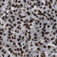
Plan MDM2 paraffin IHC around nuclear staining, using adipose adipocytes as a high-staining reference (HPA tissue IHC). The IHC-validated antibody M00054-3 has a recommended dilution range of 1:200–1:1000 (datasheet).

Evidence assembled Oct 2026 · For research use; verify linked source records and product datasheet before use
Immunohistochemistry protocol sheet for MDM2 (IHC for MDM2): expected localisation Nuclear tissue staining (HPA tissue IHC), antibody M00054-3, validated IHC image, and IHC protocol steps
Printable MDM2 IHC protocol sheet — expected localisation Nuclear tissue staining (HPA tissue IHC), antibody M00054-3, controls and protocol steps. Open the full MDM2 IHC guide →

MDM2 Immunohistochemistry Experimental Design Guide

Expected localisation, validated protocols, controls and antibodies — the at-a-glance facts below, then the full design guide.

Must know before staining
Expected localisation Nuclear tissue staining (HPA tissue IHC)
Staining pattern Nuclear staining across diverse tissue cell types (HPA tissue IHC)
Antigen retrieval Tris-EDTA pH 9.0 HIER, 95–98 °C, 20 min (rule: nuclear antigen)
Positive control ⓘ Adipose tissue+4 more · see all
Negative control ⓘ None in HPA (detected in all 45 tissues); use no-primary + isotype controls
Important caveats
Reasons your staining may differ from the expected pattern.
Fixation Keep fixation consistent across sections (standard IHC practice; not target-specific); Matched tissue-IHC evidence does not establish the fixation claim. Validate the specimen-specific method before use. (selected-SKU IHC image M00054-3)
Caveat Cancer-associated splice isoforms may affect antibody recognition (UniProt)
Regulation Expression regulation is unspecified (UniProt)
Isoform / epitope 11 isoforms; verify epitope coverage across splice forms (UniProt)
Section 1

Recommended MDM2 IHC & IF Protocols

Compare the catalog antibody’s IHC-P protocol (catalog datasheet) with published paraffin-section MDM2 methods from three articles (PMC8760962; PMC12061814; PMC10700221).

Recommended immunohistochemistry (IHC-P) protocol parameters
SampleTissue sections; selected-image fixative not specified (standard IHC workflow)
FixationImage fixative and duration unreported (datasheet M00054-3); verify before use.
Sectioning4–5 µm sections on charged slides (standard)
DeparaffinisationXylene, graded ethanol series to water (standard)
Antigen retrievalHeat-induced epitope retrieval in Tris-EDTA buffer, pH 9.0, 20 min at 95–98 °C (standard rule: nuclear antigen)
Peroxidase block3% H2O2, 10 min, room temperature (standard)
Blocking10% normal serum of the secondary host, 30 min, room temperature (standard)
Primary antibodyRabbit anti-MDM2, 1:200-1:1000 (datasheet M00054-3)
Primary incubationOvernight at 4 °C (standard)
DetectionHRP-polymer secondary, DAB chromogen 5–10 min (standard)
CounterstainHematoxylin, blue, dehydrate and mount (standard)
Expected resultMDM2-positive staining in adipocytes of adipose tissue (HPA tissue IHC: High). HPA tissue profile: Ubiquitous nuclear expression. No signal in the no-primary control.
💡Decision noteStart with Tris-EDTA pH 9.0 at 95–98 °C for 20 min (page retrieval rule: nuclear antigen); adjust retrieval for the antibody and staining platform.
Section 2

What Is the Expected MDM2 Staining Pattern?

MDM2 is a non-membrane protein expressed predominantly in the nucleoplasm, with cytoplasmic and context-dependent nucleolar localization (UniProt Q00987: localization and topology). In paraffin-section IHC, expect widespread nuclear staining across tissues; HPA describes the tissue pattern as ubiquitous and rates it Supported because staining agrees with RNA expression (HPA: tissue IHC). High staining is documented in the specific cells listed below, but intensity should not be assumed uniform across every cell (HPA: tissue IHC).

What am I looking at on my slide?
Nuclear staining in adipocytes, glandular cells, or hematopoietic cells.This fits the expected compartment and documented high staining in adipocytes of adipose tissue and breast, glandular cells of adrenal gland and appendix, and bone-marrow hematopoietic cells (HPA: tissue IHC). Compare each cell population with its surrounding tissue; widespread expression does not imply identical intensity in every nucleus (HPA: ubiquitous nuclear expression).
Nuclear staining with some cytoplasmic staining.The nuclear component fits routine tissue IHC (HPA: ubiquitous nuclear expression). A cytoplasmic component is biologically plausible because UniProt lists cytoplasm and HPA ICC-IF supports cytosol; those localization records alone do not establish the expected strength or distribution of cytoplasmic chromogenic staining in paraffin sections (UniProt Q00987: localization; HPA: subcellular ICC-IF).
Signal confined to cell borders, extracellular material, or other structures without convincing nuclei.Treat this as a questionable MDM2 pattern: UniProt reports no transmembrane segment and predominantly nucleoplasmic localization, while HPA reports ubiquitous nuclear tissue staining (UniProt Q00987: topology and localization; HPA: tissue IHC). General IHC practice: inspect morphology and control staining before interpreting an isolated non-nuclear signal as positive.
Strong staining in an unexpected cell population while documented positive cells remain unstained.Consider cross-reactivity or endogenous detection activity, then check controls and cell identity (general IHC practice). An unlisted cell type is not automatically negative: MDM2 is described as ubiquitous, and the supplied HPA tissue record has no negative-cell list (UniProt Q00987: tissue specificity; HPA: tissue IHC).
Diffuse staining across nuclei, cytoplasm, and surrounding tissue.Poor compartment contrast makes the result hard to score against HPA's nuclear tissue pattern (HPA: tissue IHC). General IHC practice: evaluate background with appropriate negative controls and review blocking, antibody concentration, washes, and detection conditions before assigning cellular positivity.
💡Expected MDM2 appearanceCall a section positive when identifiable cells show nuclear MDM2 staining, including the HPA-listed high-staining adipocytes, glandular cells, or hematopoietic cells (HPA: tissue IHC); isolated extracellular or border staining without convincing nuclei is a questionable positive against the reported nuclear pattern (HPA: tissue IHC; UniProt Q00987: topology).
How each factor affects the staining
Cell and tissue contextHPA reports ubiquitous nuclear expression with low RNA tissue specificity, and lists high staining in several distinct cell populations; choose a documented positive population for interpretation rather than assuming a negative reference tissue from this record (HPA: tissue IHC).
Subcellular location and topologyUniProt places MDM2 predominantly in the nucleoplasm, also lists cytoplasm and nucleolus, and reports no transmembrane segment; the nucleolar localization is linked to interaction with ARF(P14), so a nucleolar pattern should be interpreted in context (UniProt Q00987: localization and topology).
IsoformsUniProt lists 11 isoforms and reports several cancer-associated isoforms absent from normal tissues; this record does not identify the antibody epitope or show which isoforms a given IHC reagent detects, so staining cannot assign an isoform (UniProt Q00987: isoforms and tissue specificity).
Antibody validationHPA marks tissue IHC Supported for CAB000086, CAB016303, and CAB079977; that status supports the observed staining pattern for those antibodies, without establishing that every antibody will reproduce it (HPA: antibody validation; HPA: tissue IHC).
Fixation and retrieval evidenceTarget-specific fixation sensitivity and retrieval conditions are unreported in the supplied UniProt and HPA records; no MDM2-specific change in epitope access or signal can be inferred from the reported topology, modifications, or tissue staining (UniProt Q00987: topology and modifications; HPA: tissue IHC).
IF/ICC Q&A: where should signal appear?The separate IF/ICC guide should use nucleoplasm and cytosol as its main supported locations; HPA also reports additional locations in its ICC-IF record (HPA: subcellular ICC-IF). These observations do not provide an IF/ICC protocol for this IHC-P section.
Why is my staining missing, weak or wrong?
SituationLikely causeNext action
No staining in a documented high-staining population.The result conflicts with HPA's reported high staining in the chosen population; the cause cannot be determined from the tissue record alone (HPA: tissue IHC).General IHC practice: check tissue identity and preservation, then review primary antibody, retrieval, detection reagents, and a positive control in the same run; document which step restores nuclear signal.
Only diffuse cytoplasmic staining appears.UniProt allows cytoplasmic MDM2, but absence of convincing nuclei conflicts with the predominant nucleoplasmic and HPA tissue patterns (UniProt Q00987: localization; HPA: tissue IHC).General IHC practice: inspect nuclei and negative controls, review antibody concentration and washes, and avoid scoring diffuse cytoplasm alone as a confirmed tissue-IHC positive.
Signal follows cell edges or extracellular deposits.That distribution conflicts with HPA's nuclear tissue pattern and UniProt's lack of a transmembrane segment (HPA: tissue IHC; UniProt Q00987: topology).General IHC practice: compare with a negative control, inspect section morphology, and review blocking and detection before attributing the deposit to MDM2.
One unlisted cell type stains strongly.Unexpected cell-type staining may reflect cross-reactivity or detection background, but the record supplies no negative cell types and describes MDM2 as ubiquitous (HPA: tissue IHC; UniProt Q00987: tissue specificity).General IHC practice: verify the cell type and controls, then compare nearby documented positive populations; do not classify the unlisted cells as false positives solely because HPA did not list them.
Brown signal obscures nuclear boundaries.Excess background can prevent assessment of the nuclear pattern reported by HPA (HPA: tissue IHC).General IHC practice: review chromogen development, counterstain, antibody concentration, blocking, and washes; repeat scoring only when nuclei and surrounding background can be distinguished.
Nucleolar staining dominates the section.UniProt links nucleolar MDM2 localization to ARF(P14) interaction, whereas HPA describes the overall tissue pattern as ubiquitous nuclear expression (UniProt Q00987: localization; HPA: tissue IHC).General IHC practice: verify that the puncta are within identifiable nuclei and compare controls; report the nucleolar distribution separately instead of assuming it is the routine tissue pattern.

Sample controls for MDM2 IHC & IF

🧪Run adipose tissue first and score adipocytes, which HPA rates High for MDM2 (HPA: High in adipocytes). HPA detects MDM2 in all 45 scored tissues, so there is no supported negative tissue; use no-primary and isotype controls, and treat any unstained cells within the adipose section as a local background reference, not a validated MDM2-negative population (HPA: no negative tissue rows; detected in all 45 scored tissues).
Positive control tissue: Adipose tissue (Adipocytes, HPA High)
Negative control tissue: None in HPA: MDM2 is detected in all 45 scored tissues. Use a no-primary (secondary-only) and an isotype control instead.
ICC-IF cell lines (HPA subcellular resource): HPA ICC-IF images show MDM2 in A-431, U-251MG, U2OS, REH, RT-4, A-549, HEK293, ASC52telo, Sperm, with annotated localisation: Nucleoplasm (supported), Cytosol (supported) (HPA subcellular).
Technical controls: Include a no-primary, secondary-only slide and an isotype control matched to the primary antibody’s host species, immunoglobulin class and monoclonal or polyclonal format; use matched MDM2-knockout material as a biological specificity control where available (standard IHC practice). Quench endogenous peroxidase for chromogenic detection, and account for lipid-extraction spaces and thin adipocyte cytoplasm when judging staining in adipose tissue (standard paraffin IHC practice).
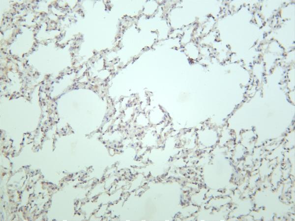
⚠️Feasibility: A target-specific fixation window and fixation effect are unreported, and the selected M00054-3 rat-lung IHC caption does not state a fixative (supplied target/application evidence; selected-SKU caption). Retrieval dependence is unreported, so optimize antigen retrieval with the IHC-validated antibody rather than inferring it from HPA staining or MDM2 features (supplied target/application evidence; standard IHC practice). The supplied evidence does not establish whether frozen sections or IF are easier for MDM2; adipocyte lipid loss during paraffin processing can complicate assignment of signal to the thin cytoplasmic rim (supplied target/application evidence; standard paraffin IHC practice).

HPA tissue IHC evidence for MDM2

Comprehensive Human Protein Atlas IHC scoring per tissue (reliability: Supported — High consistency between antibody staining and RNA expression data.). Rows are taken directly from the HPA tissue chart — click any row's HPA link to view the source.

Positive expression · recommended positive controls

TissueCell typeLevelEvidenceSource
Adipose tissue Adipocytes High Protein (IHC) HPA →
Adrenal gland Glandular cells High Protein (IHC) HPA →
Appendix Glandular cells High Protein (IHC) HPA →
Bone marrow Hematopoietic cells High Protein (IHC) HPA →
Breast Adipocytes High Protein (IHC) HPA →

Undetected expression · recommended negative controls

TissueCell typeLevelEvidenceSource
None in HPA: MDM2 is detected in all 45 scored tissues. Use a no-primary (secondary-only) and an isotype control instead.
Section 3

Advanced MDM2 IHC Tips

Troubleshoot MDM2 staining in paraffin sections by checking retrieval, nuclear localisation, antibody specificity and scoring controls before interpreting chromogenic signal.

How should I adjust retrieval when MDM2 nuclear staining is weak?
Start with Tris-EDTA pH 9.0 HIER at 95–98 °C for 20 min (page retrieval setting). Let slides cool in retrieval buffer, then compare the expected nucleoplasmic signal across a consistently processed control section (UniProt Q00987: predominantly nucleoplasmic; standard IHC practice). If signal remains weak, check section adhesion and antibody titration before testing citrate pH 6.0 as a fallback on matched sections (standard IHC practice). If tissue lifts or nuclear detail deteriorates, shorten heating while keeping the starting buffer at pH 9.0, and record the change for every comparison (standard IHC practice).
Could fixation explain weak or uneven MDM2 staining?
Target-specific fixation sensitivity is unknown from the supplied evidence; the rat lung staining caption identifies antibody M00054-3 but does not state a fixative (selected caption M00054-3). Record fixative, fixation duration and tissue thickness for each specimen before comparing chromogenic intensity (standard IHC practice). Compare sections fixed and processed together, because uneven fixation and processing can produce spatial staining differences in paraffin sections (standard IHC practice). If a fixation effect is suspected, test matched material under the same Tris-EDTA pH 9.0, 20 min retrieval setting before changing antibody concentration (page retrieval setting; standard IHC practice).
Is cytoplasmic or nucleolar MDM2 staining credible in tissue sections?
MDM2 is predominantly nucleoplasmic, with reported cytoplasmic and nucleolar localisation (UniProt Q00987: subcellular location). HPA describes ubiquitous nuclear tissue staining and supports nucleoplasm and cytosol in subcellular images (HPA tissue IHC: ubiquitous nuclear expression; HPA subcellular: supported locations). Score nuclear and cytoplasmic chromogen separately, using a hematoxylin counterstain to identify nuclear boundaries (standard IHC practice). Nucleolar enrichment can be biologically plausible when MDM2 interacts with ARF(P14), but a nucleolar pattern alone does not establish that interaction in the section (UniProt Q00987: ARF(P14)-associated localisation). Diffuse staining over tissue edges or empty spaces warrants a background check before localisation is assigned (standard IHC practice).
Can this stain distinguish full-length MDM2 from its splice isoforms?
Do not assign an isoform from chromogenic staining unless the antibody epitope and its presence in each isoform are documented (standard IHC interpretation). UniProt lists 11 MDM2 isoforms and a SWIB/MDM2 domain at residues 26–109, but the supplied antibody caption gives no epitope (UniProt Q00987: isoforms and domain; selected caption M00054-3). Map the immunogen or binding region against isoform sequences before interpreting a positive nucleus as a particular variant (standard IHC practice). Several named isoforms are reported in cancers and absent from normal tissues, yet staining intensity alone cannot identify which isoform produced the signal (UniProt Q00987: tissue specificity; standard IHC interpretation).
How can I check MDM2 localisation by multiplex IF?
For a separate IF/ICC experiment, pair MDM2 with a validated marker for the cell population being examined and include a nuclear counterstain (standard IF practice). MDM2 is reported in nucleoplasm and cytosol, so choose a permeabilisation condition that gives antibodies access to the cytoplasm and nucleus (HPA subcellular: supported locations; UniProt Q00987: no transmembrane segment). Choose spectrally separated fluorophores and inspect an unstained section in each channel before selecting the MDM2 channel, because tissue autofluorescence can resemble specific signal (standard IF practice). Optimise fixation and permeabilisation for the IF antibody independently; the selected tissue-IHC caption does not report fixation or establish an IF protocol (selected caption M00054-3; standard IF practice).
What should I check when MDM2 chromogen appears throughout the section?
First compare the stained slide with a no-primary control and inspect whether colour follows tissue edges, folds or damaged regions (standard IHC practice). Block endogenous peroxidase before an HRP–DAB detection workflow, and use an appropriate secondary-only control to assess detection-reagent background (standard IHC practice). Titrate the primary antibody and blocking conditions on matched sections while holding the Tris-EDTA pH 9.0, 20 min retrieval setting constant (page retrieval setting; standard IHC practice). Because MDM2 has broad tissue expression, widespread staining is not by itself proof of background; assess crisp nuclear signal and control-slide staining together (UniProt Q00987: ubiquitous expression; HPA tissue IHC: ubiquitous nuclear expression).
How should I quantify MDM2 across paraffin sections? ⚠ ANSWER MARKED FOR VERIFICATION
Define the cell population and score the percentage of positive nuclei plus nuclear intensity using a consistent H-score threshold across sections (standard IHC practice). Report cytoplasmic staining separately, because MDM2 has supported nucleoplasmic and cytosolic localisation, while HPA describes ubiquitous nuclear tissue expression (HPA subcellular: supported locations; HPA tissue IHC: profile). Normalise positive-cell counts to the number of evaluable cells in the same annotated region, or report positive-cell density per mm² of viable tissue (standard IHC practice). Exclude folds, necrosis and edge artefact, and keep fixation, retrieval, detection and image-acquisition settings consistent across comparison groups (standard IHC practice).
How can I distinguish genuine MDM2 positivity from artefact?
A credible result shows staining in identifiable cells with interpretable nuclear detail and a consistent pattern across comparable viable regions (standard IHC practice). Predominantly nuclear staining fits the tissue profile, while cytosolic staining can also be plausible; neither pattern alone proves antibody specificity (HPA tissue IHC: ubiquitous nuclear expression; HPA subcellular: supported nucleoplasm and cytosol). Check unexpected colour at cut edges, folds, necrotic areas and sites with endogenous peroxidase against matched no-primary controls (standard IHC practice). Compare the stained cell population with its morphology and an appropriate control section before claiming a cell-type-specific difference, since MDM2 expression is reported as ubiquitous (UniProt Q00987: tissue specificity; standard IHC practice).
Boster reagents

Best MDM2 / E3 ubiquitin-protein ligase Mdm2 IHC Antibodies

Two rabbit monoclonals have IHC tissue images (catalog IHC captions). Both list human, mouse, and rat reactivity (catalog reactivity); M00054-3 also lists IF, although no IF image is supplied (catalog applications/IF images).

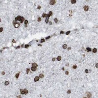
Real IHC data Rat lung was stained with anti-MDM2 rabbit antibody
Anti-MDM2 Rabbit Monoclonal Antibody
Cat # M00054-3
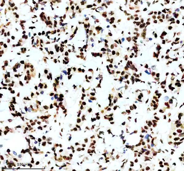
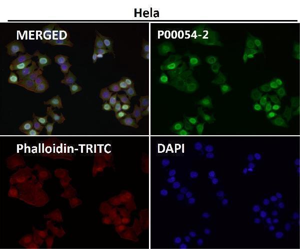
Real IHC data IHC analysis of P-MDM2 using anti-P-MDM2 antibody (P00054-2). P-MDM2 was detected in a paraffin-embedded section of human breast cancer tissue. Heat mediated antigen retrieval was performed in EDTA buffer (pH 8.0, epitope retrieval solution). The tissue section was blocked with 10% goat serum. The tissue section was then incubated with 1:50 rabbit anti-P-MDM2 Antibody (P00054-2) overnight at 4°C. Peroxidase Conjugated Goat Anti-rabbit IgG was used as secondary antibody and incubated for 30 minutes at 37°C. The tissue section was developed using HRP Conjugated Rabbit IgG Super Vision Assay Kit (Catalog # SV0002) with DAB as the chromogen.
Anti-Phospho-MDM2 (S166) Rabbit Monoclonal Antibody
Cat # P00054-2

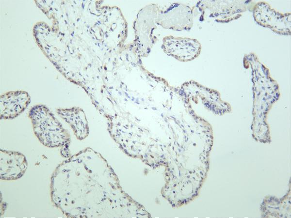
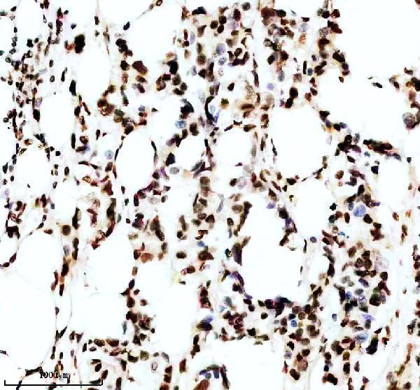
M00054-3 will render with rat lung IHC; another caption shows human placenta, and its listed applications include IHC and IF (catalog: M00054-3 IHC captions/applications). P00054-2 will render with phospho-MDM2 (S166) IHC in a paraffin section of human breast cancer; other IHC captions show human lung cancer and mouse testis (catalog: P00054-2 IHC captions).

Which to pick: For total MDM2 tissue IHC, choose M00054-3, a rabbit monoclonal whose rendered caption shows rat lung staining but reports neither section processing nor fixative (catalog: M00054-3 description/IHC caption). For phospho-MDM2 (S166) tissue IHC, choose P00054-2; its caption specifies a paraffin section, EDTA retrieval at pH 8.0, and 1:50 primary antibody, but does not report the fixative (catalog: P00054-2 title/IHC caption). For IF, choose M00054-3 at the listed 1:200–1:1000 range; ICC validation is unlisted, and both SKUs list human, mouse, and rat reactivity for cross-species IHC planning (catalog applications/dilutions/reactivity).

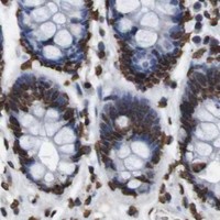
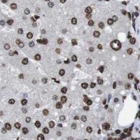
Each figure is that product's own IHC / IF validation image from its datasheet.

References

  1. UniProt Consortium. UniProt entry Q00987 (MDM2_HUMAN, E3 ubiquitin-protein ligase Mdm2).
  2. Human Protein Atlas. MDM2 tissue IHC expression (reliability: Supported).
  3. Human Protein Atlas. MDM2 subcellular location (ICC-IF): Mainly localized to the nucleoplasm and cytosol. In addition localized to the centriolar satellites, mid piece, principal piece and end piece..
  4. Human Protein Atlas. MDM2 antibody validation summary (4 antibodies).
  5. Juvenile trabecular ossifying fibroma: Immunohistochemical expression of MDM2, CDK4 and p53 compared to conventional ossifying fibroma. Journal of clinical and experimental dentistry 2022 — PMC8760962.
  6. The expression of MDM2 in gastrointestinal stromal tumors: immunohistochemical analysis of 35 cases. BMC clinical pathology 2018 — PMC5781285.
  7. Recurrent MDM2 Amplification in the Spectrum of HMGA2-Altered Pleomorphic Adenoma, Atypical Pleomorphic Adenoma and Carcinoma Ex Pleomorphic Adenoma. Head and neck pathology 2025 — PMC12061814.
  8. MDM2 amplification is rare in gastric cancer. Virchows Archiv : an international journal of pathology 2023 — PMC10700221.
  9. PubMed PMID:1614537 — UniProt-cited evidence.
  10. PubMed PMID:8705862 — UniProt-cited evidence.
  11. PubMed PMID:10597303 — UniProt-cited evidence.