MECP2 / Methyl-CpG-binding protein 2 · IHC design guide

Design Immunohistochemistry for MECP2

Plan MECP2 staining in paraffin sections around its nuclear tissue pattern (HPA tissue IHC). This guide covers fixation consistency, controls and interpretation of cell-type differences in staining intensity (HPA tissue IHC).

Evidence assembled Oct 2026 · For research use; verify linked source records and product datasheet before use
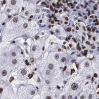
Immunohistochemistry protocol sheet for MECP2 (IHC for MECP2): expected localisation Nuclear staining across tissues (HPA tissue IHC), antibody A00047-1, validated IHC image, and IHC protocol steps
Printable MECP2 IHC protocol sheet — expected localisation Nuclear staining across tissues (HPA tissue IHC), antibody A00047-1, controls and protocol steps. Open the full MECP2 IHC guide →

MECP2 Immunohistochemistry Experimental Design Guide

Expected localisation, validated protocols, controls and antibodies — the at-a-glance facts below, then the full design guide.

Must know before staining
Expected localisation Nuclear staining across tissues (HPA tissue IHC)
Staining pattern Nuclear staining across tissue cell types (HPA tissue IHC)
Antigen retrieval EDTA pH 8.0 HIER, heat-mediated (datasheet A00047-1)
Positive control ⓘ Adrenal gland+4 more · see all
Negative control ⓘ None in HPA (detected in all 45 tissues); use no-primary + isotype controls
Important caveats
Reasons your staining may differ from the expected pattern.
Fixation Matched tissue-IHC evidence does not establish the fixation claim. Validate the specimen-specific method before use. (selected-SKU IHC image A00047-1)
Caveat Staining is low in rectal endothelial cells (HPA tissue IHC)
Regulation Broad adult somatic expression (UniProt)
Isoform / epitope 2 isoforms (A and B); epitope coverage is unspecified (UniProt)
Section 1

Recommended MECP2 IHC & IF Protocols

The catalog antibody’s IHC-P protocol is accompanied by published MECP2 protocols for ocular tissue, ampullary carcinoma, and gastric tissue (PMC2916867; PMC12971181; PMC4564377).

Recommended immunohistochemistry (IHC-P) protocol parameters
SampleParaffin-embedded mouse brain tissue; fixative not specified (datasheet A00047-1)
FixationImage fixative and duration unreported (datasheet A00047-1); verify before use.
Sectioning4–5 µm sections on charged slides (standard)
DeparaffinisationXylene, graded ethanol series to water (standard)
Antigen retrievalHeat retrieval: EDTA pH 8.0 (datasheet A00047-1); 20 min, 95–100 °C (standard)
Peroxidase block3% H2O2, 10 min, room temperature (standard)
Blocking10% goat serum (datasheet A00047-1)
Primary antibodyRabbit anti-MECP2, 2-5 μg/ml (datasheet A00047-1)
Primary incubationOvernight at 4 °C (datasheet A00047-1)
DetectionHRP-conjugated secondary, DAB chromogen (datasheet A00047-1)
CounterstainHematoxylin, blue, dehydrate and mount (standard)
Expected resultMECP2-positive staining in glandular cells of adrenal gland (HPA tissue IHC: High). HPA tissue profile: Nuclear expression in all tissues. No signal in the no-primary control.
💡Decision noteStart with EDTA pH 8.0 heat retrieval for the catalog antibody (datasheet A00047-1). The published protocols use citrate retrieval (PMC2916867; PMC12971181; PMC4564377).
Section 2

What Is the Expected MECP2 Staining Pattern?

MECP2 should appear in nuclei across tissues, consistent with its DNA-binding role and lack of a transmembrane segment (UniProt P51608; HPA: nuclear expression in all tissues). Expect staining in the relevant resident cells, including glandular cells, hematopoietic cells and glia (HPA: High in listed examples). The tissue IHC profile is rated Enhanced, reflecting high consistency between antibody staining and RNA expression (HPA: tissue reliability).

What am I looking at on my slide?
Distinct chromogenic staining is confined mainly to nuclei of the expected cells.This matches MECP2's nuclear location and the HPA tissue profile (UniProt P51608: Nucleus; HPA: nuclear expression in all tissues). Compare cells within the same section: HPA reports High staining in several cell populations but Low staining in others, so uniform intensity across every cell is unnecessary (HPA: tissue IHC).
Strong staining appears chiefly in cytoplasm or along cell borders, while nuclei remain pale.That compartment conflicts with the reported nuclear pattern (UniProt P51608: Nucleus; HPA: nuclear tissue expression). Treat it as a possible artefact and inspect the counterstain, tissue morphology and detection controls before scoring cells as MECP2 positive (general IHC practice).
An unexpected cell population stains strongly while the expected population does not.Possible explanations include antibody cross-reactivity or endogenous detection activity (general IHC practice). First check the cell identity against morphology and the HPA cell-level pattern; HPA reports, for example, High staining in breast glandular cells and Low staining in rectal endothelial cells (HPA: tissue IHC).
Weak, diffuse color covers nuclei, cytoplasm and surrounding tissue.This obscures the compartment needed to interpret MECP2 (UniProt P51608: Nucleus; HPA: nuclear tissue expression). Uneven blocking, excess detection background or poorly controlled staining can produce a similar appearance (general IHC practice); diffuse color alone is insufficient evidence of MECP2 positivity.
There is no nuclear signal in a tissue with a reported High cell population.A negative run is difficult to interpret without a working positive control (general IHC practice). Check the relevant cells: HPA reports High staining in adrenal glandular cells, bone marrow hematopoietic cells and cerebral cortex glia, among others (HPA: tissue IHC). HPA's profile does not establish fixation sensitivity.
💡Expected MECP2 appearanceA credible positive is clearly discernible nuclear staining in the relevant cells, potentially High in HPA-listed populations; dominant cytoplasmic or diffuse tissue-wide color is suspect (HPA: tissue IHC; UniProt P51608: Nucleus).
How each factor affects the staining
Cell population and tissueInterpret intensity by cell type: HPA reports High staining in several glandular, hematopoietic and glial populations, but Low staining in oral squamous epithelial and rectal endothelial cells (HPA: tissue IHC). Low is not an established negative control.
Subnuclear distributionMECP2 colocalizes with methyl-CpG and with TBL1X at heterochromatin foci (UniProt P51608: subcellular location). HPA calls the ICC-IF location nucleoplasmic; these sources support nuclear interpretation without requiring every IHC-positive nucleus to show visible foci (HPA: subcellular).
Antibody validationHPA lists Enhanced IHC status for HPA000593 and HPA001341, and Supported IHC status for CAB037264 (HPA: antibodies). Those ratings support the reported pattern; they do not prove that every antibody or staining run will reproduce it.
Isoforms and epitope coverageUniProt lists two MECP2 isoforms, A and B, and an MBD at residues 90–162 (UniProt P51608). No epitope positions are supplied, so these facts cannot establish which isoform a particular IHC antibody detects.
IF/ICC Q: What location should fluorescence show?A: Nucleoplasmic signal is the approved HPA ICC-IF location (HPA: subcellular). HPA lists A-431, U-251MG and U2OS as lines with ICC-IF images, but those images do not supply an IHC-P protocol or change the expected chromogenic nuclear pattern.
Why is my staining missing, weak or wrong?
SituationLikely causeNext action
Positive-control cells show no nuclear staining.The staining run may have failed, or the selected cells may not match the expected population (general IHC practice; HPA: cell-level tissue IHC).Confirm tissue and cell identity, then review the antibody's IHC-P instructions and run controls for retrieval, antibody incubation and detection (general IHC practice). No MECP2-specific retrieval condition or dilution is supplied here.
Only cytoplasmic color is conspicuous.The signal disagrees with MECP2's reported nuclear location (UniProt P51608: Nucleus; HPA: nuclear tissue expression). Background or nonspecific binding is possible (general IHC practice).Use the nuclear counterstain to check compartment boundaries; compare a reported High cell population with appropriate staining controls before assigning positivity (HPA: tissue IHC; general IHC practice).
Unexpected cells stain more strongly than the expected cells.Cell misidentification, cross-reactivity or endogenous detection activity could explain the pattern (general IHC practice). HPA levels describe specified cell populations, not every cell in a tissue (HPA: tissue IHC).Recheck morphology and the relevant HPA cell-level entry; inspect detection controls for endogenous activity and assess the primary antibody's validation information (HPA: tissue IHC and antibodies; general IHC practice).
Diffuse background makes nuclei hard to score.Broad color deposition can mask the nuclear pattern (general IHC practice; UniProt P51608: Nucleus). The supplied HPA data do not identify a MECP2-specific background mechanism.Review blocking, washing, primary-antibody concentration and detection-control results using the antibody's IHC-P instructions (general IHC practice). Score only nuclei that remain distinguishable from surrounding background.
Reported Low cells appear negative beside strongly stained cells.Staining intensity varies by population: HPA reports Low in oral mucosa squamous epithelial cells and rectal endothelial cells, alongside High populations elsewhere (HPA: tissue IHC).Judge those cells against their own HPA entry and a working positive population; avoid calling them a validated MECP2-negative control (HPA: tissue IHC; general IHC practice).
Fluorescence images seem inconsistent with the chromogenic section.HPA approves a nucleoplasmic ICC-IF location, while the tissue IHC profile reports nuclear staining; the methods provide different views of compartment and cell context (HPA: subcellular and tissue IHC).Compare nuclear localisation and cell identity within each method; use the ICC-IF entry to inform localisation, and the tissue IHC entries to interpret the paraffin-section pattern (HPA: subcellular and tissue IHC).

Sample controls for MECP2 IHC & IF

🧪Run adrenal gland first; glandular cells should show nuclear staining (HPA: High in adrenal gland glandular cells; UniProt P51608: nucleus). HPA detects MECP2 in all 45 scored tissues, so there is no negative tissue or validated negative cell population within the positive slide; use no-primary and isotype controls to judge whether apparently unstained cells are meaningful (HPA: detected in all 45 scored tissues).
Positive control tissue: Adrenal gland (Glandular cells, HPA High)
Negative control tissue: None in HPA: MECP2 is detected in all 45 scored tissues. Use a no-primary (secondary-only) and an isotype control instead.
ICC-IF cell lines (HPA subcellular resource): HPA ICC-IF images show MECP2 in A-431, U-251MG, U2OS, with annotated localisation: Nucleoplasm (approved) (HPA subcellular).
Technical controls: Include a no-primary, secondary-only slide and a nonspecific IgG control matched to the primary antibody’s host species and isotype; MECP2 knockout material, if available, provides a biological negative (standard IHC practice). For chromogenic detection in adrenal sections, quench endogenous peroxidase and check background on the control slides (standard IHC practice).
⚠️Feasibility: The selected A00047-1 mouse-brain paraffin-section caption uses heat antigen retrieval in EDTA at pH 8.0, but does not report a fixative (selected-SKU tissue-IHC caption). A target-specific fixation window or fixation effect is unreported, and the caption does not establish whether retrieval is required under other conditions (supplied target/application evidence). HPA shows nucleoplasmic ICC-IF localization, but the supplied evidence does not establish that IF or frozen sections are easier than paraffin IHC (HPA: subcellular localization).

HPA tissue IHC evidence for MECP2

Comprehensive Human Protein Atlas IHC scoring per tissue (reliability: Enhanced — High consistency between antibody staining and RNA expression data.). Rows are taken directly from the HPA tissue chart — click any row's HPA link to view the source.

Positive expression · recommended positive controls

TissueCell typeLevelEvidenceSource
Adrenal gland Glandular cells High Protein (IHC) HPA →
Appendix Non-germinal center cells High Protein (IHC) HPA →
Bone marrow Hematopoietic cells High Protein (IHC) HPA →
Breast Glandular cells High Protein (IHC) HPA →
Bronchus Respiratory epithelial cells High Protein (IHC) HPA →

Undetected expression · recommended negative controls

TissueCell typeLevelEvidenceSource
None in HPA: MECP2 is detected in all 45 scored tissues. Use a no-primary (secondary-only) and an isotype control instead.
Section 3

Advanced MECP2 IHC Tips

MECP2 is a nuclear DNA-binding protein (UniProt P51608); use nuclear staining as the primary readout in chromogenic IHC (HPA tissue IHC).

How should I retrieve MECP2 in paraffin sections when nuclear staining is weak?
Start with heat-mediated retrieval in EDTA at pH 8.0 (datasheet A00047-1). That condition accompanies MECP2 staining in a paraffin-embedded mouse brain section using 2 μg/ml primary antibody overnight at 4°C (datasheet A00047-1). If staining remains weak, compare retrieval duration across matched sections while keeping antibody concentration and detection conditions fixed (standard IHC practice). Score nuclear signal and tissue damage together, since excess heating can obscure morphology and make nuclear scoring unreliable (standard IHC practice). Use the same retrieval condition for all sections being compared quantitatively (standard IHC practice).
Could fixation explain weak or uneven MECP2 staining in my sections?
Target-specific sensitivity of MECP2 to fixation is unknown from the supplied evidence (datasheet A00047-1: fixative not stated). The selected image describes a paraffin-embedded mouse brain section but does not identify its fixative or fixation duration (datasheet A00047-1). Record fixative, fixation duration, tissue thickness and processing history before comparing sections, because these variables can change antigen accessibility and morphology (standard IHC practice). Compare matched material under the same EDTA pH 8.0 retrieval and detection conditions (datasheet A00047-1; standard IHC practice). A fixation effect requires a controlled comparison; the reported nuclear tissue pattern alone cannot establish one (standard IHC practice).
What MECP2 staining pattern should count as specific in chromogenic IHC?
Prioritise nuclear staining: MECP2 localises to the nucleus and colocalises with methyl-CpG in the genome (UniProt P51608). Nuclear expression is reported across tissues, with high staining in cerebral cortex glial cells and cerebellar Bergmann glia nuclei (HPA tissue IHC). Use an intact nuclear counterstain to distinguish stained nuclei from adjacent cytoplasm and to define the cells available for scoring (standard IHC practice). Diffuse cytoplasmic DAB without corresponding nuclear signal warrants review of background, section quality and antibody specificity (UniProt P51608; standard IHC practice). Chromatin-associated enrichment may be uneven within a nucleus, so assess multiple well-preserved fields (UniProt P51608; standard IHC practice).
Can this stain distinguish MECP2 isoforms or reveal epitope masking?
MECP2 has 2 listed isoforms, A and B, and its methyl-CpG-binding domain spans residues 90–162 (UniProt P51608). The supplied caption does not specify the A00047-1 immunogen or epitope, so its staining cannot be assigned to one isoform (datasheet A00047-1). Likewise, the listed modified residues include phosphoserines and a methylarginine, but their effect on this antibody's staining is untested here (UniProt P51608; datasheet A00047-1). When isoform resolution matters, obtain documented epitope mapping and validate against an independent isoform-specific assay (standard IHC practice). Keep retrieval and scoring constant while evaluating any alternative antibody (standard IHC practice).
How can I follow up a nuclear MECP2 IHC result with multiplex IF?
Use multiplex IF as a separate validation experiment, pairing MECP2 with a marker for the expected cell population and a nuclear counterstain (standard IF practice). Glial cells in cerebral cortex and Bergmann glia nuclei in cerebellum offer documented cell-context examples for marker selection (HPA tissue IHC). Choose spectrally separated fluorophores after checking tissue autofluorescence and include single-stain controls to assess channel bleed-through (standard IF practice). MECP2 is nuclear and has no transmembrane segment, so permeabilisation must allow antibody access to the nucleus (UniProt P51608; standard IF practice). The paraffin-section caption supplies no IF fixation or permeabilisation conditions to transfer (datasheet A00047-1).
How do I reduce diffuse brown signal without losing nuclear MECP2 staining?
First compare the test section with a no-primary control to identify signal from the secondary reagent, chromogen or tissue itself (standard IHC practice). The selected assay used 10% goat serum blocking, a peroxidase-conjugated secondary antibody and DAB development (datasheet A00047-1). Include a peroxidase block before detection and monitor DAB development, because endogenous enzyme activity or overdevelopment can mimic positive staining (standard IHC practice). If background persists, titrate the primary around the reported 2 μg/ml condition while retaining EDTA pH 8.0 retrieval (datasheet A00047-1; standard IHC practice). Judge improvement by nuclear contrast and preserved morphology, not brown intensity alone (UniProt P51608; standard IHC practice).
How should I score MECP2 IHC across sections with different cell mixtures? ⚠ ANSWER MARKED FOR VERIFICATION
Define the region and cell population before scoring, then report the percentage of MECP2-positive nuclei and a prespecified nuclear intensity score (standard IHC practice). An H-score can combine the percentage of cells at each intensity, while positive nuclei per mm² can describe spatial density (standard IHC practice). Normalise positive counts to the total number of intact nuclei in the same cell population, and report area separately when using density (standard IHC practice). This matters because documented staining differs by cell type, including high cerebral cortex glial signal and low rectal endothelial signal (HPA tissue IHC). Apply one threshold and imaging workflow across comparison groups (standard IHC practice).
When is an apparent MECP2-positive cell likely to be an artefact?
Treat a well-preserved stained nucleus as the expected pattern, since MECP2 is nuclear and HPA reports nuclear expression across tissues (UniProt P51608; HPA tissue IHC). Review isolated cytoplasmic deposits, especially when nuclear staining is absent, against a no-primary control and local tissue morphology (standard IHC practice). Exclude section edges, folds and necrotic areas from scoring when staining or structure is distorted (standard IHC practice). Endogenous peroxidase can produce DAB signal, so check that the peroxidase block and control section behave as expected (standard IHC practice). Confirm cell identity before calling a result discordant with documented cell-specific patterns (HPA tissue IHC; standard IHC practice).
Boster reagents

Best MECP2 / Methyl-CpG-binding protein 2 IHC Antibodies

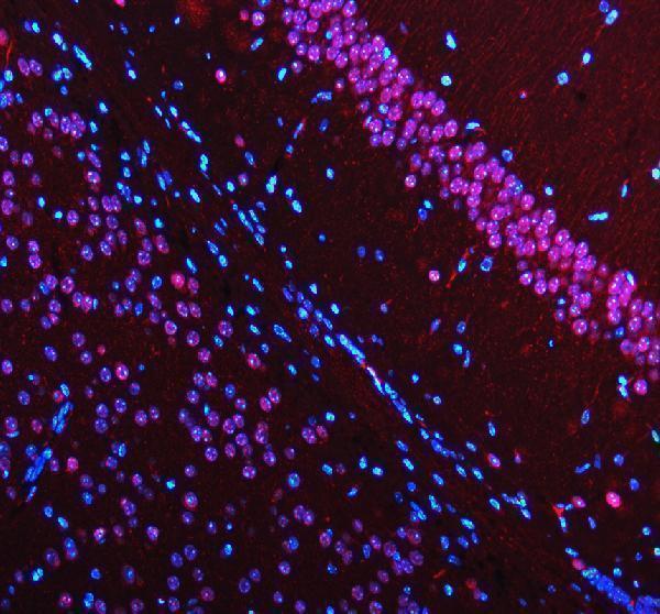
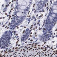
Anti-MECP2 catalog images show paraffin-section IHC in human colon and mammary cancers and mouse/rat brain, plus IF in A431 cells and mouse/rat brain (catalog IHC/IF image captions).

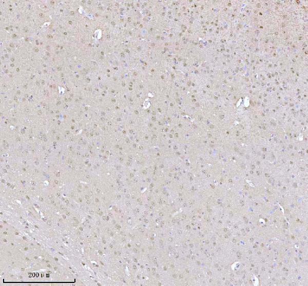
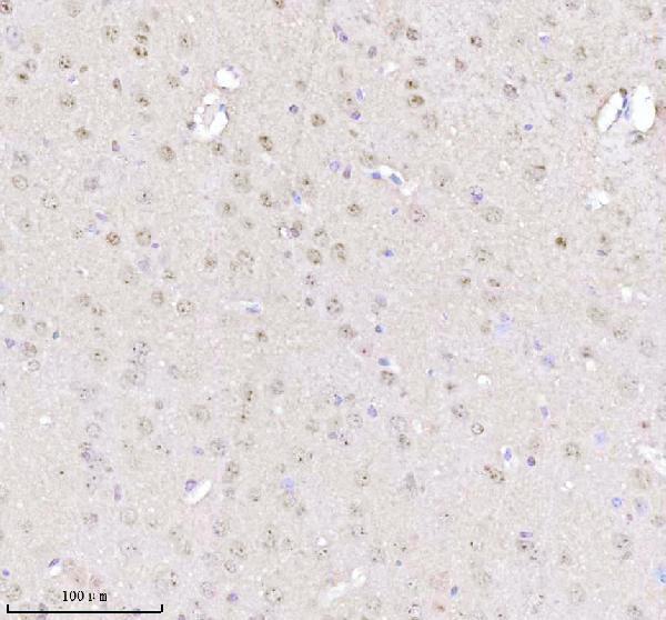
Real IHC data IHC analysis of MECP2 using anti-MECP2 antibody (A00047-1). MECP2 was detected in a paraffin-embedded section of mouse brain tissue. Heat mediated antigen retrieval was performed in EDTA buffer (pH 8.0, epitope retrieval solution). The tissue section was blocked with 10% goat serum. The tissue section was then incubated with 2 μg/ml rabbit anti-MECP2 Antibody (A00047-1) overnight at 4°C. Peroxidase Conjugated Goat Anti-rabbit IgG was used as secondary antibody and incubated for 30 minutes at 37°C. The tissue section was developed using HRP Conjugated Rabbit IgG Super Vision Assay Kit (Catalog # SV0002) with DAB as the chromogen.
Anti-MECP2 Antibody ®
Cat # A00047-1

The rendered A00047-1 card shows IHC in a paraffin-embedded mouse brain section (A00047-1 IHC image caption). Its catalog also shows rat brain IHC and mouse/rat brain IF (A00047-1 IHC/IF image captions).

Which to pick: For human tissue IHC, choose A00047: its own captions show paraffin-section staining in human colon and mammary cancers with citrate retrieval; the fixative is unreported (A00047 IHC image captions). For IF/ICC, A00047 has an A431 cell image and lists both applications, while A00047-1 has IF images of mouse and rat brain sections (A00047 applications and IF image caption; A00047-1 IF image captions). For the broadest listed species reactivity, A00047-1 includes human, monkey, mouse and rat; its IHC images document mouse and rat brain with EDTA retrieval, and clonality is unreported (A00047-1 reactivity, IHC image captions and clone field).

Each figure is that product's own IHC / IF validation image from its datasheet.

References

  1. UniProt Consortium. UniProt entry P51608 (MECP2_HUMAN, Methyl-CpG-binding protein 2).
  2. Human Protein Atlas. MECP2 tissue IHC expression (reliability: Enhanced).
  3. Human Protein Atlas. MECP2 subcellular location (ICC-IF): Localized to the nucleoplasm..
  4. Human Protein Atlas. MECP2 antibody validation summary (3 antibodies).
  5. Fluoxetine increases brain MeCP2 immuno-positive cells in a female Mecp2 heterozygous mouse model of Rett syndrome through endogenous serotonin. Scientific reports 2021 — PMC8290043.
  6. Ocular MECP2 protein expression in patients with and without Rett syndrome. Pediatric neurology 2010 — PMC2916867.
  7. Role of MeCP2 in Shaping the Histopathological Heterogeneity of Ampullary Carcinoma. Acta histochemica et cytochemica 2026 — PMC12971181.
  8. Reactivation of CDX2 in Gastric Cancer as Mark for Gene Silencing Memory. Acta histochemica et cytochemica 2015 — PMC4564377.
  9. PubMed PMID:9710633 — UniProt-cited evidence.
  10. PubMed PMID:8976388 — UniProt-cited evidence.
  11. PubMed PMID:10369871 — UniProt-cited evidence.