This website uses cookies to ensure you get the best experience on our website.


- Table of Contents
Plan MEF2B IHC in paraffin sections around selective nuclear staining in germinal center lymphocytes of tonsil and lymph node (HPA tissue IHC). The guide covers fixation consistency, interpretation of the staining pattern, and the cross-gene detection caveat (HPA tissue IHC).

Expected localisation, validated protocols, controls and antibodies — the at-a-glance facts below, then the full design guide.
| Expected localisation | Nuclear in germinal center lymphocytes (HPA tissue IHC) | |
| Staining pattern | Selective nuclear staining in tonsil and lymph node germinal centers (HPA tissue IHC) | |
| Antigen retrieval | EDTA pH 8.0 HIER, heat-mediated (datasheet A05743-1) | |
| Positive control | Lymph node+1 more · see all | |
| Negative control | Adipose tissue+4 more · see all |
| Fixation | Matched tissue-IHC evidence does not establish the fixation claim. Validate the specimen-specific method before use. (selected-SKU IHC image A05743-1) | |
| Caveat | Antibody may detect protein from more than one gene (HPA tissue IHC) | |
| Regulation | Lymphoid tissue enriched; intensity may vary by cell population (HPA tissue IHC) | |
| Isoform / epitope | 2 isoforms; epitope differences are unspecified (UniProt) |
The catalog antibody’s IHC-P protocol and one published MEF2B tumor-core IHC method provide starting conditions (PMC11143196).
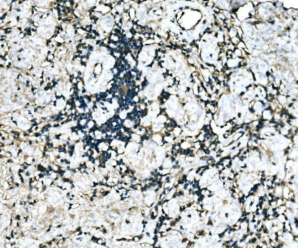
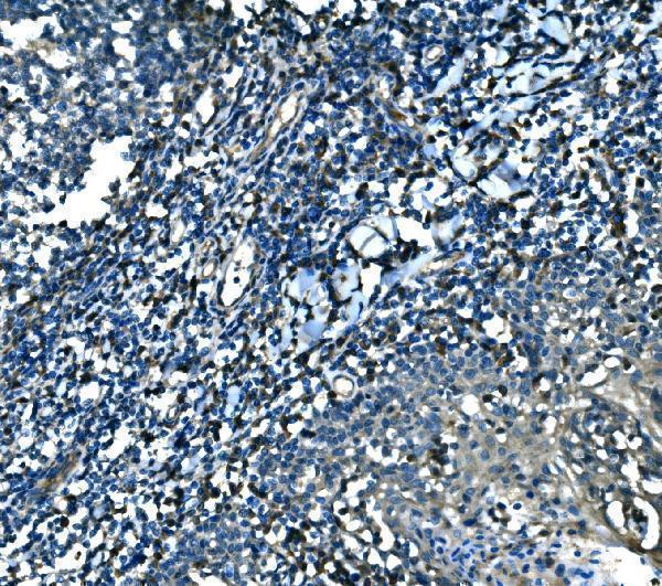
| Sample | Paraffin-embedded human hodgkin lymphoma tissue; fixative not specified (datasheet A05743-1) |
| Fixation | Image fixative and duration unreported (datasheet A05743-1); verify before use. |
| Sectioning | 4–5 µm sections on charged slides (standard) |
| Deparaffinisation | Xylene, graded ethanol series to water (standard) |
| Antigen retrieval | Heat retrieval: EDTA pH 8.0 (datasheet A05743-1); 20 min, 95–100 °C (standard) |
| Peroxidase block | 3% H2O2, 10 min, room temperature (standard) |
| Blocking | 10% goat serum (datasheet A05743-1) |
| Primary antibody | Rabbit anti-MEF2B, 2-5 μg/ml (datasheet A05743-1) |
| Primary incubation | Overnight at 4 °C (datasheet A05743-1) |
| Detection | Streptavidin-biotin complex (SABC), DAB chromogen (datasheet A05743-1) |
| Counterstain | Hematoxylin, blue, dehydrate and mount (standard) |
| Expected result | MEF2B-positive staining in germinal center cells of lymph node (HPA tissue IHC: High). HPA tissue profile: Selective nuclear expression in germinal center lymphocytes of tonsil and lymph node. No signal in the no-primary control. |
MEF2B is a nuclear transcription factor with no transmembrane segment (UniProt Q02080: localisation, topology). In paraffin-section IHC, expect selective, high nuclear staining in germinal center cells of tonsil and lymph node (HPA: tissue IHC). HPA rates the tissue pattern Supported because staining agrees with RNA expression, while cautioning that the antibody targets proteins from more than one gene (HPA: tissue IHC reliability).
| Strong, selective nuclear staining in tonsil or lymph-node germinal center cells. | This matches the reported high germinal center signal and nuclear pattern (HPA: tissue IHC). Score the cells and compartment together; a brown area alone does not establish the expected result (standard IHC interpretation). |
| Predominantly cytoplasmic or membrane-like staining in the expected germinal center cells. | This does not match the tissue IHC nuclear pattern or UniProt nuclear localisation (HPA: tissue IHC; UniProt Q02080: localisation). Check whether counterstain, pigment or detection background is being mistaken for specific signal (standard IHC practice). |
| Strong staining in cells reported as unstained, such as adipocytes or bone-marrow hematopoietic cells. | HPA reports MEF2B as not detected in those cell types (HPA: tissue IHC). Consider antibody cross-reactivity, consistent with HPA's multi-gene caution, or endogenous detection activity before calling the signal MEF2B (HPA: tissue IHC caution; standard IHC practice). |
| Diffuse colour over many cell types, extracellular areas or the whole section. | A diffuse field obscures the selective nuclear pattern reported for germinal centers (HPA: tissue IHC). Review blocking, washes and detection controls for general background; the appearance alone cannot identify its cause (standard IHC practice). |
| No convincing nuclear signal in a tonsil or lymph-node germinal center. | These are the supplied high-staining reference sites (HPA: tissue IHC). First confirm that the germinal center is present, then check antibody and detection controls; an absent signal alone cannot distinguish a technical failure from a sample difference (standard IHC practice). |
| Tissue and cell selection | Tonsil and lymph-node germinal center cells provide reported high signal; selected adipocytes and bone-marrow hematopoietic cells are not detected (HPA: tissue IHC). Interpret each cell population separately. |
| Compartment | UniProt places MEF2B in the nucleus, and tissue IHC reports selective nuclear staining (UniProt Q02080: localisation; HPA: tissue IHC). The absence of a transmembrane segment offers no basis for an expected membrane pattern (UniProt Q02080: topology). |
| Antibody evidence | The listed antibody HPA004734 has Supported IHC status; HPA also warns that its tissue evidence targets proteins from more than one gene (HPA: antibody validation; HPA: tissue IHC reliability). Treat unexpected staining as needing independent support. |
| Isoforms and processing | UniProt lists 2 isoforms and a single 1–365 chain, with no signal peptide or propeptide (UniProt Q02080: isoforms, processing). These facts alone do not predict a different IHC compartment or tissue pattern. |
| Situation | Likely cause | Next action |
|---|---|---|
| Germinal centers look negative while the rest of the slide develops colour. | The supplied positive reference is high nuclear staining in these cells, so this result is unresolved (HPA: tissue IHC). | Verify germinal center identity, inspect a concurrent positive control, and review antibody dilution and detection performance (standard IHC practice). |
| Many unrelated cells show nuclear signal. | This exceeds the selective tissue pattern; HPA cautions that the antibody targets proteins from more than one gene (HPA: tissue IHC). | Compare cell types with HPA's reported positives and negatives, then seek an independent antibody or orthogonal check before assigning identity (HPA: tissue IHC; standard IHC practice). |
| Staining is mainly cytoplasmic or outlines cell borders in paraffin IHC. | That differs from the reported nuclear tissue IHC pattern and UniProt localisation (HPA: tissue IHC; UniProt Q02080: localisation). | Check nuclear counterstain alignment and detection controls; record the discordance rather than scoring that colour as the expected IHC positive (standard IHC practice). |
| Background makes nuclear scoring difficult. | Diffuse detection colour can conceal selective nuclear staining (standard IHC practice; HPA: tissue IHC pattern). | Review blocking, wash steps, detection exposure and a negative detection control, then score only clearly localised nuclear signal (standard IHC practice). |
| Colour persists where primary antibody was omitted. | A primary-independent signal indicates background from the detection workflow, including possible endogenous activity (standard IHC practice). | Address the detection control first, then reassess whether germinal center nuclei retain selective staining (standard IHC practice; HPA: tissue IHC pattern). |
| Q: Why does an IF/ICC image show cytosol or cell junctions? | A: HPA approves nucleoplasm and cytosol as main IF/ICC locations and cell junctions as an additional location, with a multi-gene antibody caution (HPA: subcellular ICC-IF). | Interpret that IF/ICC observation in its own assay context; use the selective nuclear germinal center pattern to judge tissue IHC (HPA: subcellular ICC-IF; HPA: tissue IHC). |


Comprehensive Human Protein Atlas IHC scoring per tissue (reliability: Supported — High consistency between antibody staining and RNA expression data. Caution, targets protein from more than one gene.). Rows are taken directly from the HPA tissue chart — click any row's HPA link to view the source.
| Tissue | Cell type | Level | Evidence | Source |
|---|---|---|---|---|
| Lymph node | Germinal center cells | High | Protein (IHC) | HPA → |
| Tonsil | Germinal center cells | High | Protein (IHC) | HPA → |
| Tissue | Cell type | Level | Evidence | Source |
|---|---|---|---|---|
| Adipose tissue | Adipocytes | Not detected | Protein (IHC) | HPA → |
| Adrenal gland | Glandular cells | Not detected | Protein (IHC) | HPA → |
| Appendix | Glandular cells | Not detected | Protein (IHC) | HPA → |
| Bone marrow | Hematopoietic cells | Not detected | Protein (IHC) | HPA → |
| Breast | Adipocytes | Not detected | Protein (IHC) | HPA → |
Troubleshoot MEF2B staining in paraffin-section chromogenic IHC using the catalog antibody’s tissue example and the reported cellular expression patterns.
A05743-1 has IHC images from human paraffin-embedded Hodgkin lymphoma and tonsil sections (A05743-1 image captions). The catalog lists human, mouse, and rat reactivity (catalog: A05743-1).
A05743-1 will render with its own IHC figure from a human paraffin-embedded Hodgkin lymphoma section (A05743-1 image caption). A second caption documents human tonsil IHC, while the catalog lists IHC use and human, mouse, and rat reactivity (A05743-1 tonsil image caption; catalog: A05743-1).
Which to pick: Choose A05743-1 for paraffin-section IHC; its captions document human Hodgkin lymphoma and tonsil sections, but do not report the fixative (A05743-1 image captions). No SKU in this payload is listed for IF/ICC (catalog: A05743-1 applications). For mouse or rat IHC, A05743-1 is the listed option, although its supplied IHC images show human tissue only (catalog: A05743-1 reactivity and applications; A05743-1 image captions).