This website uses cookies to ensure you get the best experience on our website.


- Table of Contents
Plan MEGF8 paraffin IHC around the reported cytoplasmic tissue pattern (HPA tissue IHC). Compare staining with membrane topology (UniProt), and check the antibody’s epitope and documented IHC dilution before scoring (UniProt; datasheet).

Expected localisation, validated protocols, controls and antibodies — the at-a-glance facts below, then the full design guide.
| Expected localisation | Cytoplasmic tissue staining (HPA tissue IHC) | |
| Staining pattern | Cytoplasmic staining in several tissue cell types (HPA tissue IHC) | |
| Antigen retrieval | Citrate pH 6.0 HIER, 95–98 °C, 20 min (rule: cytoplasmic / membrane antigen) | |
| Positive control | Adrenal gland+4 more · see all | |
| Negative control | Adipose tissue+4 more · see all |
| Fixation | Keep fixation consistent across paraffin sections (standard IHC practice; not target-specific) | |
| Caveat | Splice/transcript discrepancy may complicate interpretation (HPA tissue IHC) | |
| Regulation | No specific expression regulator annotated (UniProt) | |
| Isoform / epitope | 2 isoforms; check whether the epitope is extracellular or cytoplasmic (UniProt) |
The catalog antibody’s IHC-P protocol is paired with one published mouse brain cryosection IHC method (PMC10778434).
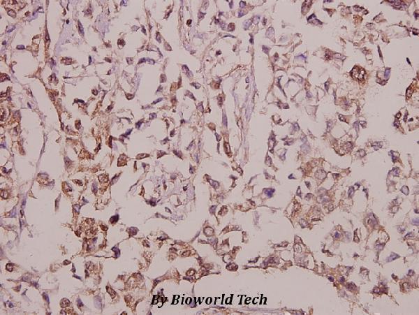
| Sample | Paraffin-embedded human breast carcinoma tissue; fixative not specified (datasheet A12840-1) |
| Fixation | Image fixative and duration unreported (datasheet A12840-1); verify before use. |
| Sectioning | 4–5 µm sections on charged slides (standard) |
| Deparaffinisation | Xylene, graded ethanol series to water (standard) |
| Antigen retrieval | Heat-induced epitope retrieval in citrate buffer, pH 6.0, 20 min at 95–98 °C (standard rule: cytoplasmic / membrane antigen) |
| Peroxidase block | 3% H2O2, 10 min, room temperature (standard) |
| Blocking | 10% normal serum of the secondary host, 30 min, room temperature (standard) |
| Primary antibody | Rabbit anti-MEGF8, 1:50-1:200 (datasheet A12840-1) |
| Primary incubation | Overnight at 4 °C (standard) |
| Detection | HRP-polymer secondary, DAB chromogen 5–10 min (standard) |
| Counterstain | Hematoxylin, blue, dehydrate and mount (standard) |
| Expected result | MEGF8-positive staining in glandular cells of adrenal gland (HPA tissue IHC: Medium). HPA tissue profile: Cytoplasmic expression in several different tissue types. No signal in the no-primary control. |
MEGF8 is a membrane protein with a large extracellular region and a cytoplasmic tail (UniProt Q7Z7M0 topology). In paraffin section IHC, expect cytoplasmic staining across several tissue types, including glandular cells, kidney tubule cells and cardiomyocytes (HPA tissue IHC: Approved; Medium). HPA flags a splice or transcript discrepancy, so assess the observed tissue pattern alongside the antibody’s validation status (HPA tissue IHC: reliability description).
| Moderate staining in colon or duodenal glandular cells, kidney tubule cells or cardiomyocytes. | These match HPA’s Medium IHC observations in the named cells (HPA tissue IHC). Compare staining within the relevant cell population; a positive result need not appear in every cell of a section (general IHC practice). |
| Predominantly crisp nuclear staining, with little staining in the expected cell population. | A nuclear-only pattern conflicts with HPA’s reported cytoplasmic tissue profile and UniProt’s membrane assignment (HPA tissue IHC; UniProt Q7Z7M0). Check counterstain, detection and primary-antibody controls before assigning it to MEGF8 (general IHC practice). |
| Strong staining in adipocytes or bone-marrow hematopoietic cells, while the expected positive cells are unstained. | HPA reports MEGF8 as Not detected in those cell populations (HPA tissue IHC). The discordance raises concern for cross-reactivity or endogenous detection activity; it does not prove either cause (general IHC practice). |
| Broad, even color over cells and surrounding section structures, with no cell-specific pattern. | This is difficult to score as MEGF8 because HPA describes a cellular staining pattern (HPA tissue IHC). Diffuse signal can reflect background from blocking, washes or detection chemistry (general IHC practice). |
| No staining in colon glandular cells or kidney tubule cells that are suitable for assessment. | Both are Medium HPA IHC examples, so an absent signal warrants a run check (HPA tissue IHC). Confirm section integrity, primary-antibody application, retrieval and detection with appropriate controls; HPA provides no MEGF8-specific retrieval condition (general IHC practice). |
| Compartment and topology | HPA describes cytoplasmic tissue staining, whereas UniProt assigns MEGF8 to the membrane, with residues 28–2647 extracellular and 2669–2845 cytoplasmic (HPA tissue IHC; UniProt Q7Z7M0 topology). These descriptions need not yield a sharply outlined membrane in chromogenic sections; do not infer the antibody’s epitope from them. |
| Antibody validation and transcript caveat | HPA labels tissue IHC Approved for HPA049248 and flags a splice or transcript discrepancy (HPA tissue IHC; HPA antibodies). Approved is the supplied status; independent-antibody reproduction or an Enhanced IHC designation is not established by this payload. |
| Isoforms and epitope knowledge | UniProt lists two MEGF8 isoforms, while the supplied antibody record gives no epitope position (UniProt Q7Z7M0; HPA antibodies). An isoform-specific staining prediction therefore cannot be made from this evidence. |
| Processing and glycosylation | UniProt lists a signal peptide at residues 1–27, a mature chain beginning at residue 28 and six glycosylation sites (UniProt Q7Z7M0). These annotations describe the protein; they do not establish shedding, an IHC staining difference or an antigen-retrieval requirement. |
| IF/ICC: what localisation is supported? | HPA summarizes MEGF8 as membrane-localized for ICC-IF but supplies no main-location assignment or cell-line images here (HPA subcellular). Treat that summary as context for IF/ICC interpretation; this IHC section supplies no IF/ICC protocol. |
| Situation | Likely cause | Next action |
|---|---|---|
| Expected positive cells show no chromogenic signal. | An IHC run or reagent problem is possible; HPA reports Medium staining in colon glands and kidney tubules (HPA tissue IHC). | Use a suitable HPA-positive tissue as a run control and verify primary-antibody application, retrieval and detection steps (HPA tissue IHC; general IHC practice). |
| The section is uniformly dark. | Background can arise from insufficient blocking or washing, or from endogenous detection activity (general IHC practice). | Review a no-primary control, blocking and wash steps; assess endogenous activity relevant to the detection chemistry (general IHC practice). |
| Only nuclei appear positive. | A nuclear-only result conflicts with the supplied cytoplasmic tissue profile and membrane annotation (HPA tissue IHC; UniProt Q7Z7M0). | Inspect counterstain and no-primary controls, then compare the expected cell populations before calling MEGF8 positive (general IHC practice; HPA tissue IHC). |
| Adipocytes or bone-marrow hematopoietic cells appear strongly positive. | HPA lists those cells as Not detected; cross-reactivity or endogenous activity is possible, but neither is proven (HPA tissue IHC; general IHC practice). | Compare an HPA Medium-positive cell population and detection controls on the same run; avoid treating the discordant color alone as MEGF8 (HPA tissue IHC; general IHC practice). |
| Staining varies between glandular samples. | HPA reports Medium staining in several glandular sites but Not detected staining in cervix, epididymis and fallopian-tube glands (HPA tissue IHC). | Identify the specific tissue and cell population before judging consistency; compare each result with its matching HPA entry (HPA tissue IHC). |
| The signal looks cytoplasmic rather than like a thin membrane rim. | That appearance can agree with HPA’s tissue IHC profile despite UniProt’s membrane topology (HPA tissue IHC; UniProt Q7Z7M0). | Score the expected cells and controls together; reserve a membrane-rim requirement until the antibody and assay establish one (HPA tissue IHC; general IHC practice). |
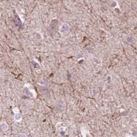


Comprehensive Human Protein Atlas IHC scoring per tissue (reliability: Approved — Caution, Splice and/or transcript discrepancy exists.). Rows are taken directly from the HPA tissue chart — click any row's HPA link to view the source.
| Tissue | Cell type | Level | Evidence | Source |
|---|---|---|---|---|
| Adrenal gland | Glandular cells | Medium | Protein (IHC) | HPA → |
| Cerebral cortex | Neuropil | Medium | Protein (IHC) | HPA → |
| Colon | Glandular cells | Medium | Protein (IHC) | HPA → |
| Duodenum | Glandular cells | Medium | Protein (IHC) | HPA → |
| Endometrium | Glandular cells | Medium | Protein (IHC) | HPA → |
| Tissue | Cell type | Level | Evidence | Source |
|---|---|---|---|---|
| Adipose tissue | Adipocytes | Not detected | Protein (IHC) | HPA → |
| Bone marrow | Hematopoietic cells | Not detected | Protein (IHC) | HPA → |
| Breast | Adipocytes | Not detected | Protein (IHC) | HPA → |
| Cervix | Glandular cells | Not detected | Protein (IHC) | HPA → |
| Epididymis | Glandular cells | Not detected | Protein (IHC) | HPA → |
Troubleshoot MEGF8 chromogenic IHC in paraffin sections using the page retrieval setting, the selected antibody image, and tissue and topology evidence.
For MEGF8, A12840-1 has an IHC image from paraffin-embedded human breast carcinoma (IHC image caption); A12840 lists IHC and IF for human, mouse and rat (catalog: A12840).
A12840-1 will render with its own IHC image of paraffin-embedded human breast carcinoma stained at 1:50 (A12840-1 IHC image caption). A12840 lists IHC and IF applications and human, mouse and rat reactivity, but has no IHC or IF image in the supplied catalog (catalog: A12840).
Which to pick: For tissue IHC, choose A12840-1 when its pictured paraffin-section example is relevant; the caption does not report the fixative (A12840-1 IHC image caption). For IF/ICC planning, choose A12840 because IF is listed at 1:50, though ICC-specific validation is unreported; A12840 is polyclonal, and A12840-1 is described as a pAb (catalog: A12840 applications and dilution; A12840-1 IHC image caption). For cross-species work, both list human, mouse and rat reactivity, while the supplied A12840-1 IHC image documents human tissue only (catalog: A12840 and A12840-1 reactivity; A12840-1 IHC image caption).