This website uses cookies to ensure you get the best experience on our website.


- Table of Contents
Plan MEP1B IHC in paraffin sections around the enterocyte brush-border pattern seen in gastrointestinal tissue (HPA tissue IHC). Use its membrane topology and potential surface shedding to interpret chromogenic staining (UniProt).

Expected localisation, validated protocols, controls and antibodies — the at-a-glance facts below, then the full design guide.
| Expected localisation | Enterocyte brush border (HPA tissue IHC) | |
| Staining pattern | Membranous staining of GI enterocyte microvilli (HPA tissue IHC) | |
| Antigen retrieval | EDTA pH 8.0 HIER, heat-mediated (datasheet A06392-1) | |
| Positive control | Colon+2 more · see all | |
| Negative control | Adipose tissue+4 more · see all |
| Fixation | Keep fixation consistent across sections (standard IHC practice; not target-specific). Selected-image fixative and duration unreported (datasheet A06392-1); verify before use. | |
| Caveat | Surface shedding may alter membrane staining (UniProt) | |
| Regulation | Expression regulation unreported (UniProt) | |
| Isoform / epitope | 0 isoforms listed; check extracellular vs cytoplasmic epitope (UniProt) |
The catalog antibody’s IHC-P protocol is paired with one published MEP1B uterine-tissue IHC protocol (PMC9990359).
| Sample | Paraffin-embedded mouse intestine tissue; fixative not specified (datasheet A06392-1) |
| Fixation | Image fixative and duration unreported (datasheet A06392-1); verify before use. |
| Sectioning | 4–5 µm sections on charged slides (standard) |
| Deparaffinisation | Xylene, graded ethanol series to water (standard) |
| Antigen retrieval | Heat retrieval: EDTA pH 8.0 (datasheet A06392-1); 20 min, 95–100 °C (standard) |
| Peroxidase block | 3% H2O2, 10 min, room temperature (standard) |
| Blocking | 10% goat serum (datasheet A06392-1) |
| Primary antibody | Rabbit anti-MEP1B, 2-5 μg/ml (datasheet A06392-1) |
| Primary incubation | Overnight at 4 °C (datasheet A06392-1) |
| Detection | HRP-conjugated secondary, DAB chromogen (datasheet A06392-1) |
| Counterstain | Hematoxylin, blue, dehydrate and mount (standard) |
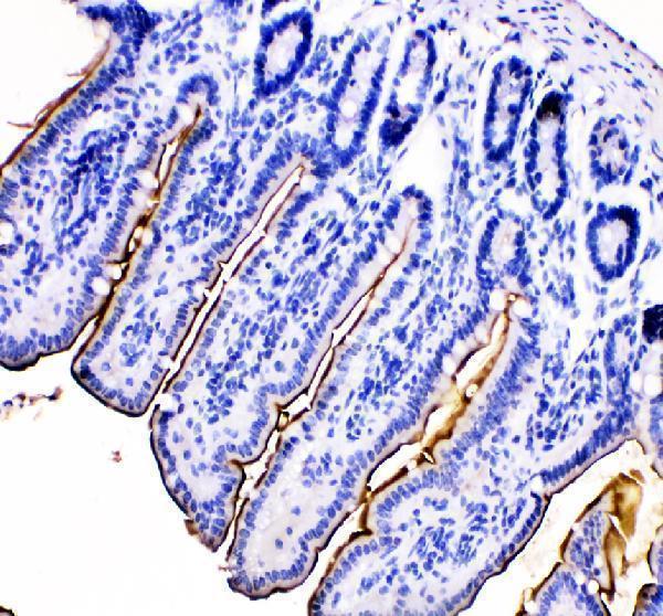
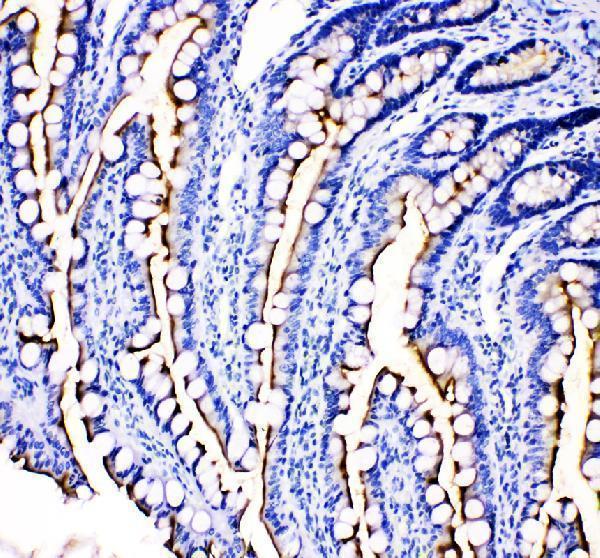
| Expected result | MEP1B-positive staining in enterocytes - Microvilli of colon (HPA tissue IHC: High). HPA tissue profile: Selective expression in brush border membrane in gastrointestinal tract. No signal in the no-primary control. |
MEP1B is a surface protein with an extracellular domain and can be shed (UniProt Q16820 topology). In paraffin IHC, expect strongest staining along enterocyte microvilli in colon, duodenum and small intestine (HPA tissue IHC: High). The selective brush border pattern has Enhanced tissue reliability, with high consistency between staining and RNA data (HPA tissue IHC).
| A crisp apical rim on intestinal enterocytes, strongest at the microvilli. | This matches the reported High microvillar staining in colon, duodenum and small intestine (HPA tissue IHC). Its surface position is consistent with the extracellular domain and membrane anchor (UniProt Q16820 topology). |
| Strong nuclear staining dominates, while the apical enterocyte rim is absent. | Nuclear localisation is unsupported by the reported membrane and secreted locations (UniProt Q16820; HPA subcellular summary). Treat a dominant nuclear pattern as suspect and check controls before assigning it to MEP1B (general IHC practice). |
| Widespread staining appears in cells outside the reported enterocyte pattern. | The HPA result is selective for gastrointestinal brush border (HPA tissue IHC). Unexpected staining may reflect antibody cross-reactivity or endogenous chromogenic activity; a negative reagent control helps separate these possibilities (general IHC practice). |
| Colour coats the section diffusely and obscures cell borders. | This cannot establish the selective microvillar pattern reported by HPA (HPA tissue IHC). Uneven reagent coverage, incomplete blocking or inadequate washing can contribute to diffuse background in chromogenic IHC (general IHC practice). |
| No apical signal appears in an otherwise intact duodenum or small intestine section. | These are reported High enterocyte microvillar sites (HPA tissue IHC). Review tissue orientation and the staining run before calling the sample negative; loss of the visible apical edge can prevent assessment (general IHC practice). |
| Tissue and cell selection | Colon, duodenum and small intestine have High enterocyte microvillar staining (HPA tissue IHC). HPA's Not detected entries describe the listed cell types, not every cell in each tissue (HPA tissue IHC). |
| Membrane topology and shedding | Residues 23–652 are extracellular and residues 653–673 span the membrane; surface homodimers may be shed (UniProt Q16820 topology). Extracellular signal therefore needs anatomical and control context (general IHC practice). |
| Antibody evidence | HPA lists HPA029119 as Enhanced for IHC, and tissue staining has Enhanced reliability (HPA antibodies; HPA tissue IHC). These ratings support the reported pattern but do not identify the cause of every unexpected signal (general IHC practice). |
| Retrieval and epitope | No antibody epitope or MEP1B-specific retrieval response is supplied (UniProt Q16820; HPA tissue IHC). If optimising retrieval, compare conditions against a known positive section and controls (general IHC practice). |
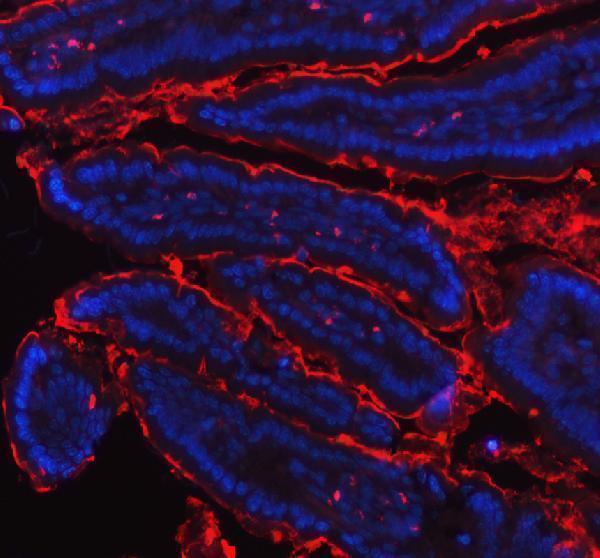
| What should IF/ICC show? | Membrane localisation and possible shedding inform interpretation (UniProt Q16820), but HPA supplies no ICC-IF images or main location assignment for MEP1B (HPA subcellular). An IF/ICC pattern is therefore unconfirmed by this evidence. |
| Situation | Likely cause | Next action |
|---|---|---|
| The expected intestinal apical rim is absent. | A staining-run failure or an apical border outside the plane of section may hide the reported High microvillar pattern (HPA tissue IHC; general IHC practice). | Inspect morphology and section orientation; repeat alongside a positive intestinal section and suitable reagent controls (general IHC practice). |
| The entire section develops a uniform brown haze. | Blocking, washing or chromogen development may be contributing background (general IHC practice). | Review blocking, washes and development time; compare a negative reagent control before interpreting faint apical colour (general IHC practice). |
| Strong colour appears in cells that lack a matching apical pattern. | Cross-reactivity or endogenous detection activity is possible (general IHC practice); HPA reports selective gastrointestinal brush border staining (HPA tissue IHC). | Compare cellular morphology, the positive tissue pattern and a negative reagent control; reassess antibody specificity if unexpected staining persists (general IHC practice). |
| Predominantly nuclear staining is scored as positive. | A nuclear pattern conflicts with the membrane and secreted locations in the supplied records (UniProt Q16820; HPA subcellular summary). | Withhold a positive call and inspect controls and the apical enterocyte compartment (general IHC practice; HPA tissue IHC). |
| Extracellular colour appears near an apical surface. | Surface MEP1B can be shed by ADAM10 or ADAM17 (UniProt Q16820), but adjacent colour alone cannot establish its identity (general IHC practice). | Assess whether a crisp enterocyte microvillar rim is also present, then compare controls before assigning extracellular signal to MEP1B (HPA tissue IHC; general IHC practice). |
| A reported HPA negative cell type stains weakly. | Not detected applies to the specified cell type and assay observation; it is not a tissue-wide exclusion (HPA tissue IHC). Weak chromogenic background is also possible (general IHC practice). | Identify the stained cell type, compare a negative reagent control and use the intestinal apical pattern as the positive reference (HPA tissue IHC; general IHC practice). |


Comprehensive Human Protein Atlas IHC scoring per tissue (reliability: Enhanced — High consistency between antibody staining and RNA expression data. At least one protein variant secreted, tissue location of RNA and protein might differ and correlation is complex.). Rows are taken directly from the HPA tissue chart — click any row's HPA link to view the source.
| Tissue | Cell type | Level | Evidence | Source |
|---|---|---|---|---|
| Colon | Enterocytes - Microvilli | High | Protein (IHC) | HPA → |
| Duodenum | Enterocytes - Microvilli | High | Protein (IHC) | HPA → |
| Small intestine | Enterocytes - Microvilli | High | Protein (IHC) | HPA → |
| Tissue | Cell type | Level | Evidence | Source |
|---|---|---|---|---|
| Adipose tissue | Adipocytes | Not detected | Protein (IHC) | HPA → |
| Adrenal gland | Glandular cells | Not detected | Protein (IHC) | HPA → |
| Appendix | Endocrine cells | Not detected | Protein (IHC) | HPA → |
| Bone marrow | Hematopoietic cells | Not detected | Protein (IHC) | HPA → |
| Breast | Adipocytes | Not detected | Protein (IHC) | HPA → |
Troubleshoot chromogenic MEP1B IHC in paraffin sections by checking retrieval, epithelial brush border staining, assay background, and compartment specific scoring.
A06392-1 has IHC images from paraffin-embedded mouse and rat intestine and IF images from paraffin-embedded mouse and rat colon; Human reactivity is listed (catalog IHC/IF image captions; catalog reactivity).
A06392-1 has IHC data from paraffin-embedded mouse and rat intestine (IHC image captions). The same SKU has IF data from paraffin-embedded mouse and rat colon and lists Human, Mouse, and Rat reactivity (IF image captions; catalog reactivity).
Which to pick: For tissue IHC, choose A06392-1: its rabbit primary was shown on paraffin-embedded mouse and rat intestine at 2 μg/ml after EDTA pH 8 retrieval; the fixative is unreported (IHC image captions). For IF, A06392-1 has mouse and rat colon images at 5 μg/ml; ICC validation and clonality are unreported (IF image captions; catalog applications and clone field). For cross-species studies, A06392-1 lists Human, Mouse, and Rat reactivity, while the supplied IHC/IF images show Mouse and Rat tissue (catalog reactivity; IHC/IF image captions).