MGAT3 / Beta-1,4-mannosyl-glycoprotein 4-beta-N-acetylglucosaminyltransferase · IHC design guide

Design Immunohistochemistry for MGAT3

Plan MGAT3 chromogenic IHC in paraffin sections using high staining in colon glandular cells and undetected staining in adipocytes as comparison tissues (HPA tissue IHC). This guide covers the catalog antibody’s 2–5 μg/ml IHC range (datasheet A08470-1) and how to interpret cytoplasmic staining (HPA tissue IHC) alongside the Golgi membrane location (UniProt).

Evidence assembled Oct 2026 · For research use; verify linked source records and product datasheet before use
Immunohistochemistry protocol sheet for MGAT3 (IHC for MGAT3): expected localisation Cytoplasmic staining (HPA tissue IHC); Golgi stack membrane expected (UniProt), antibody A08470-1, validated IHC image, and IHC protocol steps
Printable MGAT3 IHC protocol sheet — expected localisation Cytoplasmic staining (HPA tissue IHC); Golgi stack membrane expected (UniProt), antibody A08470-1, controls and protocol steps. Open the full MGAT3 IHC guide →

MGAT3 Immunohistochemistry Experimental Design Guide

Expected localisation, validated protocols, controls and antibodies — the at-a-glance facts below, then the full design guide.

Must know before staining
Expected localisation Cytoplasmic staining (HPA tissue IHC); Golgi stack membrane expected (UniProt)
Staining pattern Cytoplasmic staining in most tissues (HPA tissue IHC)
Antigen retrieval EDTA pH 8.0 HIER, heat-mediated (datasheet A08470-1)
Positive control ⓘ Adrenal gland+4 more · see all
Negative control ⓘ Adipose tissue+2 more · see all
Important caveats
Reasons your staining may differ from the expected pattern.
Fixation Keep paraffin-section fixation consistent. (standard IHC practice; not target-specific)
Caveat Staining has low consistency with RNA expression (HPA tissue IHC)
Regulation Expression regulation unreported (UniProt)
Isoform / epitope No isoforms; residues 1–7 cytosolic, 24–533 luminal (UniProt)
Section 1

Recommended MGAT3 IHC & IF Protocols

The catalog antibody’s IHC-P protocol (datasheet: A08470-1) is accompanied by published tissue IHC workflows from PMC7255015 and PMC11388069.

Recommended immunohistochemistry (IHC-P) protocol parameters
SampleParaffin-embedded human lung cancer tissue; fixative not specified (datasheet A08470-1)
FixationImage fixative and duration unreported (datasheet A08470-1); verify before use.
Sectioning4–5 µm sections on charged slides (standard)
DeparaffinisationXylene, graded ethanol series to water (standard)
Antigen retrievalHeat retrieval: EDTA pH 8.0 (datasheet A08470-1); 20 min, 95–100 °C (standard)
Peroxidase block3% H2O2, 10 min, room temperature (standard)
Blocking10% goat serum (datasheet A08470-1)
Primary antibodyRabbit anti-MGAT3, 2-5 μg/ml (datasheet A08470-1)
Primary incubationOvernight at 4 °C (datasheet A08470-1)
DetectionHRP-conjugated secondary, DAB chromogen (datasheet A08470-1)
CounterstainHematoxylin, blue, dehydrate and mount (standard)
Expected resultMGAT3-positive staining in glandular cells of adrenal gland (HPA tissue IHC: High). HPA tissue profile: Cytoplasmic expression in most tissues. No signal in the no-primary control.
💡Decision noteTry heat-mediated EDTA at pH 8.0 first (datasheet: A08470-1); citrate at pH 6.0 is a published breast cancer section alternative (PMC11388069).
Section 2

What Is the Expected MGAT3 Staining Pattern?

MGAT3 is a Golgi stack membrane enzyme with a short cytoplasmic tail and a lumenal catalytic region (UniProt Q09327 topology). In paraffin section IHC, expect cytoplasmic staining in many tissues, with high staining reported in selected glandular, respiratory epithelial, and cardiac cells (HPA: tissue IHC). Treat this as a provisional reference pattern: HPA rates its IHC staining “Approved” but reports low consistency with RNA expression and pending external verification (HPA: tissue IHC reliability).

What am I looking at on my slide?
Cytoplasmic stain in colon glandular cells, with a localized perinuclear accent where resolvable (HPA: High in colon glandular cells; UniProt Q09327: Golgi stacks).This fits the reported cell type and an intracellular Golgi location. A chromogenic section may show cytoplasmic staining without resolving individual Golgi stacks; do not require puncta to call a cell positive (HPA: tissue IHC profile; UniProt Q09327: Golgi stacks; general IHC practice).
Predominantly nuclear or sharply cell surface staining, with little cytoplasmic signal (UniProt Q09327: Golgi stack membrane).That distribution does not fit the annotated MGAT3 location. Recheck morphology, the counterstain, antibody specificity, and detection controls before interpreting the stain as MGAT3 (UniProt Q09327: Golgi stack membrane; general IHC practice).
Strong staining centered on adipocytes, liver cholangiocytes, or ovarian stromal cells (HPA: Not detected in these cell types).These cell specific results conflict with the reported IHC pattern. Consider cross reactivity or endogenous chromogenic detection activity, and inspect the corresponding control before assigning target expression; the HPA entry is pending external verification (HPA: tissue IHC reliability; general IHC practice).
Diffuse color across cells and surrounding tissue, including areas expected to have little signal (HPA: Not detected in adipocytes, cholangiocytes, and ovarian stromal cells).A loss of cell and compartment contrast favors background over a readable MGAT3 pattern. Review the negative reagent control, blocking, washes, and detection conditions as general IHC checks (HPA: tissue IHC profile; general IHC practice).
No detectable stain in a tested positive region, such as breast glandular cells or bronchial respiratory epithelium (HPA: High in these cell types).First check that the intended cell population is present and that a positive control developed. If it did, reassess the primary antibody and section workflow; a single negative slide does not establish absence of MGAT3 (HPA: tissue IHC; general IHC practice).
💡Expected MGAT3 appearanceCall a section positive when the relevant glandular or respiratory epithelial cells show clearly discernible cytoplasmic staining, potentially concentrated near the nucleus, while nuclear dominance or strong staining in HPA negative cell types prompts review (HPA: High in selected glandular and respiratory epithelial cells; HPA: Not detected in selected cell types; UniProt Q09327: Golgi stacks).
How each factor affects the staining
Cell population selected for scoring (HPA: tissue IHC).HPA reports High staining in colon and breast glandular cells and bronchial respiratory epithelial cells, but Not detected in adipocytes, cholangiocytes, and ovarian stromal cells. Score the named cells rather than assigning one result to an entire organ (HPA: tissue IHC).
Intracellular location and membrane topology (UniProt Q09327).MGAT3 has a transmembrane segment at residues 8–23, a cytoplasmic segment at 1–7, and a lumenal region at 24–533. Golgi localization supports a cytoplasmic compartment call in IHC; topology alone does not establish which epitope an antibody recognizes (UniProt Q09327 topology and location).
Evidence strength for the IHC reference pattern (HPA: tissue IHC reliability; HPA: HPA017598 validation).The listed antibody has IHC status Approved, while HPA reports low consistency between antibody staining and RNA expression and pending external verification. Use the pattern as an interpretation aid and resolve unexpected findings with controls (HPA: HPA017598 IHC Approved; HPA: tissue IHC reliability; general IHC practice).
Subcellular IF evidence (HPA: subcellular ICC-IF).HPA lists no main subcellular location and no cell lines with ICC-IF images for MGAT3. UniProt supplies the Golgi annotation, but the supplied HPA record cannot validate an IF image pattern or an IF protocol (HPA: subcellular ICC-IF; UniProt Q09327 location).
Target specific fixation sensitivity (HPA: tissue IHC; UniProt Q09327).Neither supplied record reports a target specific fixation effect for MGAT3. Choose and document antigen retrieval and detection conditions through the IHC workflow, without treating changes in staining as a documented MGAT3 fixation effect (HPA: tissue IHC; UniProt Q09327; general IHC practice).
Why is my staining missing, weak or wrong?
SituationLikely causeNext action
A named HPA High cell population has no stain (HPA: tissue IHC).The target cells may be absent from the section, or staining and detection may have failed; slide appearance alone cannot distinguish these explanations (general IHC practice).Confirm the cell population on morphology, compare a positive tissue section from the same run, and review primary antibody, retrieval, and detection records before calling the result negative (HPA: tissue IHC; general IHC practice).
Signal is mainly nuclear or outlines the cell surface (UniProt Q09327: Golgi stack membrane).The compartment conflicts with the annotated Golgi location and may reflect nonspecific staining or interpretation of the counterstain (UniProt Q09327 location; general IHC practice).Compare the primary omitted control and cell morphology, then assess whether any convincing cytoplasmic signal remains in an HPA reported positive cell type (HPA: tissue IHC; general IHC practice).
An HPA Not detected cell type stains strongly (HPA: adipocytes, cholangiocytes, or ovarian stromal cells).Cross reactivity or endogenous detection activity is possible; HPA's Approved pattern also has low RNA concordance, so a discrepancy is not resolved by the reference alone (HPA: tissue IHC reliability; general IHC practice).Check the primary omitted control and any detection specific endogenous activity control, and compare the stained cell population with a reported positive region (HPA: tissue IHC; general IHC practice).
Background obscures cell boundaries or the cytoplasmic pattern (HPA: tissue IHC profile).Nonspecific reagent binding, incomplete washing, or detection background can reduce contrast in chromogenic IHC (general IHC practice).Inspect negative reagent controls; review blocking, wash steps, and detection development, then reassess the same cell populations against the HPA reference pattern (HPA: tissue IHC; general IHC practice).
Staining varies between sampled tissues or cell populations (HPA: tissue IHC).The reported levels differ by cell type, and HPA flags low consistency with RNA expression; variation alone does not identify a technical failure (HPA: tissue IHC and reliability).Record the tissue and exact scored cell type, compare run controls, and interpret each observation against its own HPA entry rather than an organ wide expectation (HPA: tissue IHC; general IHC practice).
Can an IF/ICC image establish the expected MGAT3 pattern? (HPA: subcellular ICC-IF).HPA supplies no MGAT3 ICC-IF image bearing cell lines or main location in this record, although UniProt annotates Golgi stacks (HPA: subcellular ICC-IF; UniProt Q09327 location).Use the Golgi annotation as a localization hypothesis, and seek independent IF controls on the separate IF/ICC guide page; do not infer IF validation from this IHC pattern (UniProt Q09327 location; HPA: subcellular ICC-IF; general IF practice).

Sample controls for MGAT3 IHC & IF

🧪Run colon first: glandular cells should stain (HPA: High in colon glandular cells). Use ovarian stroma as the negative tissue (HPA: Not detected in ovarian stroma cells); on the colon slide, non-glandular cells should show little or no specific staining and serve as internal background comparators, without assuming they are MGAT3-negative.
Positive control tissue: Adrenal gland (Glandular cells, HPA High)
Negative control tissue: Adipose tissue (HPA Not detected)
ICC-IF cell lines (HPA subcellular resource): HPA carries no ICC-IF cell line for MGAT3; derive a cell-line control from the positive tissue's cell type (Glandular cells) and confirm it by RNA or western blot first.
Technical controls: Include no-primary (secondary-only) and host-matched rabbit IgG isotype controls (caption: rabbit anti-MGAT3 antibody). For biological specificity, use an MGAT3 knockout sample if available or immunizing-peptide competition if the peptide is known; quench endogenous peroxidase and check inflammatory cells separately when interpreting DAB staining in colon sections (standard IHC practice).
⚠️Feasibility: A target-specific fixation window is unreported, and the selected A08470-1 paraffin-section caption does not state a fixative. That caption uses heat-mediated EDTA retrieval at pH 8.0, but whether retrieval is required relative to other conditions is unreported (A08470-1 tissue-IHC caption). There is no supplied evidence that frozen sections or IF are easier, and HPA lists no ICC-IF images for MGAT3 (HPA subcellular payload); endogenous peroxidase in inflammatory cells can complicate colon DAB interpretation (standard IHC practice).

HPA tissue IHC evidence for MGAT3

Comprehensive Human Protein Atlas IHC scoring per tissue (reliability: Approved — Low consistency between antibody staining and RNA expression data. Pending external verification.). Rows are taken directly from the HPA tissue chart — click any row's HPA link to view the source.

Positive expression · recommended positive controls

TissueCell typeLevelEvidenceSource
Adrenal gland Glandular cells High Protein (IHC) HPA →
Breast Glandular cells High Protein (IHC) HPA →
Bronchus Respiratory epithelial cells High Protein (IHC) HPA →
Colon Glandular cells High Protein (IHC) HPA →
Gallbladder Glandular cells High Protein (IHC) HPA →

Undetected expression · recommended negative controls

TissueCell typeLevelEvidenceSource
Adipose tissue Adipocytes Not detected Protein (IHC) HPA →
Liver Cholangiocytes Not detected Protein (IHC) HPA →
Ovary Ovarian stroma cells Not detected Protein (IHC) HPA →
Section 3

Advanced MGAT3 IHC Tips

These questions focus on chromogenic MGAT3 staining in paraffin sections, using the catalog antibody’s tissue image and known Golgi localisation to guide troubleshooting.

How should I optimise retrieval when MGAT3 staining is weak in paraffin sections?
Start with heat mediated retrieval in EDTA at pH 8.0 for the catalog antibody (datasheet A08470-1). The selected tissue image used that retrieval, followed by 10% goat serum blocking and 2 μg/ml primary antibody overnight at 4°C (caption A08470-1). If staining remains weak, compare retrieval times on adjacent sections while holding antibody concentration and detection conditions constant, and check that tissue morphology remains intact (standard IHC practice). Score the expected cytoplasmic, preferably perinuclear, signal rather than treating every diffuse deposit as recovered antigen (HPA: cytoplasmic expression; UniProt Q09327: Golgi stack membrane).
Does fixation explain variable MGAT3 staining across paraffin sections?
The selected MGAT3 tissue caption identifies a paraffin section but does not report its fixative, so target specific fixation sensitivity is unknown (caption A08470-1). Record the fixative, fixation duration, tissue thickness and processing history for each specimen before comparing staining intensity (standard IHC practice). Process matched control and test tissue together, then apply the documented EDTA pH 8.0 retrieval and 2 μg/ml antibody conditions consistently (datasheet A08470-1; caption A08470-1). If signal varies, inspect morphology and compare adjacent sections before attributing the difference to MGAT3 biology; HPA staining patterns cannot establish a fixation effect (standard IHC practice; HPA: tissue IHC profile).
Where should a credible MGAT3 signal appear in tissue sections?
Expect staining within the cytoplasm, with a perinuclear Golgi pattern where cellular detail permits, because MGAT3 resides in the Golgi stack membrane (HPA: cytoplasmic expression; UniProt Q09327: Golgi stack membrane). Its 8–23 transmembrane segment anchors the protein, leaving residues 24–533 lumenal and residues 1–7 cytoplasmic (UniProt Q09327 topology). Use a high power view and a counterstain to distinguish a compact perinuclear signal from diffuse cytoplasmic chromogen (standard IHC practice). Strong nuclear only or continuous plasma membrane staining warrants review of controls and detection background before assigning it to MGAT3 (UniProt Q09327 localisation; standard IHC practice).
How does epitope position affect interpretation of this MGAT3 antibody?
MGAT3 has no annotated isoforms in the supplied record, so an isoform specific staining claim is unsupported here (UniProt Q09327 isoforms). Its short cytoplasmic tail spans residues 1–7, the membrane anchor spans 8–23, and the lumenal region spans 24–533 (UniProt Q09327 topology). Four annotated glycosylation sites lie at residues 141, 241, 259 and 397; these positions matter when evaluating a mapped epitope, but the antibody epitope is not supplied (UniProt Q09327 glycosylation; caption A08470-1). Compare retrieval conditions and appropriate controls empirically, and avoid assigning an epitope side or glycosylation dependent effect from staining alone (standard IHC practice).
How can IF help assess the MGAT3 pattern seen by chromogenic IHC?
Use IF on a matched specimen as an orthogonal localisation check while retaining the documented paraffin IHC conditions for the primary readout (caption A08470-1; standard IHC practice). For bronchial tissue, multiplex MGAT3 with an epithelial marker and assess signal within respiratory epithelial cells, a reported high staining population (HPA: bronchus respiratory epithelial cells, High). Choose spectrally separated fluorophores, favouring a red or far red MGAT3 channel if tissue autofluorescence obscures shorter wavelengths (standard IF practice). If the antibody epitope is lumenal, residues 24–533 require access across cellular membranes; optimise permeabilisation while preserving Golgi structure, but do not assume that epitope position is known (UniProt Q09327 topology; standard IF practice).
What should I check when MGAT3 DAB staining looks widespread?
First compare a no primary control with the stained section to identify secondary reagent, endogenous peroxidase or chromogen background (standard chromogenic IHC practice). The selected image used 10% goat serum blocking, a peroxidase linked secondary antibody and DAB development; it does not establish that every brown deposit is MGAT3 (caption A08470-1). Include an appropriate peroxidase block and check whether excessive DAB development or primary concentration obscures cellular boundaries (standard chromogenic IHC practice). Interpret broad staining cautiously because HPA reports cytoplasmic expression in most tissues yet notes low agreement with RNA data and pending external verification (HPA: tissue IHC reliability and profile).
How should I score MGAT3 staining across tissue samples? ⚠ ANSWER MARKED FOR VERIFICATION
Define the eligible cell population and compartment before scoring, then report the percentage of positive cells and an intensity weighted H-score for cytoplasmic staining (standard IHC scoring practice; HPA: cytoplasmic expression). Keep section area, counterstain, exposure to DAB and scoring thresholds consistent across the comparison (standard chromogenic IHC practice). Normalise counts to the number of eligible cells, or positive cell density to evaluable tissue area in mm², while excluding folds, edges and necrosis (standard IHC scoring practice). Stratify by cell type because bronchial respiratory epithelial cells are reported High whereas lung macrophages are Low, and avoid treating mixed cell averages as one expression level (HPA: bronchus and lung tissue IHC).
How can I distinguish genuine MGAT3 staining from tissue artefact?
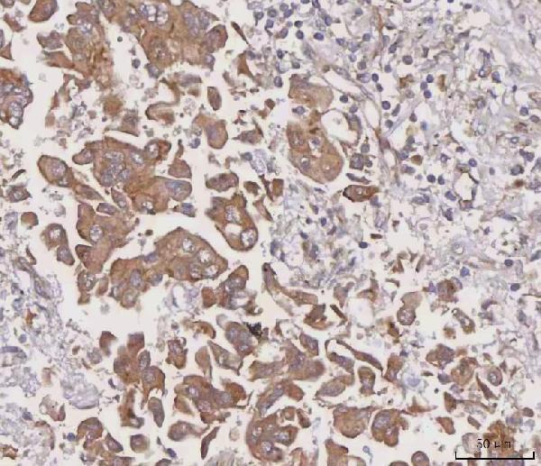
Give greatest weight to cytoplasmic, potentially perinuclear staining in intact cells, consistent with MGAT3 localisation to Golgi stack membranes (HPA: cytoplasmic expression; UniProt Q09327 localisation). Check whether positive cells match the sampled tissue: bronchial respiratory epithelial cells are reported High, while lung macrophages are reported Low (HPA: bronchus and lung tissue IHC). Treat edge staining, necrotic deposits, nuclear only signal and staining reproduced in a no primary control as reasons to investigate artefact or endogenous peroxidase (UniProt Q09327 localisation; standard chromogenic IHC practice). The catalog image demonstrates detection in paraffin embedded human lung cancer tissue, but HPA flags low RNA agreement and pending external verification, so interpret any new pattern with controls (caption A08470-1; HPA: reliability).
Boster reagents

Best MGAT3 / Beta-1,4-mannosyl-glycoprotein 4-beta-N-acetylglucosaminyltransferase IHC Antibodies

The catalog antibody has human IHC validation in a paraffin-embedded lung cancer section (A08470-1 image caption); no IF image is supplied (catalog payload).

Real IHC data IHC analysis of MGAT3 using anti-MGAT3 antibody (A08470-1). MGAT3 was detected in a paraffin-embedded section of human lung cancer tissue. Heat mediated antigen retrieval was performed in EDTA buffer (pH 8.0, epitope retrieval solution). The tissue section was blocked with 10% goat serum. The tissue section was then incubated with 2 μg/ml rabbit anti-MGAT3 Antibody (A08470-1) overnight at 4°C. Peroxidase Conjugated Goat Anti-rabbit IgG was used as secondary antibody and incubated for 30 minutes at 37°C. The tissue section was developed using HRP Conjugated Rabbit IgG Super Vision Assay Kit (Catalog # SV0002) with DAB as the chromogen.
Anti-MGAT3 Antibody ®
Cat # A08470-1

A08470-1 is listed for human IHC (catalog applications and reactivity). Its IHC image shows a paraffin-embedded human lung cancer section (A08470-1 image caption); the fixative is unreported (A08470-1 image caption).

Which to pick: Choose A08470-1 for human paraffin-section IHC: it is a rabbit polyclonal antibody listed for IHC, with an image from that sample type (catalog payload; A08470-1 image caption). No SKU is listed for IF/ICC or cross-species use (catalog applications and reactivity).

Each figure is that product's own IHC / IF validation image from its datasheet.

References

  1. UniProt Consortium. UniProt entry Q09327 (MGAT3_HUMAN, Beta-1,4-mannosyl-glycoprotein 4-beta-N-acetylglucosaminyltransferase).
  2. Human Protein Atlas. MGAT3 tissue IHC expression (reliability: Approved).
  3. Human Protein Atlas. MGAT3 subcellular location (ICC-IF): Highest expression in OE19: 62.6 nTPM.
  4. Human Protein Atlas. MGAT3 antibody validation summary (1 antibodies).
  5. Discovery of specific metastasis-related N-glycan alterations in epithelial ovarian cancer based on quantitative glycomics. PloS one 2014 — PMC3916363.
  6. Global analysis of human glycosyltransferases reveals novel targets for pancreatic cancer pathogenesis. British journal of cancer 2020 — PMC7251111.
  7. MGAT3-mediated glycosylation of tetraspanin CD82 at asparagine 157 suppresses ovarian cancer metastasis by inhibiting the integrin signaling pathway. Theranostics 2020 — PMC7255015.
  8. Bisecting GlcNAc modification reverses the chemoresistance via attenuating the function of P-gp. Theranostics 2024 — PMC11388069.
  9. PubMed PMID:8370666 — UniProt-cited evidence.
  10. PubMed PMID:15461802 — UniProt-cited evidence.
  11. PubMed PMID:10591208 — UniProt-cited evidence.