This website uses cookies to ensure you get the best experience on our website.
- Table of Contents
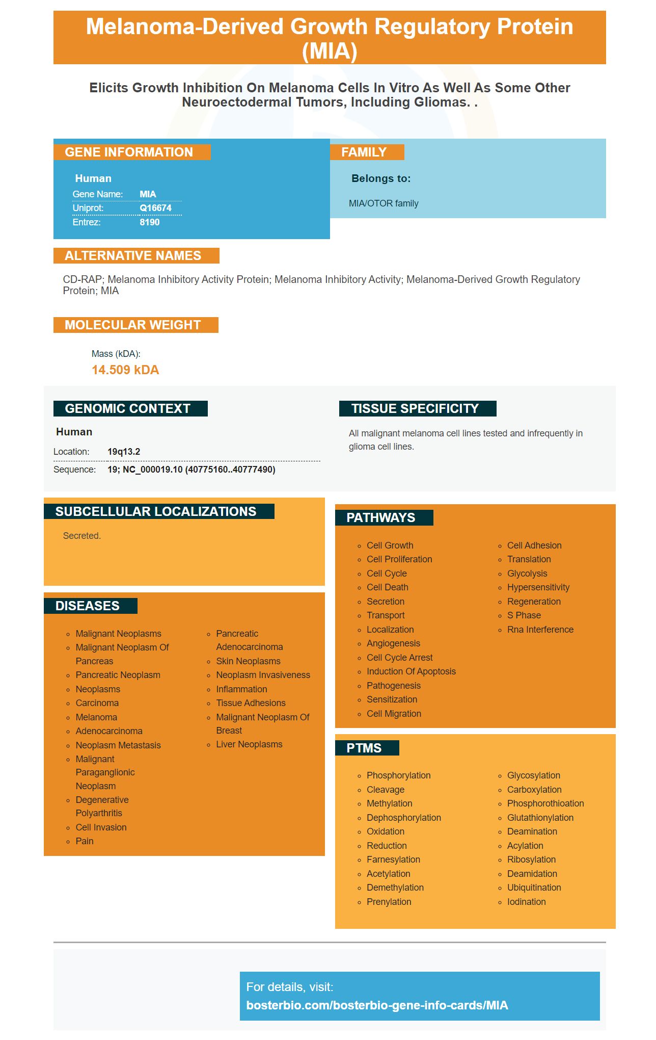
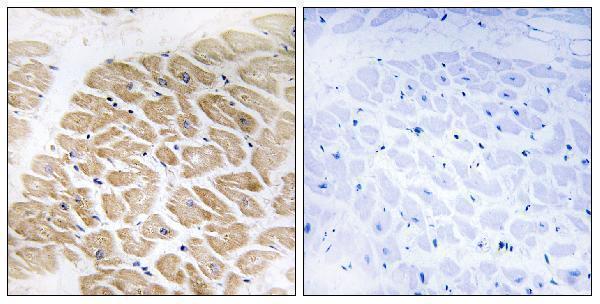
Facts about Melanoma-derived growth regulatory protein.
Human | |
---|---|
Gene Name: | MIA |
Uniprot: | Q16674 |
Entrez: | 8190 |
Belongs to: |
---|
MIA/OTOR family |
CD-RAP; Melanoma inhibitory activity protein; melanoma inhibitory activity; melanoma-derived growth regulatory protein; MIA
Mass (kDA):
14.509 kDA
Human | |
---|---|
Location: | 19q13.2 |
Sequence: | 19; NC_000019.10 (40775160..40777490) |
All malignant melanoma cell lines tested and infrequently in glioma cell lines.
Secreted.
There are many uses for the MIA marker. This article will discuss how the MIA marker is used to study microglia and synaptogenesis, spine interactions, and morphology. It will also explain the crucial role of brain IB cells. Continue reading to learn more! This Boster Biography article will answer your questions regarding this new marker and its best applications.
The uninjured brain side plays an important role in synaptogenesis. The uninjured side of the brain had previously been used to internalize drug effects. It has now been demonstrated that drug manipulations could affect the normal synaptogenesis for the MIA marker. Synaptogenesis, which is the process in which the brain creates new connections between neurons to allow these cells to communicate with one other, is what we call synaptogenesis.
The MIA genes in offspring induce an increased level of TNFa and TNFR1 during synaptogenesis. These two molecules were significantly expressed more in P14-MIA offspring than the controls. The cerebellum of MIA offspring showed an increase in TNFa levels. Both groups had statistically significant values. TNFa levels and MIP-1g expression were upregulated, whereas ICAM-1 IL-10 expression was lower.
To identify the synaptophysin immunoreactivity, we used a Nikon Eclipse 90i microscope to visualize the barrelette region of the PrV. We used a 40-mm objective and a 1,310,720-pixel-per-inch density to acquire the images. The NIS elements software was used to analyze and quantify synaptophysin-puncta density using thresholds.
These findings indicate that ASD and SZ are influenced by synaptic proteins. These proteins are thought to play a significant role in the behavioral problems seen in MIA-exposed offspring. These alterations could have an effect on peripheral immune cells and the central nervous system. MIA could affect the neurons' ability to learn new functions.
Microglial cells' morphological characteristics are classified according to their size. Microglia that have a small size soma can be considered rod-like. These cells have a positive correlation with their size in terms of the area of the cell and its soma. Ramified cells have smaller cell perimeters and a smaller soma. The length of the skeleton correlates with their size.
Female microglia display more homoeostatic genes, but not as many as their male counterparts. Interestingly, APP/PS1 mice display a reduced shift towards glycolysis. This is consistent with their decreased neuroprotective capacity and greater activation in neurodegenerative conditions. Therefore, there is likely to be some genetic basis for the sex-related differences in microglial morphology.
The BV2 microglia cell line is widely used by researchers as a model for studying microglial activation. Unfortunately, this cell line does not yield high-quality microglia due to the time-consuming process. Microglia made from BV2 cell lines are not very pure and have low viability. Researchers have used BV2 Microglia in many applications, but their low yield makes them unsuitable.
The findings of this study demonstrate that microglia in male and female mice show preferential activation of a group of genes that promote apoptosis. These mice also have higher levels for a gene associated to microglial activation. These results are consistent to those of postmortem AD victims. Whether or not these findings are representative of sex-based AD models remains to be determined.
The neurons found in the brains and bodies of animals, microglia (or microglia), show sex-related variations in metabolism, function, morphology, and morphology. Also, their morphology was and is different in female mice from male mice. We are currently trying understand the biological causes of these differences. This article describes our findings. This article will help you to understand the differences in microglial branching, metabolism, and behavior between male and female mice.
We analyzed the morphological features of microglia using 3D surface plot reconstructions. We found that male WT mice with the APP/PS1 genes had significantly more microglia than their female counterparts. This was due to a larger cellular area and perimeter. However, we found significant decreases in microglial branching morphology in female mice. The changes were most evident in female microglia.
We used a microscope to take images of the morphological change of microglia. We used an oil-immersion 63x lens and open-lab software. Digital zooming into the microscope was used to take time-lapse photos. Each image was recorded with a 50x magnification. We also focused through cranial open to obtain Z/time-lapse images. To align the time-lapse/Z-stack images, we used the ImageJ plugin StackReg to align each Z-stack. We manually counted the neuronal components in each image.
The expression of genes that are associated with the sensory pathway changes as we age. Older adults tend to have higher levels of genes associated the sensome, and lower levels of genes involved in detecting ligands endogenous to their bodies. The latter changes in the brain are thought to play a role in neurodegeneration. Three types of microglia have been identified that exhibit a similar trend.
The microglial-spine interaction in Bostern Bio was measured by comparing the measured co-localization of the two cell types to the Offset image calculated by horizontally translating the stack of images by half the number of pixels. This resulted is a biased calculation of the microglial–spine interaction. The authors could improve their analysis by comparing co-localization of microglia with that of other colocalizing objects.
The interactions between neurons, microglia, and neurons might be at a supraspinal degree. In this study, glial inhibitors attenuated behavioral hypersensitivity caused by hindpaw inflammation in mice. Additionally, carrageenan-induced hindpaw inflammation induced an increase in immunolabeling of astrocytes and microglia in the RVM, a region that has been linked to inflammatory pain. CX3CR1 knockout mice had decreased neuropathic and inflammatory nociceptive response.
Microglial activity in development is affected by the nutrient regulation environment of the mother's milk. Microglia need an n-3 PUFA environment to function properly in the developing brain. Therefore, dietary n-3 PUFAs affect the neurodevelopmental trajectory of the offspring as well as the microglial-spine interaction. In addition to microglial activity, maternal n-3PUFAs influence the development the offspring’s spine.
The microglial-spine interaction in BoSTER Bio is a novel finding that could explain the underlying mechanisms of the interaction between the spine and the microglia. This finding identifies a novel role for ePS in instructing microglia during critical stages in development to engage in engulfment. This is an important discovery that highlights how important it is to understand the interactions between microglia, spines, and human development.
Multiple studies have shown that curcumin has antiinflammatory activity and inhibits the microglia over-reaction. Additionally, curcumin reduces migration and phagocytosis. The MIA marker is a therapeutic tool for neurodegenerative and inflammation, and may be used as an anti-inflammatory drug. Curcumin may also prove useful in diagnosing and treating neurological diseases, such as Alzheimer's.
Vacciniumlucidum has been used in Asia for many years. Its polysaccharides have been described as having the ability to regulate the immune response, suppress tumour growth, and modulate cell proliferation. Despite its wide application, Ganodermalucidum is still not understood to play a role in microglial synchronogenesis. However, it's been demonstrated that it can inhibit the production of neurotransmitters, such as nitric oxygen.
PMID: 7923218 by Blesch A., et al. Cloning of a novel malignant melanoma-derived growth-regulatory protein, MIA.
PMID: 8550608 by Bosserhoff A.-K., et al. Structure and promoter analysis of the gene encoding the human melanoma-inhibiting protein MIA.