MICALL2 / MICAL-like protein 2 · IHC design guide

Design Immunohistochemistry for MICALL2

Plan MICALL2 IHC in paraffin sections using glandular tissue staining as a reference (HPA tissue IHC). This guide covers fixation consistency, expected cytoplasmic and membranous signal, and interpretation across tissues (HPA tissue IHC).

Evidence assembled Oct 2026 · For research use; verify linked source records and product datasheet before use
Immunohistochemistry protocol sheet for MICALL2 (IHC for MICALL2): expected localisation Cytoplasmic and membranous staining (HPA tissue IHC), antibody A12852-1, validated IHC image, and IHC protocol steps
Printable MICALL2 IHC protocol sheet — expected localisation Cytoplasmic and membranous staining (HPA tissue IHC), antibody A12852-1, controls and protocol steps. Open the full MICALL2 IHC guide →

MICALL2 Immunohistochemistry Experimental Design Guide

Expected localisation, validated protocols, controls and antibodies — the at-a-glance facts below, then the full design guide.

Must know before staining
Expected localisation Cytoplasmic and membranous staining (HPA tissue IHC)
Staining pattern Glandular cells: cytoplasmic and membranous signal (HPA tissue IHC)
Antigen retrieval EDTA pH 8.0 HIER, heat-mediated (datasheet A12852-1)
Positive control ⓘ Colon+4 more · see all
Negative control ⓘ Bone marrow+4 more · see all
Important caveats
Reasons your staining may differ from the expected pattern.
Fixation Matched tissue-IHC evidence does not establish the fixation claim. Validate the specimen-specific method before use. (selected-SKU IHC image A12852-1)
Caveat Prostate glandular cells show no staining (HPA tissue IHC)
Regulation Low tissue specificity by RNA (HPA tissue RNA)
Isoform / epitope 5 isoforms; check epitope coverage across variants (UniProt)
Section 1

Recommended MICALL2 IHC & IF Protocols

The catalog antibody protocol uses EDTA pH 8.0 retrieval (datasheet: A12852-1). The published IHC protocols below cover ovarian, kidney, and colorectal cancer tissue (PMC10778810; PMC8899381; PMC9615392).

Recommended immunohistochemistry (IHC-P) protocol parameters
SampleParaffin-embedded human colorectal adenocarcinoma tissue; fixative not specified (datasheet A12852-1)
FixationImage fixative and duration unreported (datasheet A12852-1); verify before use.
Sectioning4–5 µm sections on charged slides (standard)
DeparaffinisationXylene, graded ethanol series to water (standard)
Antigen retrievalHeat retrieval: EDTA pH 8.0 (datasheet A12852-1); 20 min, 95–100 °C (standard)
Peroxidase block3% H2O2, 10 min, room temperature (standard)
Blocking10% goat serum (datasheet A12852-1)
Primary antibodyRabbit anti-MICALL2, 2-5 μg/ml (datasheet A12852-1)
Primary incubationOvernight at 4 °C (datasheet A12852-1)
DetectionHRP-conjugated secondary, DAB chromogen (datasheet A12852-1)
CounterstainHematoxylin, blue, dehydrate and mount (standard)
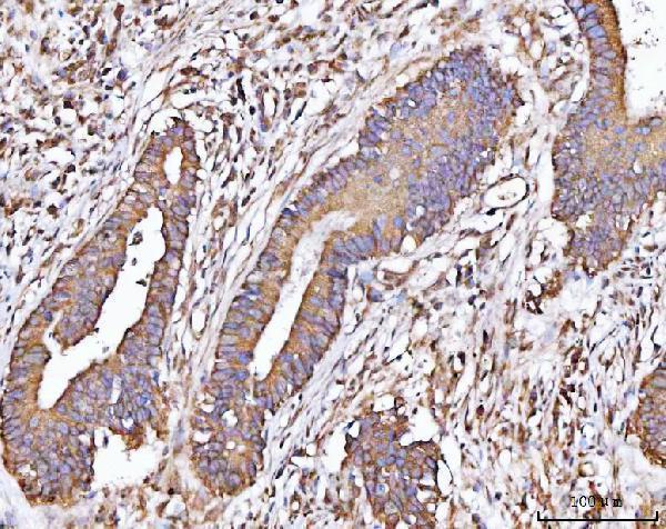
Expected resultMICALL2-positive staining in glandular cells of colon (HPA tissue IHC: High). HPA tissue profile: Cytoplasmic and membranous expression in several tissues. No signal in the no-primary control.
💡Decision noteStart with heat-mediated EDTA pH 8.0 retrieval (datasheet: A12852-1); optimize against paired tumor and adjacent tissue where available (PMC10778810; PMC8899381; PMC9615392).
Section 2

What Is the Expected MICALL2 Staining Pattern?

MICALL2 should appear chiefly in cytoplasm and along cell borders in paraffin-section IHC (HPA: cytoplasmic and membranous expression; UniProt Q8IY33: cell membrane, junctions and cytoskeleton). Colon, duodenum and small-intestine glandular cells are strong reference sites (HPA: High). HPA rates tissue IHC reliability Enhanced, while reporting medium consistency between antibody staining and RNA expression (HPA: reliability description). MICALL2 has no transmembrane segment (UniProt Q8IY33 topology).

What am I looking at on my slide?
Cytoplasmic stain with cell-border accentuation in intestinal glands.This fits the reported compartment pattern and High staining in colon, duodenum and small-intestine glandular cells (HPA: tissue IHC). Compare cells within the same section before judging intensity: a visible border component is plausible, but every positive cell need not show an equally sharp junctional line (UniProt Q8IY33: junction and cytoskeleton locations).
Predominantly nuclear stain, with little cytoplasmic or border signal.Treat a nucleus-only pattern as suspect because the listed locations are membrane, junction, endosome, projection and cytoskeleton (UniProt Q8IY33 subcellular location). Check counterstain overlap and antibody-omission controls before assigning MICALL2 positivity; an unexpected compartment alone does not identify the source of the signal (general IHC interpretation).
Strong staining in a cell population listed as undetected.Hematopoietic cells in bone marrow and prostate glandular cells are reported Not detected (HPA: tissue IHC). Persistent staining there warrants review for cross-reactivity or endogenous chromogen activity (general IHC practice). HPA categories describe observed staining, so they should guide comparison rather than serve as absolute proof that every specimen is negative.
Uniform color across cells, stroma and spaces between cells.A widespread haze does not match the reported cytoplasmic and membranous cellular pattern (HPA: tissue IHC). Review antibody-omission and detection-only controls for nonspecific background or endogenous enzyme activity; assess cellular staining only after background is controlled (general chromogenic IHC practice).
No detectable stain in colon or duodenal glandular cells.Both are High reference populations (HPA: tissue IHC), making an entirely blank run a reason to check section quality, reagent performance and detection controls (general IHC practice). A blank result does not by itself establish biological absence; compare another documented positive section under the same conditions.
💡Expected MICALL2 appearanceA convincing positive is High cytoplasmic staining with plausible cell-border staining in colon, duodenal or small-intestine glandular cells (HPA: tissue IHC; UniProt Q8IY33: membrane and junction locations); nucleus-only color or uniform background is suspect (general IHC interpretation).
How each factor affects the staining
Cellular location and topologyMICALL2 is listed at cell membrane, tight junction, recycling endosome, projection and cytoskeleton, with no transmembrane segment (UniProt Q8IY33). Cytoplasmic signal can therefore coexist with border staining; the record does not require a continuous membrane outline in every cell.
Choice of comparison tissueColon, duodenum and small-intestine glandular cells are High; skin keratinocytes are Medium, while prostate glandular and bone-marrow hematopoietic cells are Not detected (HPA: tissue IHC). Score the specified cell population, since a whole-section impression can obscure the relevant comparison.
Strength of the IHC evidenceThe tissue profile is cytoplasmic and membranous, with Enhanced reliability but medium staining-to-RNA consistency (HPA: tissue IHC). HPA025695 has Enhanced IHC validation (HPA: antibody record). These summaries support a reference pattern; they do not validate every antibody or specimen.
Isoforms and antibody recognitionUniProt lists 5 MICALL2 isoforms and a full-length chain spanning residues 1–904 (UniProt Q8IY33). The supplied records give no epitope for the antibody being used, so isoform coverage cannot be predicted; consult its documented immunogen before interpreting a discordant pattern.
Retrieval and fixation evidenceThe supplied records report no MICALL2-specific retrieval condition or fixation sensitivity (UniProt Q8IY33; HPA: tissue IHC). Use the antibody's documented IHC-P conditions where available; any retrieval adjustment is a general method check, not an established MICALL2-specific effect.
IF/ICC question: what pattern is established?No main ICC-IF location or cell-line image is provided (HPA: subcellular record). UniProt lists membrane, junction, endosome and cytoskeleton locations (UniProt Q8IY33), but those annotations do not establish an observed IF pattern. Evaluate IF/ICC on its own guide page.
Why is my staining missing, weak or wrong?
SituationLikely causeNext action
Positive control tissue is blank.Colon and duodenal glandular cells are reported High (HPA: tissue IHC); a blank control suggests a run or specimen problem (general IHC practice).Check section integrity, primary-antibody application, documented IHC-P conditions and detection reagents; repeat with a documented positive section (general IHC practice).
Only faint glandular staining appears in an expected strong site.The result falls below the High reference staining in colon, duodenum or small intestine (HPA: tissue IHC). The supplied sources cannot assign that difference to fixation or retrieval.Compare matched control sections and the antibody's documented IHC-P conditions before changing one general workflow variable at a time (general IHC practice).
Nuclear staining dominates.A predominantly nuclear pattern conflicts with the listed MICALL2 locations (UniProt Q8IY33 subcellular location); its cause remains undetermined.Inspect the hematoxylin counterstain and compare primary-omission and positive-tissue controls before calling nuclei positive (general chromogenic IHC practice).
Prostate glands or bone-marrow hematopoietic cells stain strongly.Those cell populations are Not detected in the HPA tissue record (HPA: tissue IHC); cross-reactivity or endogenous detection activity is possible (general IHC practice).Verify the cell identity, review primary-omission and detection-only controls, and compare a documented positive tissue in the same run (general IHC practice).
Color is diffuse across tissue and empty spaces.Noncellular haze conflicts with the reported cytoplasmic and membranous pattern (HPA: tissue IHC) and can reflect general staining background (general IHC practice).Review washes, blocking and detection-only controls, then reassess whether signal follows cells and borders (general IHC practice).
Intestinal glands stain, but borders are not sharply outlined.HPA reports both cytoplasmic and membranous staining, without requiring a continuous junctional line (HPA: tissue IHC). UniProt lists several intracellular locations (UniProt Q8IY33).Score the cytoplasmic component and any reproducible border accentuation against controls; avoid rejecting an otherwise plausible positive solely for an indistinct border (general IHC interpretation).

Sample controls for MICALL2 IHC & IF

🧪Run colon first: glandular cells should stain (HPA: High in colon glandular cells). Use bone marrow hematopoietic cells as the negative tissue (HPA: Not detected); on the colon slide, treat non-glandular cells without signal above the no-primary background as internal negative comparators, rather than assuming every non-glandular cell is negative (HPA: High is reported for glandular cells).
Positive control tissue: Colon (Glandular cells, HPA High)
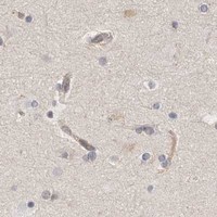
Negative control tissue: Bone marrow (HPA Not detected)
ICC-IF cell lines (HPA subcellular resource): HPA carries no ICC-IF cell line for MICALL2; derive a cell-line control from the positive tissue's cell type (Glandular cells) and confirm it by RNA or western blot first.
Technical controls: Include a no-primary, secondary-only control, a host-matched rabbit IgG isotype control, and a MICALL2-knockout sample if available to assess specificity (caption: rabbit primary; standard IHC controls). Quench endogenous peroxidase in colon sections before HRP/DAB detection (caption: HRP/DAB; standard IHC practice).
⚠️Feasibility: A MICALL2-specific fixation window or fixation effect is unreported, and the selected A12852-1 paraffin-section caption does not state the fixative (caption: fixative unreported). The caption uses heat-mediated EDTA retrieval at pH 8.0, but does not establish whether retrieval is required; no supplied matched evidence establishes that frozen sections or IF are easier (caption: EDTA retrieval; supplied application evidence). In colon, assess glandular staining against background in adjacent cells and the no-primary control, especially where endogenous peroxidase could affect HRP/DAB interpretation (HPA: High in colon glandular cells; caption: HRP/DAB; standard IHC practice).

HPA tissue IHC evidence for MICALL2

Comprehensive Human Protein Atlas IHC scoring per tissue (reliability: Enhanced — Medium consistency between antibody staining and RNA expression data.). Rows are taken directly from the HPA tissue chart — click any row's HPA link to view the source.

Positive expression · recommended positive controls

TissueCell typeLevelEvidenceSource
Colon Glandular cells High Protein (IHC) HPA →
Duodenum Glandular cells High Protein (IHC) HPA →
Small intestine Glandular cells High Protein (IHC) HPA →
Adrenal gland Glandular cells Medium Protein (IHC) HPA →
Appendix Glandular cells Medium Protein (IHC) HPA →

Undetected expression · recommended negative controls

TissueCell typeLevelEvidenceSource
Bone marrow Hematopoietic cells Not detected Protein (IHC) HPA →
Caudate Glial cells Not detected Protein (IHC) HPA →
Cerebellum Cells in granular layer Not detected Protein (IHC) HPA →
Cerebral cortex Glial cells Not detected Protein (IHC) HPA →
Hippocampus Glial cells Not detected Protein (IHC) HPA →
Section 3

Advanced MICALL2 IHC Tips

Use the catalog antibody’s tissue section conditions as the starting point, then check staining against MICALL2’s reported localisation and tissue pattern.

What retrieval conditions should I try when MICALL2 staining is weak in paraffin sections?
Start with heat mediated retrieval in EDTA at pH 8.0 (datasheet A12852-1). The illustrated paraffin section used 2 μg/ml primary antibody overnight at 4°C, so compare retrieval runs while holding primary incubation and detection constant (datasheet A12852-1; standard IHC practice). Include a glandular cell rich positive control, since colon and duodenum glandular cells show high staining (HPA tissue IHC). If signal remains weak, adjust heating duration on adjacent sections and check tissue integrity before considering another retrieval buffer; the supplied product evidence establishes EDTA at pH 8.0 as the starting condition (datasheet A12852-1; standard IHC practice).
Could fixation explain inconsistent MICALL2 staining across sections?
Target specific fixation sensitivity is unknown: the selected antibody caption identifies a paraffin section but does not state its fixative (datasheet A12852-1). Record each specimen’s fixative and processing history, then compare sections processed together before attributing a signal difference to MICALL2 expression (standard IHC practice). Use a consistent EDTA retrieval at pH 8.0 and the reported 2 μg/ml primary concentration when evaluating those sections (datasheet A12852-1). Compare tissue preservation, staining in viable glandular cells, and a matched negative control; neither the reported tissue pattern nor MICALL2’s phosphorylation sites establish how fixation affects this antibody (HPA tissue IHC; UniProt Q8IY33; standard IHC practice).
Where should convincing MICALL2 staining appear in a tissue section?
Assess cytoplasmic and membranous staining first, the pattern reported across several tissues (HPA tissue IHC). Junction associated or peripheral staining is plausible because MICALL2 is annotated at the cell membrane, tight junctions, recycling endosomes, and cytoskeleton (UniProt Q8IY33 localisation). It has no transmembrane segment, so an apparent membrane rim alone does not show that MICALL2 spans the membrane (UniProt Q8IY33 topology). Compare the pattern within intact glandular cells and across replicate sections, especially in colon or duodenum where glandular staining is high (HPA tissue IHC; standard IHC practice). Treat isolated nuclear signal or staining confined to damaged edges cautiously and inspect the negative control (UniProt Q8IY33 localisation; standard IHC practice).
Can this IHC antibody distinguish MICALL2 isoforms or phosphorylation states?
Do not assign an isoform from this stain without an antibody epitope map and evidence that the epitope differs among variants (standard IHC interpretation). MICALL2 has 5 annotated isoforms and CH, LIM, and bMERB regions, but the supplied product caption gives no epitope location (UniProt Q8IY33; datasheet A12852-1). Its annotated phosphoserines include positions 110, 143, and 153; their presence does not establish that this antibody detects a particular phosphorylation state (UniProt Q8IY33; standard IHC interpretation). When discordant staining raises an epitope question, seek sequence level antibody information and compare independently validated reagents on adjacent sections before making an isoform or modification claim (standard IHC practice).
How should I assess MICALL2 by IF alongside its chromogenic IHC result?
Treat IF as a separate validation exercise: the supplied product image documents chromogenic staining of a paraffin section, while HPA lists no cell lines with MICALL2 ICC/IF images (datasheet A12852-1; HPA subcellular). Multiplex a MICALL2 channel with an epithelial or glandular cell marker to test whether signal falls in the expected cells (HPA tissue IHC; standard IF practice). Select a spectrally separated, preferably far red fluorophore after checking the specimen’s autofluorescence and single color controls (standard IF practice). Because MICALL2 lacks a transmembrane segment and is also annotated in cytoskeletal and endosomal compartments, use permeabilisation when the antibody epitope is intracellular; its precise epitope is unspecified here (UniProt Q8IY33 topology and localisation; datasheet A12852-1).
How can I separate MICALL2 signal from nonspecific DAB staining?
Run a section without primary antibody and inspect it alongside the stained section for secondary reagent or detection background (standard IHC practice). The illustrated assay used a peroxidase linked anti rabbit secondary followed by DAB, so include a peroxidase blocking step and check whether pigment or endogenous enzyme activity survives it (datasheet A12852-1; standard IHC practice). If diffuse staining persists, reassess blocking and washes before increasing confidence in faint cytoplasmic signal; the illustrated section used 10% goat serum for blocking (datasheet A12852-1; standard IHC practice). Prioritise reproducible cytoplasmic or membranous staining in intact glandular cells over deposits at cut edges or necrotic areas (HPA tissue IHC; standard IHC interpretation).
How should I score MICALL2 staining across tissue samples? ⚠ ANSWER MARKED FOR VERIFICATION
Define the cell compartment and region of interest before scoring, then record the percentage of positive viable cells and staining intensity using one consistent threshold (standard IHC practice). An H score combines percentages at intensity grades and ranges from 0 to 300; retain compartment specific scores if cytoplasmic and membranous signals differ (standard IHC scoring; HPA tissue IHC). For heterogeneous sections, normalise positive cell counts to the number of viable cells of the same type, or report positive cell density per mm² of viable tissue (standard IHC practice). Keep acquisition, counterstain assessment, and scoring rules consistent, and exclude necrosis, cut edges, and obvious DAB deposits (standard IHC practice).
When should an apparent MICALL2 positive result be considered an artefact?
A credible result should recur in intact cells with cytoplasmic or membranous signal and should be judged against a primary omitted control (HPA tissue IHC; standard IHC practice). High staining in colon or duodenum glandular cells supports a positive control, whereas bone marrow hematopoietic cells are reported as not detected and need cautious interpretation if strongly stained (HPA tissue IHC). Question isolated nuclear staining because the supplied localisation annotations emphasise junctions, endosomes, membrane, and cytoskeleton (UniProt Q8IY33 localisation). Staining restricted to section edges, necrosis, pigment, or residual endogenous peroxidase can mimic positivity; check morphology and controls before scoring it (standard IHC practice). HPA describes only medium consistency between antibody staining and RNA expression, so interpret discordant samples conservatively (HPA tissue IHC).
Boster reagents

Best MICALL2 / MICAL-like protein 2 IHC Antibodies

A12852-1 has real IHC data from a human paraffin-embedded colorectal adenocarcinoma section (A12852-1 image caption). No IF image is provided (catalog image inventory).

Real IHC data IHC analysis of MICALL2 using anti-MICALL2 antibody (A12852-1). MICALL2 was detected in a paraffin-embedded section of human colorectal adenocarcinoma tissue. Heat mediated antigen retrieval was performed in EDTA buffer (pH 8.0, epitope retrieval solution). The tissue section was blocked with 10% goat serum. The tissue section was then incubated with 2 μg/ml rabbit anti-MICALL2 Antibody (A12852-1) overnight at 4°C. Peroxidase Conjugated Goat Anti-rabbit IgG was used as secondary antibody and incubated for 30 minutes at 37°C. The tissue section was developed using HRP Conjugated Rabbit IgG Super Vision Assay Kit (Catalog # SV0002) with DAB as the chromogen.
Anti-MICALL2 Antibody ®
Cat # A12852-1

A12852-1 is listed for human IHC (catalog applications and reactivity). Its own image documents staining in a paraffin-embedded human colorectal adenocarcinoma section (A12852-1 image caption).

Which to pick: Choose A12852-1 for human paraffin-section IHC: its own caption reports EDTA pH 8.0 retrieval and 2 μg/ml primary antibody; the fixative is unreported (A12852-1 image caption). No listed SKU has IF/ICC validation or reactivity beyond human (catalog applications, reactivity and image inventory). A12852-1 is rabbit-hosted, and its clonality is unreported (catalog host and clone fields).

Each figure is that product's own IHC / IF validation image from its datasheet.

References

  1. UniProt Consortium. UniProt entry Q8IY33 (MILK2_HUMAN, MICAL-like protein 2).
  2. Human Protein Atlas. MICALL2 tissue IHC expression (reliability: Enhanced).
  3. Human Protein Atlas. MICALL2 subcellular location (ICC-IF): Highest expression in OE19: 37.0 nTPM.
  4. Human Protein Atlas. MICALL2 antibody validation summary (1 antibodies).
  5. Comprehensive Analysis of MICALL2 Reveals Its Potential Roles in EGFR Stabilization and Ovarian Cancer Cell Invasion. International journal of molecular sciences 2023 — PMC10778810.
  6. DUSP4-mediated dephosphorylation of PSMD14 enhances PSMD14-dependent deubiquitination of MICALL2 and promotes malignant progression in clear cell renal cell carcinoma. Journal of translational medicine 2026 — PMC13281508.
  7. Identification of MICALL2 as a Novel Prognostic Biomarker Correlating with Inflammation and T Cell Exhaustion of Kidney Renal Clear Cell Carcinoma. Journal of Cancer 2022 — PMC8899381.
  8. MICALL2 as a substrate of ubiquitinase TRIM21 regulates tumorigenesis of colorectal cancer. Cell communication and signaling : CCS 2022 — PMC9615392.
  9. PubMed PMID:14702039 — UniProt-cited evidence.
  10. PubMed PMID:17974005 — UniProt-cited evidence.
  11. PubMed PMID:12853948 — UniProt-cited evidence.