MID1 / E3 ubiquitin-protein ligase MID1 · IHC design guide

Design Immunohistochemistry for MID1

Plan MID1 paraffin-section IHC around cytoplasmic staining reported in several tissues (HPA tissue IHC). Compare staining with appropriate tissue controls, since antibody staining shows low consistency with RNA expression (HPA tissue IHC).

Evidence assembled Oct 2026 · For research use; verify linked source records and product datasheet before use
Immunohistochemistry protocol sheet for MID1 (IHC for MID1): expected localisation Cytoplasmic staining in several tissues (HPA tissue IHC), antibody A01774-1, validated IHC image, and IHC protocol steps
Printable MID1 IHC protocol sheet — expected localisation Cytoplasmic staining in several tissues (HPA tissue IHC), antibody A01774-1, controls and protocol steps. Open the full MID1 IHC guide →

MID1 Immunohistochemistry Experimental Design Guide

Expected localisation, validated protocols, controls and antibodies — the at-a-glance facts below, then the full design guide.

Must know before staining
Expected localisation Cytoplasmic staining in several tissues (HPA tissue IHC)
Staining pattern Cytoplasmic staining in glandular, glial and tubular cells (HPA tissue IHC)
Antigen retrieval Citrate pH 6 HIER, heat-mediated (datasheet A01774-1)
Positive control ⓘ Appendix+4 more · see all
Negative control ⓘ Adipose tissue+4 more · see all
Important caveats
Reasons your staining may differ from the expected pattern.
Fixation Keep fixation conditions consistent across sections. (standard IHC practice; not target-specific)
Caveat Low agreement between staining and RNA expression (HPA tissue IHC)
Regulation Adult heart, placenta, brain most abundant (UniProt)
Isoform / epitope 2 isoforms; epitope coverage needs checking (UniProt)
Section 1

Recommended MID1 IHC & IF Protocols

The catalog antibody’s IHC-P protocol (datasheet: A01774-1) is followed by three published MID1 IHC protocols (PMC5653760; PMC4074869; PMC11978649).

Recommended immunohistochemistry (IHC-P) protocol parameters
SampleParaffin-embedded human placenta tissues; fixative not specified (datasheet A01774-1)
FixationImage fixative and duration unreported (datasheet A01774-1); verify before use.
Sectioning4–5 µm sections on charged slides (standard)
DeparaffinisationXylene, graded ethanol series to water (standard)
Antigen retrievalHeat retrieval: Citrate pH 6, 20 min (datasheet A01774-1)
Peroxidase block3% H2O2, 10 min, room temperature (standard)
Blocking10% goat serum (datasheet A01774-1)
Primary antibodyRabbit anti-MID1, 0.5-1μg/ml (datasheet A01774-1)
Primary incubationOvernight at 4 °C (datasheet A01774-1)
DetectionStreptavidin-biotin complex (SABC), DAB chromogen (datasheet A01774-1)
CounterstainHematoxylin, blue, dehydrate and mount (standard)
Expected resultMID1-positive staining in glandular cells of appendix (HPA tissue IHC: Medium). HPA tissue profile: Cytoplasmic expression in several tissues. No signal in the no-primary control.
💡Decision noteStart with citrate pH 6 heat retrieval (datasheet: A01774-1); the published protocols provide pepsin and Tris-borate alternatives (PMC5653760; PMC4074869).
Section 2

What Is the Expected MID1 Staining Pattern?

MID1 is a cytoplasmic, microtubule-associated protein with no transmembrane segment (UniProt O15344). In paraffin-section IHC, expect cytoplasmic staining in selected glandular cells, glial cells and kidney tubule cells (HPA tissue IHC). HPA rates its tissue IHC evidence Approved but reports low consistency between antibody staining and RNA expression, so interpret tissue patterns with that limitation (HPA tissue IHC).

What am I looking at on my slide?
Medium cytoplasmic staining in colon or appendix glandular cells, or kidney tubule cells (HPA tissue IHC).This matches reported positive cell populations and MID1's cytoplasmic location (HPA tissue IHC; UniProt O15344). Score the named cells and compartment rather than treating the whole section as uniformly positive; HPA reports low consistency between staining and RNA expression (HPA tissue IHC).
Staining is confined to nuclei or outlines cell membranes, with little cytoplasmic signal.A predominantly nuclear or membrane pattern conflicts with UniProt's cytoplasmic, microtubule-associated annotation and lack of a transmembrane segment (UniProt O15344). Treat the pattern as suspect; compare it with a reported positive cell population and appropriate detection controls (HPA tissue IHC; standard IHC practice).
Adipocytes or bone-marrow hematopoietic cells stain strongly while expected positive cells do not (HPA tissue IHC).HPA reports those cell populations as not detected (HPA tissue IHC). Check for cross-reactivity or endogenous detection activity before assigning the signal to MID1 (standard IHC practice). A negative HPA observation is a reference pattern, not proof that every specimen must be negative.
Brown signal spreads across stroma, blank areas or many cell types without a clear cytoplasmic pattern.Diffuse background makes cell-specific interpretation unreliable (standard IHC practice). Assess a no-primary control, endogenous peroxidase blocking, antibody concentration and washing before scoring the section (standard chromogenic IHC practice). HPA's reported pattern is cytoplasmic expression in several tissues (HPA tissue IHC).
No cytoplasmic signal appears in colon glandular cells or kidney tubule cells (HPA tissue IHC).These are reported medium-staining reference populations, so a blank result warrants a run-quality check (HPA tissue IHC). Confirm tissue morphology, reagent function, retrieval and detection using established controls (standard IHC practice); do not infer a MID1-specific fixation effect from the supplied evidence.
💡Expected MID1 appearanceCall a positive result when selected glandular, glial or kidney tubule cells show medium cytoplasmic staining (HPA tissue IHC); predominantly nuclear, membrane-only or widespread cell-independent signal is suspect against MID1's cytoplasmic annotation and the HPA cell pattern (UniProt O15344; HPA tissue IHC).
How each factor affects the staining
Cell and tissue selectionHPA reports medium staining in glandular cells of appendix, colon, duodenum and endometrium; glial cells of caudate, cerebral cortex and hippocampus; and kidney tubule cells (HPA tissue IHC). Its not-detected calls apply to the specified cell populations, such as adipocytes and lung alveolar cells, rather than every cell in those tissues (HPA tissue IHC).
Subcellular resolutionUniProt places MID1 on cytoplasmic microtubules, including interphase fibers, mitotic spindles and midbodies (UniProt O15344). HPA tissue IHC supports a cytoplasmic readout, but its tissue summary does not require those fine structures to be resolved in chromogenic sections (HPA tissue IHC).
Evidence strength and antibody choiceHPA marks tissue IHC Approved while noting low consistency with RNA expression; HPA003715 has IHC Approved status (HPA tissue IHC; HPA antibody validation). Use that status as validation context, not as a guarantee that every tissue or specimen reproduces the reported intensity (HPA tissue IHC).
Isoforms and epitope interpretationUniProt lists 2 MID1 isoforms and a COS, fibronectin type-III and B30.2/SPRY region (UniProt O15344). The supplied record does not locate either antibody's epitope, so it cannot establish whether both isoforms are detected or predict an epitope-specific IHC pattern.
Antigen retrieval and fixationAntigen retrieval is a standard paraffin-section IHC variable to check when a run fails (standard IHC practice). Neither UniProt nor HPA supplies a MID1-specific fixation or retrieval effect here; do not attribute weak staining, epitope masking or a tissue discrepancy to one without experimental evidence.
IF/ICC Q: should it reproduce the IHC pattern?IF/ICC offers a separate localisation readout: HPA reports centriolar satellites as uncertain, Golgi localisation as uncertain and cytosol as supported (HPA subcellular ICC-IF). HPA002878 has ICC Supported status, whereas HPA003715 has IHC Approved status (HPA antibody validation); do not equate their validation or require a satellite pattern in paraffin IHC.
Why is my staining missing, weak or wrong?
SituationLikely causeNext action
A reported positive reference population is blank.The run may have failed at retrieval, antibody incubation or detection; HPA reports medium staining in colon glandular cells and kidney tubule cells (standard IHC practice; HPA tissue IHC).Check a known working positive section and detection control, then review the documented retrieval and antibody conditions (standard IHC practice). The supplied sources give no MID1-specific fixation-sensitivity claim.
Only nuclei or cell borders stain.That distribution does not match MID1's cytoplasmic annotation or lack of a transmembrane segment (UniProt O15344).Compare the same run with a reported cytoplasmic positive cell population, verify morphology and review no-primary and antibody controls before calling it MID1 (HPA tissue IHC; standard IHC practice).
Many cells show uniform brown haze.Diffuse deposition can obscure compartment and cell identity (standard chromogenic IHC practice).Inspect the no-primary control; review endogenous peroxidase blocking, antibody concentration and washes, then score only interpretable cellular staining (standard IHC practice).
A HPA not-detected cell population stains strongly.For example, HPA reports adipocytes and bone-marrow hematopoietic cells as not detected; unexpected signal could reflect cross-reactivity or endogenous activity (HPA tissue IHC; standard IHC practice).Check the exact cell type against the HPA entry, assess no-primary and reagent controls, and compare with a reported positive population on a controlled run (HPA tissue IHC; standard IHC practice).
Staining is weaker than the HPA reference.HPA reports medium signal in specified positive cells but also notes low consistency between antibody staining and RNA expression (HPA tissue IHC). The record cannot identify a specimen-specific cause.Confirm the same cell population is being compared, check run controls and report the observed intensity; avoid claiming a MID1-specific fixation or retrieval mechanism without evidence (HPA tissue IHC; standard IHC practice).
An IF/ICC pattern differs from chromogenic tissue IHC.HPA describes cytosol plus uncertain satellite and Golgi localisation in ICC-IF, while tissue IHC is summarized as cytoplasmic expression; the two antibody validation entries also differ (HPA subcellular ICC-IF; HPA tissue IHC; HPA antibody validation).Interpret each assay against its own HPA validation and cellular resolution; use the separate IF/ICC guide for IF design, and do not turn its localisation findings into a required paraffin-section pattern (HPA subcellular ICC-IF; HPA tissue IHC).

Sample controls for MID1 IHC & IF

🧪Run appendix first and look for staining in its glandular cells (HPA: Appendix glandular cells, Medium). Use adipose tissue adipocytes as the negative tissue (HPA: Adipose tissue adipocytes, Not detected); on the appendix slide, neighboring non-glandular cells should show background-level staining to serve as internal comparators, but should not be assumed MID1-negative without validation.
Positive control tissue: Appendix (Glandular cells, HPA Medium)
Negative control tissue: Adipose tissue (HPA Not detected)
ICC-IF cell lines (HPA subcellular resource): HPA ICC-IF images show MID1 in A-549, GAMG, U2OS, with annotated localisation: Centriolar satellite (uncertain) (HPA subcellular).
Technical controls: Include a no-primary, secondary-only control; nonimmune IgG matched to the primary antibody’s host species and clonality; and a validated MID1-knockout sample or immunizing-peptide competition if the peptide is available (standard IHC controls). Quench endogenous peroxidase and check for endogenous biotin background in appendix with the biotin-based DAB detection system (caption: SABC/DAB).
⚠️Feasibility: A MID1-specific fixation window or fixation effect is unreported, and the selected A01774-1 paraffin-section caption does not state the fixative (caption: fixative not stated). Start with heat-mediated citrate retrieval at pH 6 for 20 minutes, as reported for the catalog antibody, then optimize against the controls (caption: citrate retrieval). Frozen sections are not established as easier; IF/ICC may help assess cytoplasmic localization, but requires separate validation (UniProt O15344 subcellular localization; HPA: ICC-IF images).

HPA tissue IHC evidence for MID1

Comprehensive Human Protein Atlas IHC scoring per tissue (reliability: Approved — Low consistency between antibody staining and RNA expression data.). Rows are taken directly from the HPA tissue chart — click any row's HPA link to view the source.

Positive expression · recommended positive controls

TissueCell typeLevelEvidenceSource
Appendix Glandular cells Medium Protein (IHC) HPA →
Caudate Glial cells Medium Protein (IHC) HPA →
Cerebral cortex Glial cells Medium Protein (IHC) HPA →
Colon Glandular cells Medium Protein (IHC) HPA →
Duodenum Glandular cells Medium Protein (IHC) HPA →

Undetected expression · recommended negative controls

TissueCell typeLevelEvidenceSource
Adipose tissue Adipocytes Not detected Protein (IHC) HPA →
Adrenal gland Glandular cells Not detected Protein (IHC) HPA →
Bone marrow Hematopoietic cells Not detected Protein (IHC) HPA →
Cervix Glandular cells Not detected Protein (IHC) HPA →
Esophagus Squamous epithelial cells Not detected Protein (IHC) HPA →
Section 3

Advanced MID1 IHC Tips

Troubleshoot MID1 staining in paraffin sections by checking retrieval, controls, cytoplasmic localisation and cell type before interpreting chromogenic signal.

Which retrieval conditions should I start with for MID1 in paraffin sections?
Start with heat-mediated retrieval in citrate buffer at pH 6 for 20 minutes (datasheet A01774-1). The catalog antibody stained a paraffin section of human placenta after this retrieval, followed by 1 µg/ml primary antibody overnight at 4°C (datasheet A01774-1). If staining is weak, compare retrieval durations on adjacent sections while holding antibody concentration and detection conditions constant (standard IHC practice). Include a section processed without primary antibody to assess detection background, and examine tissue preservation alongside signal intensity (standard IHC practice). The caption does not identify the fixative, so its result cannot establish retrieval performance across different fixation conditions (datasheet A01774-1).
How should I troubleshoot fixation-related loss of MID1 staining?
Target-specific sensitivity of MID1 staining to fixative type or fixation duration is unknown from the supplied evidence (datasheet A01774-1: fixative not stated). Record the fixative, time to fixation, fixation duration and processing conditions for each specimen before comparing staining (standard IHC practice). If signal differs between batches, stain sections together using the same citrate pH 6, 20-minute retrieval and detection workflow (datasheet A01774-1; standard IHC practice). Examine morphology and staining in an internal control region, where available, before changing antibody concentration (standard IHC practice). Neither the reported tissue patterns nor MID1's cytoplasmic localisation establishes a fixation effect (HPA: tissue IHC; UniProt O15344: subcellular location).
Where should convincing MID1 staining appear in a paraffin section?
Assess signal primarily in the cytoplasm, since MID1 is reported as cytoplasmic and microtubule-associated (UniProt O15344: subcellular location). In dividing cells, its reported association with the spindle and midbody provides context for a structured cytoplasmic pattern, although routine sections may not resolve those structures (UniProt O15344: subcellular location; standard IHC practice). Compare staining with cell morphology and an adjacent section processed without primary antibody before calling isolated dark deposits positive (standard IHC practice). HPA reports cytoplasmic expression across several tissues but also low consistency between antibody staining and RNA data (HPA: tissue IHC). A predominantly nuclear DAB pattern therefore warrants scrutiny rather than automatic assignment to MID1 (UniProt O15344: subcellular location; standard IHC practice).
Can this stain distinguish MID1 isoforms or a modified epitope?
Do not score isoform 1 and isoform 2 separately without evidence that the antibody distinguishes them (UniProt O15344: isoforms; standard IHC practice). MID1 includes COS, fibronectin type-III and B30.2/SPRY regions, but the supplied caption does not specify this antibody's epitope (UniProt O15344: domains; datasheet A01774-1). Phosphoserines at positions 92, 96 and 511 are recorded, but this IHC result does not establish sensitivity to their phosphorylation state (UniProt O15344: modified residues; datasheet A01774-1). If an isoform-specific or modification-specific conclusion matters, obtain epitope and validation information before interpreting a staining difference that way (standard IHC practice). Keep retrieval and detection conditions matched when comparing specimens (standard IHC practice).
How can IF help assess an ambiguous MID1 IHC pattern?
Use IF/ICC as a separate localisation check, with a marker for the expected cell type so MID1 signal can be assigned to the correct cells (standard IF practice). Choose spectrally separated fluorophores and inspect an unstained tissue control before selecting channels where tissue autofluorescence is low (standard IF practice). MID1 has no transmembrane segment and is reported in the cytoplasm, so permeabilise sufficiently to access an intracellular epitope while checking cell morphology (UniProt O15344: topology and subcellular location; standard IF practice). The antibody epitope is unspecified, so optimise permeabilisation empirically rather than assuming its precise accessibility (datasheet A01774-1; standard IF practice). Compare IF localisation with the IHC cytoplasmic pattern without treating IF conditions as a validated paraffin protocol (UniProt O15344: subcellular location; standard IHC/IF practice).
What should I check when MID1 DAB staining is diffuse or granular?
First compare the stained section with a no-primary control to separate antibody-dependent signal from detection background (standard IHC practice). The reported placenta workflow used 10% goat serum blocking, a biotinylated secondary antibody, streptavidin–biotin detection and DAB (datasheet A01774-1). Check whether blocking and wash steps were applied consistently, and assess endogenous peroxidase or biotin contributions with appropriate controls for this detection chemistry (standard IHC practice). Inspect tissue edges, folds and damaged areas before interpreting concentrated deposits as cellular staining (standard IHC practice). Genuine MID1 signal should be evaluated against its reported cytoplasmic localisation and the cells visible in the section (UniProt O15344: subcellular location; standard IHC practice).
How should I quantify MID1 staining across paraffin sections? ⚠ ANSWER MARKED FOR VERIFICATION
Define the cell population and tissue region before scoring, then record cytoplasmic intensity and the percentage of positive cells for an H-score (standard IHC practice; UniProt O15344: subcellular location). Alternatively, report positive-cell density per mm² when cell counts and sampled area are reliable (standard IHC practice). Normalise comparisons to the same eligible cell population or analysed tissue area, and hold retrieval, antibody incubation and DAB development constant (standard IHC practice). The reported placenta protocol used citrate pH 6 retrieval for 20 minutes and 1 µg/ml primary antibody overnight at 4°C (datasheet A01774-1). Report control staining and exclude folds or necrotic regions from the predefined scoring area (standard IHC practice).
How can I distinguish true MID1 staining from a staining artefact?
Prioritise cytoplasmic staining within identifiable cells, consistent with MID1's reported cytoplasmic and cytoskeletal association (UniProt O15344: subcellular location). Compare the signal with a no-primary control and inspect section edges, folds and necrotic regions for deposits that follow tissue damage rather than cell boundaries (standard IHC practice). Check endogenous peroxidase background because the reported detection workflow used DAB with a biotin-based system (datasheet A01774-1; standard IHC practice). Cell type also matters: HPA reports medium staining in kidney tubule cells and no detection in adipocytes, while rating tissue IHC reliability Approved with low staining–RNA consistency (HPA: tissue IHC). Treat an unexpected nuclear pattern or an isolated unexpected cell population as provisional until supported by controls or independent evidence (UniProt O15344: subcellular location; standard IHC practice).
Boster reagents

Best MID1 / E3 ubiquitin-protein ligase MID1 IHC Antibodies

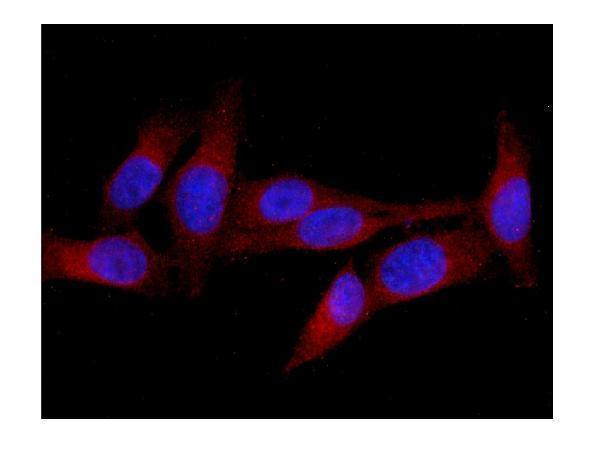
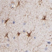
The catalog antibody has IHC images from human placenta and mouse brain paraffin sections (A01774-1 IHC captions) and an IF image from U20S cells (A01774-1 IF caption).

Real IHC data IHC analysis of MID1 using anti-MID1 antibody (A01774-1). MID1 was detected in paraffin-embedded section of human placenta tissues. Heat mediated antigen retrieval was performed in citrate buffer (pH6, epitope retrieval solution) for 20 mins. The tissue section was blocked with 10% goat serum. The tissue section was then incubated with 1μg/ml rabbit anti-MID1 Antibody (A01774-1) overnight at 4°C. Biotinylated goat anti-rabbit IgG was used as secondary antibody and incubated for 30 minutes at 37°C. The tissue section was developed using Strepavidin-Biotin-Complex (SABC)(Catalog # SA1022) with DAB as the chromogen.
Anti-MID1 Antibody ®
Cat # A01774-1

A01774-1 has IHC images from human placenta and mouse brain paraffin sections (A01774-1 IHC captions). The same SKU has an IF image from U20S cells (A01774-1 IF caption) and lists human and mouse reactivity (catalog: A01774-1 reactivity).

Which to pick: Choose A01774-1 for paraffin-section IHC: its own captions document staining in human placenta and mouse brain after citrate-buffer retrieval (A01774-1 IHC captions); the fixative is unreported. For IF/ICC, the same rabbit antibody lists both applications (catalog: A01774-1 host and applications) and has an IF image from U20S cells (A01774-1 IF caption). It is also the catalog choice for human and mouse samples (catalog: A01774-1 reactivity); the IHC captions show staining in a paraffin section from each species (A01774-1 IHC captions).

Each figure is that product's own IHC / IF validation image from its datasheet.

References

  1. UniProt Consortium. UniProt entry O15344 (TRI18_HUMAN, E3 ubiquitin-protein ligase MID1).
  2. Human Protein Atlas. MID1 tissue IHC expression (reliability: Approved).
  3. Human Protein Atlas. MID1 subcellular location (ICC-IF): Mainly localized to the centriolar satellites. In addition localized to the Golgi apparatus and cytosol..
  4. Human Protein Atlas. MID1 antibody validation summary (2 antibodies).
  5. Resveratrol induces dephosphorylation of Tau by interfering with the MID1-PP2A complex. Scientific reports 2017 — PMC5653760.
  6. A hormone-dependent feedback-loop controls androgen receptor levels by limiting MID1, a novel translation enhancer and promoter of oncogenic signaling. Molecular cancer 2014 — PMC4074869.
  7. Midline-1 inhibited high glucose-induced epithelial-mesenchymal transition, fibrosis and inflammation through WNT/β-catenin signaling in benign prostatic hyperplasia. Frontiers in endocrinology 2025 — PMC11978649.
  8. Exosomes derived from myeloid-derived suppressor cells facilitate castration-resistant prostate cancer progression via S100A9/circMID1/miR-506-3p/MID1. Journal of translational medicine 2022 — PMC9344715.
  9. PubMed PMID:9354791 — UniProt-cited evidence.
  10. PubMed PMID:9425238 — UniProt-cited evidence.
  11. PubMed PMID:9722948 — UniProt-cited evidence.