MIOX / Inositol oxygenase · IHC design guide

Design Immunohistochemistry for MIOX

Plan chromogenic IHC for MIOX in paraffin sections using kidney proximal tubules as a positive reference (HPA tissue IHC). This guide covers fixation, staining controls, and interpretation of the expected cytoplasmic pattern (HPA tissue IHC).

Evidence assembled Oct 2026 · For research use; verify linked source records and product datasheet before use
Immunohistochemistry protocol sheet for MIOX (IHC for MIOX): expected localisation Cytoplasmic staining in renal tubules (HPA tissue IHC), antibody A06957, validated IHC image, and IHC protocol steps
Printable MIOX IHC protocol sheet — expected localisation Cytoplasmic staining in renal tubules (HPA tissue IHC), antibody A06957, controls and protocol steps. Open the full MIOX IHC guide →

MIOX Immunohistochemistry Experimental Design Guide

Expected localisation, validated protocols, controls and antibodies — the at-a-glance facts below, then the full design guide.

Must know before staining
Expected localisation Cytoplasmic staining in renal tubules (HPA tissue IHC)
Staining pattern Proximal tubule cell bodies show cytoplasmic staining (HPA tissue IHC)
Antigen retrieval EDTA pH 8.0 HIER, heat-mediated (datasheet A06957)
Positive control ⓘ Kidney
Negative control ⓘ Adipose tissue+4 more · see all
Important caveats
Reasons your staining may differ from the expected pattern.
Fixation Keep fixation consistent across sections (standard IHC practice; not target-specific). Selected-image fixative and duration unreported (datasheet A06957); verify before use.
Caveat Endogenous peroxidase may mimic renal DAB signal (standard IHC practice)
Regulation Kidney-enriched expression (HPA tissue IHC)
Isoform / epitope Two isoforms; epitope coverage is unspecified (UniProt)
Section 1

Recommended MIOX IHC & IF Protocols

The catalog antibody protocol is accompanied by published MIOX IHC methods for human kidney and mouse xenograft tissue (PMC4128578; PMC9433379).

Recommended immunohistochemistry (IHC-P) protocol parameters
SampleParaffin-embedded rat kidney tissue; fixative not specified (datasheet A06957)
FixationImage fixative and duration unreported (datasheet A06957); verify before use.
Sectioning4–5 µm sections on charged slides (standard)
DeparaffinisationXylene, graded ethanol series to water (standard)
Antigen retrievalHeat retrieval: EDTA pH 8.0 (datasheet A06957); 20 min, 95–100 °C (standard)
Peroxidase block3% H2O2, 10 min, room temperature (standard)
Blocking10% goat serum (datasheet A06957)
Primary antibodyRabbit anti-MIOX, 2-5 μg/ml (datasheet A06957)
Primary incubationOvernight at 4 °C (datasheet A06957)
DetectionHRP-conjugated secondary, DAB chromogen (datasheet A06957)
CounterstainHematoxylin, blue, dehydrate and mount (standard)
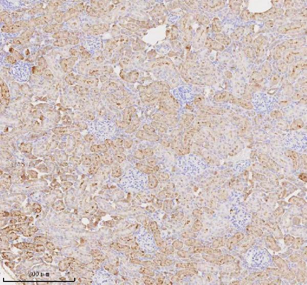
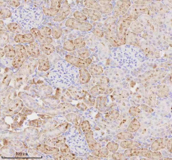
Expected resultMIOX-positive staining in proximal tubules (cell body) of kidney (HPA tissue IHC: Medium). HPA tissue profile: Selective cytoplasmic expression in renal tubules. No signal in the no-primary control.
💡Decision noteStart with heat-mediated EDTA pH 8.0 retrieval for the catalog antibody (datasheet A06957); sodium citrate retrieval was reported for human kidney (PMC4128578).
Section 2

What Is the Expected MIOX Staining Pattern?

MIOX is annotated as cytoplasmic and has no transmembrane segment (UniProt Q9UGB7: cytoplasm, topology). In paraffin-section IHC, expect selective staining of renal proximal tubule cell bodies at a medium level (HPA tissue IHC: kidney, proximal tubules, Medium). HPA rates its tissue IHC evidence Enhanced while describing medium consistency between antibody staining and RNA expression (HPA tissue IHC: reliability).

What am I looking at on my slide?
Selective cytoplasmic staining in renal proximal tubule cell bodies.This matches the reported cell type, compartment and medium staining level (HPA tissue IHC: kidney, proximal tubules, Medium; UniProt Q9UGB7: cytoplasm). Compare the distribution across tubules before scoring: a positive result depends on both where the stain appears and which cells contain it (HPA tissue IHC: selective cytoplasmic expression in renal tubules).
Predominantly nuclear or sharply membrane-restricted staining.These compartments do not match the annotated cytoplasmic location or lack of a transmembrane segment (UniProt Q9UGB7: cytoplasm, topology). Treat an isolated wrong-compartment pattern as suspect and check controls and detection conditions before assigning it to MIOX (general IHC interpretation practice).
Strong staining in cells outside the reported renal tubular pattern.A widespread or unrelated cell-type pattern conflicts with HPA's selective renal tubular staining (HPA tissue IHC: tissue profile). Cross-reactivity or endogenous detection activity are possible explanations; neither can be distinguished from the slide alone (general IHC interpretation practice). Check a no-primary control and antibody specificity evidence (general IHC practice).
Diffuse chromogen across many tissue compartments or the slide background.Diffuse background prevents confident assignment of signal to proximal tubule cytoplasm (HPA tissue IHC: selective cytoplasmic renal tubular pattern). Review the no-primary control, blocking, wash stringency and detection development before scoring cells as positive (general IHC practice).
No visible staining in a kidney section containing proximal tubules.This disagrees with the reported medium proximal tubular staining, but one negative run does not establish absence of MIOX (HPA tissue IHC: kidney, proximal tubules, Medium; general IHC interpretation practice). Verify tissue identity and assay controls, then review the antibody's stated IHC-P conditions and detection steps (general IHC practice).
💡Expected MIOX appearanceCall a slide positive when renal proximal tubule cell bodies show selective, approximately medium cytoplasmic staining (HPA tissue IHC: kidney, proximal tubules, Medium; UniProt Q9UGB7: cytoplasm); predominant nuclear, membrane-only or broadly distributed staining is a suspect pattern requiring control review (general IHC interpretation practice).
How each factor affects the staining
Which tissue best anchors interpretation?Kidney is tissue-enriched by RNA and is the reported IHC-positive tissue (HPA: tissue-enriched RNA; kidney proximal tubules, Medium). UniProt also describes kidney-specific expression (UniProt Q9UGB7: tissue specificity).
What does the protein record predict about compartment?UniProt annotates cytoplasm, no transmembrane segment, no signal peptide and a single 1–285 chain (UniProt Q9UGB7: location, topology, processing). These annotations support assessing cytoplasmic cell-body staining; they do not establish an IHC retrieval condition (UniProt Q9UGB7: annotations; general IHC practice).
How strong is the tissue-pattern evidence?HPA rates tissue IHC Enhanced and lists two rabbit polyclonal antibodies with Enhanced IHC status, HPA039451 and HPA039562 (HPA: tissue reliability; antibody validation). HPA also reports medium staining–RNA consistency, so preserve that caveat when interpreting discordant slides (HPA: reliability description).
Do isoforms or modifications explain a different stain?Two isoforms and phosphoserine at residue 33 are annotated (UniProt Q9UGB7: isoforms, modified residues). The supplied records do not map antibody epitopes or link either feature to a distinct tissue pattern (UniProt Q9UGB7: supplied annotations; HPA: supplied IHC profile).
IF/ICC Q&A: Is a cellular image pattern established?No ICC-IF images or main subcellular location are supplied by HPA (HPA subcellular: images absent, location unavailable). Cytoplasm is a UniProt annotation, not an HPA ICC-IF image result (UniProt Q9UGB7: cytoplasm; HPA subcellular: location unavailable).
Why is my staining missing, weak or wrong?
SituationLikely causeNext action
Kidney proximal tubules show no signal.A missed assay step, unsuitable antibody conditions or weak detection may produce an apparent negative (general IHC practice).Confirm proximal tubules are present, inspect run controls and follow the IHC-validated antibody's stated IHC-P conditions; assess retrieval with a positive kidney section (HPA: proximal tubules, Medium; general IHC practice). No MIOX-specific retrieval requirement is supplied (HPA and UniProt: supplied records).
Nuclei or cell borders dominate the stain.The compartment differs from MIOX's cytoplasmic annotation and lack of a transmembrane segment (UniProt Q9UGB7: location, topology).Review the counterstain and a no-primary control, then compare antibody and detection conditions on the same tissue (general IHC practice). Score the pattern as unresolved until cytoplasmic localization can be distinguished (UniProt Q9UGB7: cytoplasm).
Adipocytes or adrenal glandular cells stain prominently.HPA reports these sampled cells as not detected; cross-reactivity or endogenous detection activity may account for unexpected signal (HPA tissue IHC: adipocytes, adrenal glandular cells, Not detected; general IHC practice).Check a no-primary control and, where relevant, endogenous-enzyme blocking; confirm antibody specificity before interpreting these cells as MIOX-positive (general IHC practice).
Background is diffuse across the section.Nonspecific binding, incomplete washing or excess chromogen development can obscure cell-level localization (general IHC practice).Compare no-primary and positive-tissue controls, then review blocking, washes, antibody concentration and development time under the chosen IHC-P method (general IHC practice). Reassess the selective tubular pattern after background is controlled (HPA tissue IHC: profile).
A proposed negative tissue also appears positive.HPA reports no detection only in the listed sampled cell types; an unexpected stain alone does not establish a new MIOX distribution (HPA tissue IHC: negative entries; general IHC interpretation practice).Identify the stained cell type, compare it with HPA's specified cells, and review assay controls and antibody specificity (HPA tissue IHC: negative entries; general IHC practice). Avoid extending a cell-specific HPA result to every cell in that tissue.
IF/ICC fluorescence suggests a different location.The supplied HPA subcellular record has no ICC-IF images or main-location call, while UniProt annotates cytoplasm (HPA subcellular: images absent, location unavailable; UniProt Q9UGB7: cytoplasm).Treat the fluorescent location as unconfirmed by these sources; evaluate IF/ICC controls in its own guide before comparing it with the paraffin-section IHC pattern (HPA subcellular: location unavailable; general IF/ICC practice).

Sample controls for MIOX IHC & IF

🧪Run kidney first: proximal tubule cell bodies should stain at a medium level (HPA: Kidney, proximal tubules, Medium). Use adipose tissue as the negative tissue because its adipocytes are not detected; on the kidney slide, other structures should show only background staining, but treat them as internal comparators rather than confirmed MIOX-negative cells (HPA: Adipose tissue, adipocytes, Not detected; HPA: Kidney, proximal tubules, Medium).
Positive control tissue: Kidney (Proximal tubules (cell body), HPA Medium)
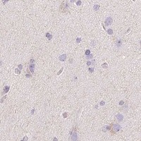
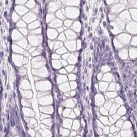
Negative control tissue: Adipose tissue (HPA Not detected)
ICC-IF cell lines (HPA subcellular resource): HPA carries no ICC-IF cell line for MIOX; derive a cell-line control from the positive tissue's cell type (Proximal tubules (cell body)) and confirm it by RNA or western blot first.
Technical controls: Include a no-primary, secondary-only control, a concentration-matched rabbit IgG isotype control, and a MIOX knockout tissue control if available (A06957 tissue-IHC caption: rabbit primary antibody; standard IHC practice). Quench endogenous peroxidase and check the no-primary kidney section for background before interpreting HRP/DAB staining (A06957 tissue-IHC caption: HRP/DAB; standard IHC practice).
⚠️Feasibility: The A06957 rat kidney paraffin-section caption reports heat-mediated EDTA retrieval at pH 8.0 and HRP/DAB detection; its fixative, a target-specific fixation window, and any fixation effect are unreported (A06957 tissue-IHC caption). Retrieval was used, but whether MIOX staining requires it is unreported; the supplied evidence does not establish that frozen sections or IF are easier than paraffin IHC (A06957 tissue-IHC caption; HPA subcellular: no ICC-IF image cell lines). Endogenous peroxidase can contribute kidney background in chromogenic detection, so assess it on the no-primary slide (standard IHC practice).

HPA tissue IHC evidence for MIOX

Comprehensive Human Protein Atlas IHC scoring per tissue (reliability: Enhanced — Medium consistency between antibody staining and RNA expression data.). Rows are taken directly from the HPA tissue chart — click any row's HPA link to view the source.

Positive expression · recommended positive controls

TissueCell typeLevelEvidenceSource
Kidney Proximal tubules (cell body) Medium Protein (IHC) HPA →

Undetected expression · recommended negative controls

TissueCell typeLevelEvidenceSource
Adipose tissue Adipocytes Not detected Protein (IHC) HPA →
Adrenal gland Glandular cells Not detected Protein (IHC) HPA →
Appendix Glandular cells Not detected Protein (IHC) HPA →
Bone marrow Hematopoietic cells Not detected Protein (IHC) HPA →
Breast Adipocytes Not detected Protein (IHC) HPA →
Section 3

Advanced MIOX IHC Tips

Troubleshoot MIOX staining in paraffin sections using the catalog antibody’s kidney IHC result and the expected proximal tubule cytoplasmic pattern.

What retrieval should I try first for weak MIOX staining in paraffin sections?
Start with heat mediated retrieval in EDTA, pH 8.0 (datasheet A06957). That condition preceded MIOX detection in a paraffin embedded rat kidney section, although the caption does not report the fixative (datasheet A06957). Keep heating, cooling, section thickness, and antibody incubation consistent while comparing retrieval runs, so a change in staining has a clear cause (standard IHC practice). Use proximal tubules as the expected positive compartment and assess cytoplasmic signal against nearby tissue background (HPA: medium staining in proximal tubule cell bodies; UniProt Q9UGB7: cytoplasm). If staining remains weak, adjust retrieval duration before considering another buffer as a fallback (standard IHC practice).
Could fixation explain weak or uneven MIOX staining?
Target specific fixation sensitivity is unknown: the selected paraffin section caption does not state a fixative (datasheet A06957). Record fixative identity and fixation duration for each specimen, then compare sections processed and stained together before attributing a difference to MIOX abundance (standard IHC practice). Uneven preservation, detached epithelium, or stronger staining at section edges warrants a review of tissue handling and retrieval consistency (standard IHC practice). Include a kidney control in each run and judge whether proximal tubule cytoplasmic staining is retained under the same processing conditions (HPA: proximal tubule cell body staining; UniProt Q9UGB7: cytoplasm). Do not infer fixation tolerance from that expression pattern (datasheet A06957: fixative unreported).
Where should a convincing MIOX signal appear in kidney sections?
Look for cytoplasmic staining in renal tubules, particularly proximal tubule cell bodies (HPA: selective cytoplasmic expression in renal tubules; HPA: medium staining in proximal tubule cell bodies). MIOX is annotated as cytoplasmic and has no transmembrane segment, so a predominantly crisp membrane rim would need independent validation (UniProt Q9UGB7: cytoplasm; no transmembrane segment). Compare the distribution with intact tubular morphology and a counterstain, rather than scoring brown material in the tubular lumen as cellular signal (standard IHC practice). A kidney section provides a positive tissue reference, while HPA reports no detection in adipocytes of adipose tissue under its assay conditions (HPA: kidney positive; adipose tissue adipocytes not detected).
Can this stain distinguish MIOX isoforms or a modified epitope?
Do not assign an isoform from this IHC result alone: the record lists 2 isoforms, but the selected caption supplies no antibody epitope map (UniProt Q9UGB7: isoforms 1 and 2; datasheet A06957: epitope unspecified). The record also lists phosphoserine at position 33, without establishing whether this antibody recognizes that modification (UniProt Q9UGB7: modified residue 33; datasheet A06957: epitope unspecified). Check the antibody’s mapped immunogen and isoform sequences before interpreting differential staining as splice specific (standard IHC practice). If the comparison matters, use an independently validated isoform or modification specific method on matched material (standard IHC practice). Keep the IHC conclusion at the level supported by the antibody’s documented specificity (standard IHC practice).
How should I assess MIOX in a multiplex IF experiment?
Treat IF as a separate assay requiring its own validation, because the selected antibody evidence describes chromogenic IHC in a paraffin embedded rat kidney section (datasheet A06957). Pair MIOX with an independently validated proximal tubule marker and check whether signals coincide within cell bodies (HPA: proximal tubule cell body expression; standard IF practice). Select fluorophores and imaging channels after measuring kidney tissue autofluorescence in an unstained control, and include single stain controls for spectral overlap (standard IF practice). For a cytoplasmic antigen, test post fixation permeabilisation while preserving morphology; no membrane sided epitope can be assigned from the available record (UniProt Q9UGB7: cytoplasm, no transmembrane segment; standard IF practice).
How can I separate MIOX staining from kidney background?
Compare stained kidney sections with a no primary control and inspect both proximal tubules and surrounding tissue (standard IHC practice; HPA: selective cytoplasmic renal tubule expression). The selected method used 10% goat serum blocking, 2 µg/ml primary antibody overnight at 4°C, and a peroxidase secondary for 30 minutes at 37°C (datasheet A06957). A peroxidase block and careful DAB development help assess enzymatic background in chromogenic IHC; they are general workflow measures (standard IHC practice). If diffuse staining persists, compare no primary and full stain sections before adjusting blocking, antibody concentration, or wash stringency one variable at a time (standard IHC practice).
How should I quantify MIOX staining across kidney sections? ⚠ ANSWER MARKED FOR VERIFICATION
Define proximal tubules on the counterstained section before scoring, because the reported signal is selective in renal tubules (HPA: selective cytoplasmic renal tubule expression; standard IHC practice). Report the percentage of positive proximal tubule cells and, when intensity matters, an H score from 0–300 using the same thresholds across slides (standard IHC practice). Normalize counts to the number of evaluable proximal tubule cells, or stained area to evaluable proximal tubule area, rather than total kidney area (standard IHC practice). Keep acquisition, DAB development, and scoring rules consistent, and exclude folds, damaged tissue, and poorly preserved tubules before comparing groups (standard IHC practice).
When is a brown signal likely to be artefact rather than MIOX?
A convincing result follows proximal tubule cell bodies with predominantly cytoplasmic staining (HPA: medium proximal tubule cell body staining; UniProt Q9UGB7: cytoplasm). Isolated nuclear, membrane rim, or luminal staining needs further validation against morphology and controls, given the annotated cytoplasmic location and lack of a transmembrane segment (UniProt Q9UGB7: cytoplasm, no transmembrane segment; standard IHC practice). Discount staining confined to section edges, folds, or necrotic areas when scoring preserved tubules (standard IHC practice). Check a no primary control for residual endogenous peroxidase or nonspecific DAB signal before calling unexpected cells positive (standard IHC practice). Interpret a failed kidney control as an assay problem before interpreting a study section as biologically negative (HPA: kidney expression; standard IHC practice).
Boster reagents

Best MIOX / Inositol oxygenase IHC Antibodies

The IHC-validated anti-MIOX antibody has real paraffin-section kidney images from rat and human samples (A06957 image captions). Its listed reactivity covers human, mouse and rat (A06957 catalog).

Real IHC data IHC analysis of MIOX using anti-MIOX antibody (A06957). MIOX was detected in a paraffin-embedded section of rat kidney tissue. Heat mediated antigen retrieval was performed in EDTA buffer (pH 8.0, epitope retrieval solution). The tissue section was blocked with 10% goat serum. The tissue section was then incubated with 2 μg/ml rabbit anti-MIOX Antibody (A06957) overnight at 4°C. Peroxidase Conjugated Goat Anti-rabbit IgG was used as secondary antibody and incubated for 30 minutes at 37°C. The tissue section was developed using HRP Conjugated Rabbit IgG Super Vision Assay Kit (Catalog # SV0002) with DAB as the chromogen.
Anti-MIOX Antibody ®
Cat # A06957

A06957 will render with a rat kidney paraffin-section IHC figure (A06957 figure caption). Its additional image caption shows human kidney paraffin-section IHC, while the application list includes IHC and lists human, mouse and rat reactivity (A06957 image caption; A06957 catalog).

Which to pick: Choose A06957 for paraffin-section tissue IHC: its rat and human kidney captions describe EDTA pH 8.0 retrieval and 2 μg/ml primary antibody (A06957 image captions). A06957 is the only listed cross-species candidate, with human, mouse and rat reactivity; the supplied IHC images show rat and human tissue, and the fixative is unreported (A06957 catalog; A06957 image captions). There is no IF/ICC-validated option in the supplied catalog because A06957 has no IF/ICC application or IF image (A06957 catalog).

Each figure is that product's own IHC / IF validation image from its datasheet.

References

  1. UniProt Consortium. UniProt entry Q9UGB7 (MIOX_HUMAN, Inositol oxygenase).
  2. Human Protein Atlas. MIOX tissue IHC expression (reliability: Enhanced).
  3. Human Protein Atlas. MIOX subcellular location (ICC-IF): Highest expression in HDLM-2: 25.6 nTPM.
  4. Human Protein Atlas. MIOX antibody validation summary (2 antibodies).
  5. Development of an immunoassay for the kidney-specific protein myo-inositol oxygenase, a potential biomarker of acute kidney injury. Clinical chemistry 2014 — PMC4128578.
  6. Long noncoding RNA NEAT1 promotes ferroptosis by modulating the miR-362-3p/MIOX axis as a ceRNA. Cell death and differentiation 2022 — PMC9433379.
  7. Gene regulatory networks orchestrating oocyte fate bifurcation in primordial follicles revealed by single-cell transcriptomics. Communications biology 2026 — PMC13376924.
  8. PubMed PMID:10944187 — UniProt-cited evidence.
  9. PubMed PMID:15504367 — UniProt-cited evidence.
  10. PubMed PMID:14702039 — UniProt-cited evidence.