This website uses cookies to ensure you get the best experience on our website.
- Table of Contents
5 Citations 1 Q&As
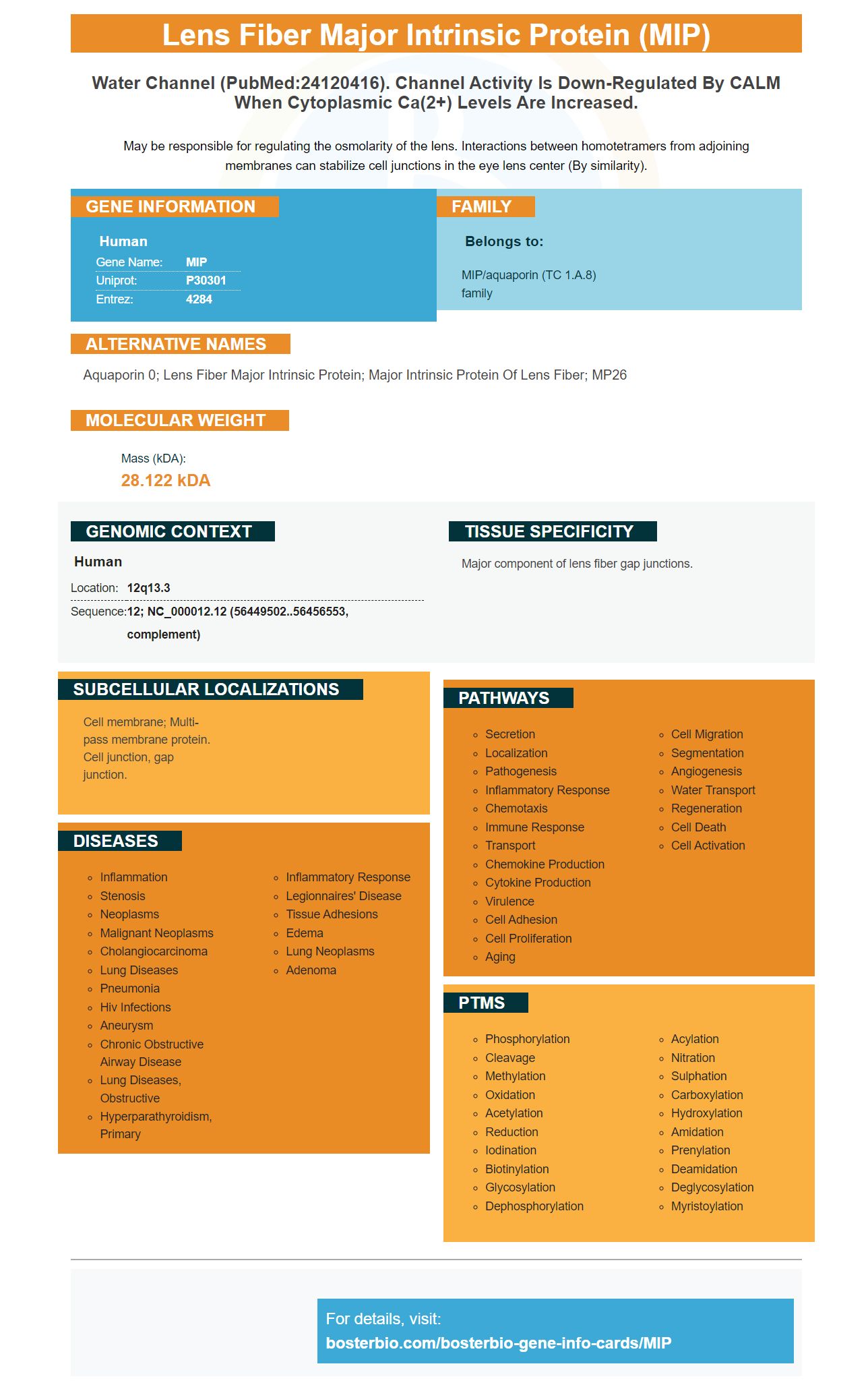
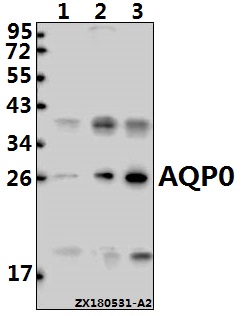
Facts about Lens fiber major intrinsic protein.
May be responsible for regulating the osmolarity of the lens. Interactions between homotetramers from adjoining membranes can stabilize cell junctions in the eye lens center (By similarity).
Human | |
---|---|
Gene Name: | MIP |
Uniprot: | P30301 |
Entrez: | 4284 |
Belongs to: |
---|
MIP/aquaporin (TC 1.A.8) family |
aquaporin 0; lens fiber major intrinsic protein; major intrinsic protein of lens fiber; MP26
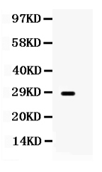
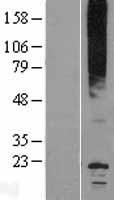
Mass (kDA):
28.122 kDA
Human | |
---|---|
Location: | 12q13.3 |
Sequence: | 12; NC_000012.12 (56449502..56456553, complement) |
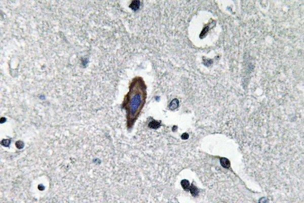
Major component of lens fiber gap junctions.
Cell membrane; Multi-pass membrane protein. Cell junction, gap junction.
Boster Bio, an antibody manufacturing company is in operation since 1993. They specialize in signature products, including antibodies and ELISA kits but recently expanded their services to include molecular biology-related PCR. Boster Bio's technical staff is able to answer any questions about their products or services. Boster Bio provides 24 hour support and technical support to scientists.
CD68 is a glycosylated multidrug resistant glycoprotein found on the surface macrophages and monocytes. This antigen is utilized as a cytochemical marker of these cells, and is utilized in immunostaining tests. It can also be useful in diagnosing cancer patients. However, it is not expressed in lymphoid cells, tumor cells and non-hematopoietic tissues. A complex interplay between transcriptional factors determines expression levels.
CD68 is part of the Scavenger receptor family. It attaches to modified LDL, phosphatidylserine as well as other inflammatory stimuli once activated. It also has the capability of binding to cells that are dying. Although its role in the immune system is undefined but it has been linked as a contributor to atherogenesis. It has been shown that CD68 expression on macrophages may affect the development and progression of atherosclerosis.
Anti-CD68 mAbs were also examined in a variety of studies. These included immunohistochemistry, flow cytometry, and fibroblast cultures. These studies involved staining cells with CD68 and analyzing them for CD80 and CD206 expression. The anti-CD68 antibodies were also utilized in flow cytometry. The analysis of flow cytometry revealed that a high percentage of SFMCs expressed CD68. This supports the notion that Mf cells are highly concentrated in OJIA-SF.
Macrophage inflammatory proteins-3 beta, also known as CCL19 is a chemokine involved in immune responses, T cell activation, and homing. It connects to the chemokine C-C receptor 7 and has a potent chemotactic activity against B cells, T cells, granulocytes, and granulocytes. Although the protein is present in various tissues, it is particularly essential in the immune system as a low level of CCL19 is associated with autoimmune diseases such as lupus, psoriasis, and arthritis.
The anti-MIP-3 beta antibody from Boster Bio is tested in ELISA and immunohistochemistry. It detects mAb3b in serum, plasma, cell culture fluid, and other biological fluids. It reacts with MIP-3 Beta in humans, in addition to many other known positive and negative samples. It is easy to use and highly effective for research purposes.
Monocytes secrete primary chemokines including human CCL19/MIP-3 Beta. It has six cysteine residues. It binds to CCR7 on mature dendritic cell cells and participates in the T cell immune response. This chemokine is associated with allergies, inflammation, and other diseases.
Boster Bio's anti-MIP 3 beta/Ccl19 antibody has been validated using ELISA assays, immunohistochemistry (IHC), and immunohistochemistry. It reacts with Human CCR7 and Mouse CCR7 and has a sensitivity level of 10pg/ml. It was also mentioned in a publication. Here are the results. You can determine whether this antibody is suitable to you by performing an experiment using a positive or negative sample.
The MIP-3 protein is a CC chemical, is CCR1 receptor. It acts as a chemoattractant neutrophils, resting T-lymphocytes and monocytes. It also inhibits the development of colony-forming myeloid immature progenitors within the bone marrow. Recombinant MIP-3, a 11.3 kDa protein, contains 99 amino acids and four conserved cysteine residues.
The antibody originates from human monoclonal antibodies to CCL19 and MIP-3 beta. CCL19 and MIP-3beta are asymmetrically expressed proteins and act in different ways in immune response induction. The MIP-3beta/Ccl19 boster bio antibody is a monoclonal antibody which targets mouse antigens. It is also a powerful inhibitor of MIP-3beta/CCL19.
The diagnosis of inflammation is the most important application of the MIP marker. Inflammatory diseases are recognized by elevated levels of the MIP marker. MIP-1a/CCL3 a precursor protein, which prematurely splits at multiple sites into two biologically active mature protein. This marker is extremely sensitive to proteoglycans. It could be enhanced by the interaction between MIP-1a/heparin.
The MIP-1a/CCL3 proteins could be a biomarker of inflammatory diseases. This article explains the protein's biological functions and discusses various diseases that are related to this protein, including periodontitis. These results indicate that MIP markers are effective biomarkers of inflammation. Further research is needed to confirm the diagnostic value of this marker. Here are some examples of the MIP marker.
This study confirmed the MIP marker by measuring its differential expression in cancer patients. This test is correlated with the absence of distant metastasis. The MIP marker was also highly predictive of survival in patients with metastatic disease. However, further research is needed to confirm this correlation. A separate control group is needed to increase the reliability of MIP markers. Before implementing the assay in clinical trials, it is essential that it be validated.
MIP assays can be used for the generation of 1,000-25,000 specific SNPs. MIP technology is also used in human and bovine HapMap projects and has been successfully employed in wild mouse populations to determine susceptibility to diseases such as Type I diabetes, colorectal cancer, and prostate cancer. It is now being utilized more frequently in research studies to determine disease susceptibility. The MIP test is also available as a kit comprising tag microarrays and probes.
This assay cannot substitute for human immunohistochemical testing, but it can provide more accurate prognostication results to patients suffering from stage II-III Melanoma. To validate the MIP Marker, more research is required in well-curated cohorts taken from samples of cooperative groups. It is important to remember that the MIP is an objective metric that could translate research on melanoma into patient treatment.
In addition to offering comprehensive IHC services, Boster Bio also offers immunohistochemistry resources that can aid research and facilitate the transfer of assays from bench-top research into diagnostic tests. This comprehensive guide to IHC offers troubleshooting tips for many problems, including weak staining as well as high background. Boster Bio has chosen key techniques and protocols from various sources.
Boster Bio's antibodies are endorsed for multiple applications including IHC. They are also available for FC, ELISA, WB and ELISA. Boster IHC antibody collections comprise rabbit polyclonal and monoclonal antibodies from mice, as well as human monoclonal antibodies. Boster provides a second antibody for free with every kit purchased. This is a significant benefit when working with samples from many clinical conditions.
The primary-secondary-ABC system is a popular method for protein detection. Avidins that are conjugated with signal molecules such as Horseradish Peroxide all noresearchers to identify proteins with high specificity. Other detection systems employ organic polymers or polysaccharides. Boster Bio's Super Vision Detection Kits are an excellent example. Boster Bio's IHC kit is a great choice.
A specific antibody against MIP was created for this study. The antibody was used to study the subcellular location of MIP in Hepatocellular Carcinoma (HCC) cells. The results revealed that MIP protein was expressed in less than 10% HCC cells, in contrast to a median of 58% in adjacent tissues. This antibody can detect MIP proteins in both HCC cells and adjacent tissues.
Anti-MIP polyclonal antibody were used to prepare antibodies. The rabbit antisera recognized his MIP protein in whole-lysates of nontransformed and non-induced bacteria. The antibody had a specificity over 150,000. Boster Bio's MIP marker for immunofluorescence was created to all noresearchers to analyze MIP protein expression in various types of tissues.
PCR amplification was used to obtain the full-length open reading frame for the MIP gene. The product was measured at 375 billion base pairs. Double-enzyme digestion of the recombinant plasmid proved that the fragment was successfully inserted into the pET28a expression vector. Sequencing the resultant recombinant virus confirmed the sequence as well as the reading frame.
PMID: 1840563 by Pisano M.M., et al. Genomic cloning, complete nucleotide sequence, and structure of the human gene encoding the major intrinsic protein (MIP) of the lens.
PMID: 10634618 by Schey K.L., et al. Characterization of human lens major intrinsic protein structure.
*More publications can be found for each product on its corresponding product page