This website uses cookies to ensure you get the best experience on our website.


- Table of Contents
Plan chromogenic IHC for MIPEP in paraffin sections using the catalog antibody’s 2–5 μg/ml range (datasheet A05926-1). Compare granular cytoplasmic staining in kidney tubule cells with adipocytes, where staining was not detected (HPA tissue IHC).

Expected localisation, validated protocols, controls and antibodies — the at-a-glance facts below, then the full design guide.
| Expected localisation | Granular cytoplasm (HPA tissue IHC); mitochondrial matrix (UniProt) | |
| Staining pattern | General granular cytoplasmic staining (HPA tissue IHC) | |
| Antigen retrieval | EDTA pH 8.0 HIER, heat-mediated (datasheet A05926-1) | |
| Positive control | Appendix+4 more · see all | |
| Negative control | Adipose tissue+2 more · see all |
| Fixation | Keep fixation consistent across sections (standard IHC practice; not target-specific) | |
| Caveat | Staining and RNA show medium consistency (HPA tissue IHC) | |
| Regulation | Low tissue RNA specificity (HPA tissue RNA) | |
| Isoform / epitope | No isoforms annotated; mature chain begins at 36 (UniProt) |
Compare the catalog antibody’s IHC-P protocol (datasheet: A05926-1) with the published canine mammary tumour IHC protocol below (PMC3750412).
| Sample | Paraffin-embedded human placenta tissue; fixative not specified (datasheet A05926-1) |
| Fixation | Image fixative and duration unreported (datasheet A05926-1); verify before use. |
| Sectioning | 4–5 µm sections on charged slides (standard) |
| Deparaffinisation | Xylene, graded ethanol series to water (standard) |
| Antigen retrieval | Heat retrieval: EDTA pH 8.0 (datasheet A05926-1); 20 min, 95–100 °C (standard) |
| Peroxidase block | 3% H2O2, 10 min, room temperature (standard) |
| Blocking | 10% goat serum (datasheet A05926-1) |
| Primary antibody | Rabbit anti-MIPEP, 2-5 μg/ml (datasheet A05926-1) |
| Primary incubation | Overnight at 4 °C (datasheet A05926-1) |
| Detection | HRP-conjugated secondary, DAB chromogen (datasheet A05926-1) |
| Counterstain | Hematoxylin, blue, dehydrate and mount (standard) |
| Expected result | MIPEP-positive staining in glandular cells of appendix (HPA tissue IHC: High). HPA tissue profile: General cytoplasmic expression with a granular pattern. No signal in the no-primary control. |
MIPEP is a mitochondrial matrix protein with no transmembrane segment (UniProt Q99797: localization and topology). In paraffin-section IHC, expect granular cytoplasmic staining (HPA: tissue IHC profile), especially in kidney tubular cells, intestinal glandular cells, and bone marrow hematopoietic cells (HPA: High). HPA rates the tissue IHC profile Approved, with medium consistency between antibody staining and RNA expression (HPA: reliability).
| Granular cytoplasmic signal in kidney tubules or duodenal glands (HPA: High; granular cytoplasmic profile). | This fits the reported IHC pattern and mitochondrial matrix location (HPA: tissue IHC profile; UniProt Q99797: localization). Score the named cells and the granular compartment together; a tissue-wide positive label alone loses that distinction (general IHC interpretation). |
| Dominant nuclear or cell-surface signal, with little granular cytoplasmic staining. | That compartment conflicts with the reported mitochondrial localization and granular IHC profile (UniProt Q99797: localization; HPA: tissue IHC profile). Treat it as suspect staining and compare it with a known-positive tissue and detection controls before assigning MIPEP (general IHC practice). |
| Strong staining in adipocytes, ovarian stromal cells, or soft-tissue fibroblasts (HPA: Not detected). | These are reported negative cell populations, so strong signal warrants a specificity check (HPA: tissue IHC). Cross-reactivity or endogenous chromogen activity are possibilities, not diagnoses from appearance alone; compare antibody and detection controls (general IHC practice). |
| Diffuse, uniform color obscures cell borders and any granular pattern. | The expected tissue profile is granular cytoplasmic staining (HPA: tissue IHC profile). Uniform haze limits compartment scoring; inspect background in adjacent cells and a no-primary control, then review blocking, washes, and detection chemistry (general IHC practice). |
| No signal in kidney tubular cells or small-intestinal glandular cells (HPA: High). | An absent signal in a reported high-staining population makes that run hard to interpret (HPA: tissue IHC). Check tissue preservation, primary-antibody and detection steps, and an established positive control before calling the specimen negative (general IHC practice). |
| Compartment and topology | MIPEP resides in the mitochondrial matrix and has no transmembrane segment (UniProt Q99797). Granular cytoplasmic IHC is therefore coherent with its location (HPA: tissue IHC profile); a membrane outline is not the reported pattern. |
| Cell-specific tissue expression | HPA reports High staining in several glandular populations, kidney tubules, and bone marrow hematopoietic cells, but Not detected in adipocytes, ovarian stromal cells, and soft-tissue fibroblasts (HPA: tissue IHC). Interpret each cell population separately. |
| Strength of IHC evidence | The tissue IHC profile is Approved, with medium consistency between antibody staining and RNA expression (HPA: reliability). The listed staining levels guide control selection; they do not make every discrepant specimen a technical failure. |
| Processing and epitope position | The annotated mature chain spans residues 36–713 (UniProt Q99797: processing). The payload gives no antibody epitope, so it cannot establish whether maturation affects recognition by the IHC-validated antibody; avoid an epitope-specific prediction. |
| Does IF/ICC support the compartment call? | Yes: HPA reports mitochondrial localization by ICC-IF and lists A-431, U-251MG, and U2OS image lines (HPA: subcellular). This supports the location interpretation; it does not supply an IF protocol or establish how any paraffin section will stain. |
| Situation | Likely cause | Next action |
|---|---|---|
| A reported high-staining cell population is blank (HPA: High). | The run may have a primary-antibody, detection, or tissue-quality problem (general IHC practice); HPA staining levels alone cannot identify which. | Check an established positive section processed in the same run, reagent steps, and tissue integrity before scoring the specimen negative (general IHC practice). |
| Color is concentrated in nuclei or along cell borders. | This differs from mitochondrial matrix localization and the granular cytoplasmic profile (UniProt Q99797; HPA: tissue IHC). | Compare compartment staining in a known-positive section and review antibody and detection controls; withhold a MIPEP-positive call if the discordant pattern persists (general IHC practice). |
| Staining is prominent in HPA-listed negative cell populations. | Specificity or endogenous detection activity may be involved (general IHC practice); HPA reports these populations as Not detected (HPA: tissue IHC). | Run a no-primary detection control and compare the suspected cells with reported high-staining cells in the same workflow (general IHC practice; HPA: tissue IHC). |
| Diffuse chromogen obscures the expected granules (HPA: granular profile). | Background from the staining workflow can mask cell-level localization (general IHC practice). The appearance alone does not identify its source. | Inspect the no-primary control, then review blocking, wash steps, and detection development against the validated workflow (general IHC practice). |
| Some glandular cells stain while neighboring cells do not. | HPA reports cell-specific levels across tissues, with low tissue RNA specificity overall (HPA: tissue IHC); uneven color alone does not establish assay failure. | Score the named cell population and compartment, and compare an internal or parallel positive population before changing assay conditions (HPA: tissue IHC; general IHC practice). |
| The observer wants to infer a paraffin-section result from IF images. | HPA ICC-IF supports mitochondrial localization, while HPA tissue IHC separately describes granular cytoplasmic staining (HPA: subcellular; tissue IHC). | Use the IF result to check compartment plausibility, then judge IHC against its own tissue profile and same-run controls (HPA: subcellular; tissue IHC; general IHC practice). |


Comprehensive Human Protein Atlas IHC scoring per tissue (reliability: Approved — Medium consistency between antibody staining and RNA expression data.). Rows are taken directly from the HPA tissue chart — click any row's HPA link to view the source.
| Tissue | Cell type | Level | Evidence | Source |
|---|---|---|---|---|
| Appendix | Glandular cells | High | Protein (IHC) | HPA → |
| Bone marrow | Hematopoietic cells | High | Protein (IHC) | HPA → |
| Breast | Glandular cells | High | Protein (IHC) | HPA → |
| Duodenum | Glandular cells | High | Protein (IHC) | HPA → |
| Kidney | Cells in tubules | High | Protein (IHC) | HPA → |
These questions address chromogenic MIPEP staining in paraffin sections, with one companion question on IF/ICC and its separate validation needs.
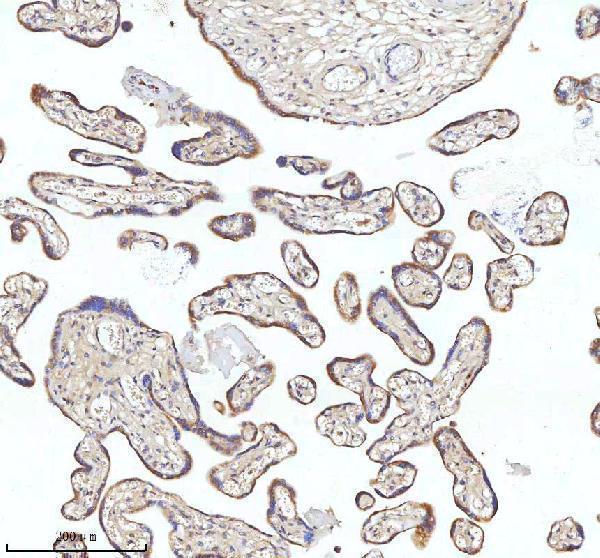
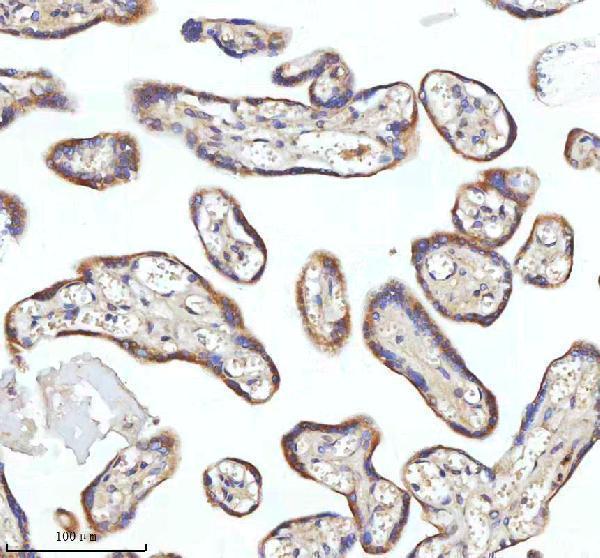
The catalog antibody A05926-1 has real IHC data from paraffin sections of human placenta and thyroid papillary carcinoma (IHC image captions). No IF data are shown (catalog: IF images absent).
A05926-1 was used for IHC on paraffin sections of human placenta and thyroid papillary carcinoma (IHC image captions). Its listed reactivity is human, mouse and rat, while the supplied IHC images show human tissue only (catalog reactivity; IHC image captions).
Which to pick: Choose A05926-1 for paraffin-section IHC: its rabbit antibody has an IHC application listing and was used at 2 μg/ml after EDTA pH 8.0 heat retrieval in the supplied images; the fixative is unreported (catalog; IHC image captions). There is no IF/ICC-validated SKU in this payload (catalog applications; IF images absent). For mouse or rat tissue, A05926-1 lists reactivity with those species, but its supplied IHC evidence is human tissue, so validate staining in the intended species (catalog reactivity; IHC image captions).