This website uses cookies to ensure you get the best experience on our website.


- Table of Contents
Plan MKNK1 staining in paraffin sections using its general cytoplasmic tissue pattern (HPA tissue IHC). Compare staining across consistently fixed sections and account for possible isoform-dependent localisation (standard IHC practice; HPA tissue IHC).

Expected localisation, validated protocols, controls and antibodies — the at-a-glance facts below, then the full design guide.
| Expected localisation | Cytoplasmic tissue staining (HPA tissue IHC); nuclear location annotated (UniProt) | |
| Staining pattern | General cytoplasmic staining across tissue cell types (HPA tissue IHC) | |
| Antigen retrieval | Tris-EDTA pH 9.0 HIER, 95–98 °C, 20 min (rule: nuclear antigen) | |
| Positive control | Appendix+4 more · see all | |
| Negative control | None in HPA (detected in all 45 tissues); use no-primary + isotype controls |
| Fixation | Keep fixation consistent across sections. (standard IHC practice; not target-specific) | |
| Caveat | Isoform-dependent localisation may alter staining (HPA tissue IHC) | |
| Regulation | Expression regulation unreported (UniProt) | |
| Isoform / epitope | 3 isoforms; epitope coverage is unspecified (UniProt; datasheet) |
The catalog antibody protocol is followed by published MKNK1 IHC protocols for tissue sections (PMC9957794) and mouse primary tumors (PMC11397505).
| Sample | Tissue sections; selected-image fixative not specified (standard IHC workflow) |
| Fixation | Image fixative and duration unreported (datasheet A03783-2); verify before use. |
| Sectioning | 4–5 µm sections on charged slides (standard) |
| Deparaffinisation | Xylene, graded ethanol series to water (standard) |
| Antigen retrieval | Heat-induced epitope retrieval in Tris-EDTA buffer, pH 9.0, 20 min at 95–98 °C (standard rule: nuclear antigen) |
| Peroxidase block | 3% H2O2, 10 min, room temperature (standard) |
| Blocking | 10% normal serum of the secondary host, 30 min, room temperature (standard) |
| Primary antibody | Rabbit anti-MKNK1, 1:50-1:200 (datasheet A03783-2) |
| Primary incubation | Overnight at 4 °C (standard) |
| Detection | HRP-polymer secondary, DAB chromogen 5–10 min (standard) |
| Counterstain | Hematoxylin, blue, dehydrate and mount (standard) |
| Expected result | MKNK1-positive staining in glandular cells of appendix (HPA tissue IHC: High). HPA tissue profile: General cytoplasmic expression. No signal in the no-primary control. |
MKNK1 is reported in the cytoplasm and nucleus and has no transmembrane segment (UniProt Q9BUB5). In tissue IHC, expect a general cytoplasmic pattern, including strong staining in the cell populations listed by HPA (HPA: tissue IHC). HPA rates the tissue profile Approved, with medium consistency between antibody staining and RNA expression; its annotation is based on CAB037310 (HPA: tissue IHC).
| Cytoplasmic staining in appendix or colon glandular cells, or bronchial respiratory epithelial cells. | This fits the general tissue pattern and named high staining populations (HPA: tissue IHC). Compare intensity with neighboring cells on the same section; HPA levels describe observed staining, not a numerical scoring cutoff. |
| Predominantly nuclear staining, or a nuclear signal alongside cytoplasmic staining. | Check the assay context before rejecting it: UniProt lists both compartments, while HPA tissue IHC describes general cytoplasmic expression (UniProt Q9BUB5; HPA: tissue IHC). HPA reports supported nucleoplasmic localization by ICC-IF and cautions that localization may depend on isoform (HPA: subcellular; HPA: tissue IHC). |
| A crisp membrane rim or extracellular deposit dominates the signal. | This is outside the reported cytoplasmic and nuclear locations and is unexplained by MKNK1 topology (UniProt Q9BUB5). Treat it as suspect staining; check the detection control and compare a second IHC-validated antibody before assigning it to MKNK1 (standard IHC practice). |
| Strong staining appears in an unexpected cell population while expected cells remain weak. | Possible causes include cross-reactivity or endogenous detection activity (standard IHC practice). HPA lists low staining in spleen red pulp cells, but its tissue list is not an exhaustive map of negative cells (HPA: tissue IHC). Confirm the cell identity and staining controls before interpreting the difference. |
| No visible signal in a selected high staining reference tissue. | For example, HPA reports high staining in appendix glandular cells and bone marrow hematopoietic cells (HPA: tissue IHC). A blank result there warrants checks of section quality, antibody dilution, retrieval, and detection reagents (standard IHC practice); it does not alone establish absent MKNK1. |
| Tissue and cell selection | HPA reports high staining in specified glandular, epithelial, hematopoietic, neuronal, Purkinje, and endothelial populations; spleen red pulp cells are low (HPA: tissue IHC). Select a named high staining population as a reference without treating other cells as established negatives. |
| Antibody validation | CAB027355 and CAB037310 have Approved IHC status; HPA's tissue profile is based on CAB037310 and has medium staining-to-RNA consistency (HPA: antibodies; HPA: tissue IHC). Interpret discrepancies in light of that qualification. |
| Isoforms and compartment | UniProt records three isoforms and both cytoplasmic and nuclear locations (UniProt Q9BUB5). HPA notes possible isoform-dependent localization, so an IHC–ICC compartment difference needs antibody and assay context (HPA: tissue IHC). |
| IF/ICC Q: What should appear? | A: HPA reports supported nucleoplasmic localization in ICC-IF, with images from HaCaT, RT-4, and U2OS (HPA: subcellular). That observation does not replace the general cytoplasmic tissue IHC pattern (HPA: tissue IHC). |
| Situation | Likely cause | Next action |
|---|---|---|
| High staining reference cells are blank. | The HPA reference predicts staining, but a negative slide can reflect an assay failure (HPA: tissue IHC; standard IHC practice). | Check section integrity and controls, then review retrieval, catalog antibody dilution, and detection steps; no target-specific retrieval condition is supplied (standard IHC practice). |
| Brown signal covers most tissue structures without cell boundaries. | Diffuse background can arise from detection reagents, blocking, or excess primary antibody (standard IHC practice). | Examine a no-primary control, review blocking and washes, and titrate the primary antibody while retaining a high staining reference section (standard IHC practice; HPA: tissue IHC). |
| Signal follows vessel or cell membranes as a sharp outline. | A membrane-dominant pattern is inconsistent with the listed intracellular locations and lack of a transmembrane segment (UniProt Q9BUB5). | Review morphology and detection controls; repeat with an independently IHC-validated antibody if available (standard IHC practice; HPA: antibodies). |
| Nuclear signal exceeds cytoplasmic signal. | The tissue profile is generally cytoplasmic, while supported ICC-IF localization is nucleoplasmic; HPA flags possible isoform dependence (HPA: tissue IHC; HPA: subcellular). | Record both compartments and the antibody used, then compare with a named high staining tissue population before calling the signal nonspecific (HPA: tissue IHC; standard IHC practice). |
| Unexpected cells stain more strongly than nearby expected cells. | Cell identification, cross-reactivity, or endogenous detection activity may explain the contrast (standard IHC practice). HPA's named levels do not define every cell as positive or negative (HPA: tissue IHC). | Recheck cell morphology and a no-primary control, and compare an independent IHC-validated antibody where available (standard IHC practice; HPA: antibodies). |
| IHC appears cytoplasmic but IF/ICC appears nucleoplasmic. | These are the respective reported patterns, and localization may be isoform dependent (HPA: tissue IHC; HPA: subcellular). | Keep the readouts assay-specific; record antibody identity and compartment scoring, and consult the separate IF/ICC guide for its workflow (HPA: antibodies; standard IHC/IF practice). |
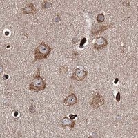
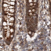
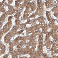
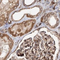


Comprehensive Human Protein Atlas IHC scoring per tissue (reliability: Approved — Medium consistency between antibody staining and RNA expression data. External characterization data suggest that subcellular localization is isoform dependent. Annotated expression profile is based on CAB037310.). Rows are taken directly from the HPA tissue chart — click any row's HPA link to view the source.
| Tissue | Cell type | Level | Evidence | Source |
|---|---|---|---|---|
| Appendix | Glandular cells | High | Protein (IHC) | HPA → |
| Bone marrow | Hematopoietic cells | High | Protein (IHC) | HPA → |
| Bronchus | Respiratory epithelial cells | High | Protein (IHC) | HPA → |
| Caudate | Neuronal cells | High | Protein (IHC) | HPA → |
| Cerebellum | Purkinje cells | High | Protein (IHC) | HPA → |
| Tissue | Cell type | Level | Evidence | Source |
|---|---|---|---|---|
| None in HPA: MKNK1 is detected in all 45 scored tissues. Use a no-primary (secondary-only) and an isotype control instead. | ||||
Troubleshoot MKNK1 staining in chromogenic IHC of paraffin sections, with one entry on translating the analysis to IF.
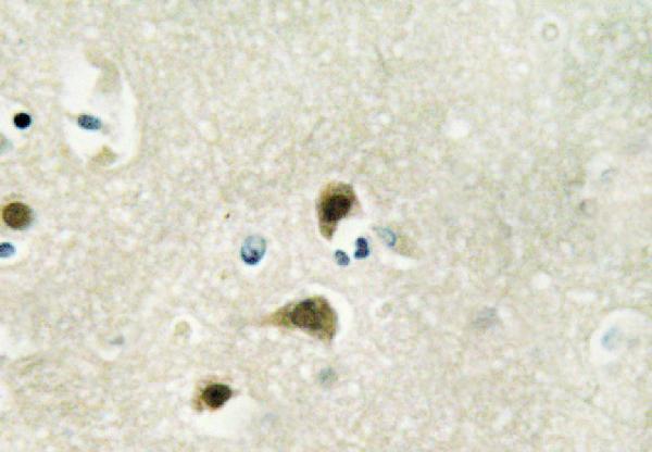
The catalog lists one IHC antibody with a human brain tissue image and stated human, mouse, and rat reactivity (catalog: A03783-2; IHC image caption). No IF image is supplied (catalog: A03783-2).
A03783-2 is the only SKU that will render; its IHC image shows human brain tissue (IHC image caption: A03783-2). The catalog lists IHC and WB applications and human, mouse, and rat reactivity (catalog: A03783-2).
Which to pick: For tissue IHC, choose A03783-2: it lists IHC and has a human brain tissue image (catalog and IHC image caption: A03783-2). For cross-species work, A03783-2 lists human, mouse, and rat reactivity, though its supplied IHC image shows only human tissue (catalog and IHC image caption: A03783-2). No IF/ICC application or image is listed, so an IF/ICC choice is unsupported; the IHC caption does not report section processing or fixative (catalog and IHC image caption: A03783-2).