MLPH / Melanophilin · IHC design guide

Design Immunohistochemistry for MLPH

Plan chromogenic MLPH IHC around the cytoplasmic staining reported in most tissues (HPA tissue IHC). Prostate glandular cells show high staining (HPA tissue IHC); consider the reported splice/transcript discrepancy when interpreting results (HPA tissue IHC).

Evidence assembled Oct 2026 · For research use; verify linked source records and product datasheet before use
Immunohistochemistry protocol sheet for MLPH (IHC for MLPH): expected localisation Cytoplasmic staining in most tissues (HPA tissue IHC), antibody A06081-2, validated IHC image, and IHC protocol steps
Printable MLPH IHC protocol sheet — expected localisation Cytoplasmic staining in most tissues (HPA tissue IHC), antibody A06081-2, controls and protocol steps. Open the full MLPH IHC guide →

MLPH Immunohistochemistry Experimental Design Guide

Expected localisation, validated protocols, controls and antibodies — the at-a-glance facts below, then the full design guide.

Must know before staining
Expected localisation Cytoplasmic staining in most tissues (HPA tissue IHC)
Staining pattern Cytoplasmic staining in prostate glandular cells (HPA tissue IHC)
Antigen retrieval EDTA pH 8.0 HIER, heat-mediated (datasheet A06081-2)
Positive control ⓘ Colon+4 more · see all
Negative control ⓘ Hippocampus+1 more · see all
Important caveats
Reasons your staining may differ from the expected pattern.
Fixation Keep fixation consistent across paraffin sections. (standard IHC practice; not target-specific)
Caveat Staining–RNA agreement is medium; splice/transcript discrepancy noted (HPA tissue IHC)
Regulation Expression regulation not annotated (UniProt)
Isoform / epitope 5 isoforms; assess antibody epitope coverage (UniProt)
Section 1

Recommended MLPH IHC & IF Protocols

The catalog antibody's IHC-P protocol (datasheet A06081-2) is accompanied by published protocols for prostate, rectal tumor, and breast carcinoma sections (PMC4715509; PMC8621396; PMC3783308).

Recommended immunohistochemistry (IHC-P) protocol parameters
SampleParaffin-embedded human prostate cancer tissue; fixative not specified (datasheet A06081-2)
FixationImage fixative and duration unreported (datasheet A06081-2); verify before use.
Sectioning4–5 µm sections on charged slides (standard)
DeparaffinisationXylene, graded ethanol series to water (standard)
Antigen retrievalHeat retrieval: EDTA pH 8.0 (datasheet A06081-2); 20 min, 95–100 °C (standard)
Peroxidase block3% H2O2, 10 min, room temperature (standard)
Blocking10% goat serum (datasheet A06081-2)
Primary antibodyRabbit anti-MLPH, 1:50 recommended; image 1:100 (datasheet A06081-2)
Primary incubationOvernight at 4 °C (datasheet A06081-2)
DetectionHRP-conjugated secondary, DAB chromogen (datasheet A06081-2)
CounterstainHematoxylin, blue, dehydrate and mount (standard)
Expected resultMLPH-positive staining in endothelial cells of colon (HPA tissue IHC: High). HPA tissue profile: Cytoplasmic expression in most tissues. No signal in the no-primary control.
💡Decision noteStart with EDTA pH 8.0 heat retrieval (datasheet A06081-2); the breast carcinoma study used an unspecified retrieval solution at 95 °C for 20 min (PMC3783308).
Section 2

What Is the Expected MLPH Staining Pattern?

MLPH is a cytoplasmic, non-transmembrane protein involved in melanosome transport (UniProt Q9BV36). In paraffin-section IHC, expect cytoplasmic staining in several epithelial and glandular cell populations, and in colon endothelial cells (HPA tissue IHC). HPA describes cytoplasmic expression in most tissues, with Enhanced IHC reliability but medium agreement between staining and RNA data and a splice or transcript discrepancy caution (HPA tissue IHC).

What am I looking at on my slide?
Cytoplasmic chromogen in duodenal, gallbladder, prostate, small-intestinal or stomach glandular cells.This matches the cell compartment (UniProt Q9BV36; HPA tissue IHC) and cell types scored High by HPA (HPA tissue IHC). Judge the signal within the named cells; a tissue-wide score can hide differences among its cell populations (general IHC interpretation).
Predominantly nuclear or membrane-outlined staining, without a convincing cytoplasmic component.That distribution conflicts with the reported cytoplasmic location and lack of a transmembrane segment (UniProt Q9BV36). Treat it as an unresolved staining pattern; inspect controls and detection background before assigning it to MLPH (general IHC practice).
Strong staining in hippocampal glial cells or spleen red-pulp cells.HPA reports MLPH as not detected in those specific cell populations (HPA tissue IHC). Unexpected signal may reflect cross-reactivity or endogenous detection activity; it does not make either entire tissue a universal negative control (general IHC interpretation).
Uniform haze across cells, stroma and slide, with little cell-specific contrast.This does not resemble HPA's cytoplasmic, cell-associated pattern (HPA tissue IHC). Diffuse background can arise from nonspecific reagent binding, insufficient blocking or detection background (general IHC practice); assess it against a negative detection control (general IHC practice).
No signal in an adequately preserved prostate glandular-cell or duodenal glandular-cell area.Both are HPA High populations (HPA tissue IHC), so an absent result warrants checking section quality, assay controls and reagent performance (general IHC practice). HPA notes a splice or transcript discrepancy, so one failed sample alone does not establish absent MLPH expression (HPA tissue IHC).
💡Expected MLPH appearanceCall a positive result when cytoplasmic chromogen is discernible in an HPA High population such as prostate glandular cells or esophageal squamous epithelial cells (UniProt Q9BV36; HPA tissue IHC); predominantly nuclear signal or staining that overwhelms cell boundaries is suspect (general IHC interpretation).
How each factor affects the staining
Compartment and topologyMLPH is cytoplasmic and has no transmembrane segment (UniProt Q9BV36). Interpret cell-associated cytoplasmic staining as the expected compartment; membrane-only staining needs independent scrutiny (general IHC interpretation).
Cell-specific tissue contextHPA scores colon endothelial cells and several glandular or epithelial populations High, but adrenal glandular cells Medium (HPA tissue IHC). Compare like cell types when judging intensity; do not treat all cells in a positive tissue as equally positive (general IHC interpretation).
Isoforms and evidence limitsUniProt lists 5 isoforms (UniProt Q9BV36). HPA flags a splice or transcript discrepancy and medium agreement with RNA despite Enhanced IHC reliability (HPA tissue IHC). The supplied evidence does not identify the assay epitope or establish isoform-specific staining.
IHC validation versus ICC/IF evidenceHPA lists antibody HPA014685 as IHC Enhanced, with no ICC validation status or ICC/IF images (HPA antibodies; HPA subcellular). IHC tissue staining therefore supplies a compartment expectation, not an independently confirmed ICC/IF staining pattern.
Fixation sensitivityTarget-specific effects of fixation, cross-linking and antigen retrieval on MLPH staining are unreported in the supplied UniProt and HPA records. Choose and document an IHC retrieval condition using ordinary assay controls (general IHC practice); no MLPH-specific optimum is established here.
Why is my staining missing, weak or wrong?
SituationLikely causeNext action
The positive tissue shows little or no cytoplasmic signal.The assay may have failed, or the chosen area may lack the HPA-positive cell population (general IHC interpretation; HPA tissue IHC).Inspect prostate or duodenal glandular cells scored High by HPA, then review section quality, antibody incubation and detection controls (HPA tissue IHC; general IHC practice).
Signal is chiefly nuclear or forms a sharp membrane rim.This disagrees with MLPH's cytoplasmic location and non-transmembrane topology (UniProt Q9BV36); background or off-target staining is possible (general IHC interpretation).Compare a matched detection control and an HPA-positive cell population; score MLPH only where a credible cytoplasmic component remains (general IHC practice; HPA tissue IHC).
The slide has widespread brown haze.Nonspecific binding or endogenous chromogen-generating activity can obscure cell-specific signal (general chromogenic IHC practice).Check a negative detection control, blocking and wash conditions; evaluate whether the haze persists outside the expected cytoplasmic cell pattern (general IHC practice; HPA tissue IHC).
Hippocampal glia or spleen red-pulp cells stain strongly.Those cells are scored not detected by HPA (HPA tissue IHC); cross-reactivity or endogenous detection activity is possible (general IHC interpretation).Verify the cell identity and compare negative detection controls before calling the unexpected staining MLPH-positive (general IHC practice).
A weak cell population looks negative beside a strong one.HPA reports different cell-specific levels, including Low in lung alveolar cells and High in prostate glandular cells (HPA tissue IHC).Score intensity within each identified cell population and record the comparison tissue; avoid a single threshold inferred from one strong field (general IHC interpretation).
Can this IHC result establish the expected ICC/IF image?HPA provides no ICC/IF images or ICC validation status for the listed antibody (HPA subcellular; HPA antibodies).Use cytoplasm as a provisional compartment expectation from UniProt, and assess any ICC/IF result with its own controls and validation (UniProt Q9BV36; general ICC/IF practice).

Sample controls for MLPH IHC & IF

🧪Run prostate first and expect staining in glandular cells (HPA: High in prostate glandular cells). Use hippocampus glial cells as the negative tissue (HPA: Not detected in hippocampus glial cells); on the prostate slide, assess adjacent nonglandular cells as candidate internal negatives rather than assuming they lack MLPH.
Positive control tissue: Colon (Endothelial cells, HPA High)
Negative control tissue: Hippocampus (HPA Not detected)
ICC-IF cell lines (HPA subcellular resource): HPA carries no ICC-IF cell line for MLPH; derive a cell-line control from the positive tissue's cell type (Endothelial cells) and confirm it by RNA or western blot first.
Technical controls: Include a no-primary, secondary-only control, an isotype-matched rabbit IgG control, and an MLPH knockout biological negative (caption: rabbit primary antibody; standard IHC controls). Block endogenous peroxidase and check for nonspecific DAB signal in the prostate section (caption: prostate cancer section detected with HRP/DAB).
⚠️Feasibility: No target-specific fixation window or fixation effect is reported, and the selected A06081-2 paraffin-section caption does not state a fixative (caption: fixative not stated). The illustrated IHC uses heat-mediated retrieval in EDTA, pH 8.0; compare with a section without retrieval to assess dependence (caption: EDTA retrieval). The supplied evidence does not establish that frozen sections or IF/ICC are easier, and it reports no prostate-specific artefact.

HPA tissue IHC evidence for MLPH

Comprehensive Human Protein Atlas IHC scoring per tissue (reliability: Enhanced — Medium consistency between antibody staining and RNA expression data. Caution, Splice and/or transcript discrepancy exists.). Rows are taken directly from the HPA tissue chart — click any row's HPA link to view the source.

Positive expression · recommended positive controls

TissueCell typeLevelEvidenceSource
Colon Endothelial cells High Protein (IHC) HPA →
Duodenum Glandular cells High Protein (IHC) HPA →
Esophagus Squamous epithelial cells High Protein (IHC) HPA →
Gallbladder Glandular cells High Protein (IHC) HPA →
Prostate Glandular cells High Protein (IHC) HPA →

Undetected expression · recommended negative controls

TissueCell typeLevelEvidenceSource
Hippocampus Glial cells Not detected Protein (IHC) HPA →
Spleen Cells in red pulp Not detected Protein (IHC) HPA →
Section 3

Advanced MLPH IHC Tips

Troubleshoot MLPH staining in paraffin sections by checking retrieval, cytoplasmic localisation, cell identity and assay controls before interpreting intensity.

Which retrieval conditions should I use when MLPH staining is weak?
Use heat-mediated retrieval in EDTA at pH 8.0 for paraffin sections stained with A06081-2 (datasheet A06081-2). The selected tissue image used this retrieval before a 1:100 primary antibody incubation overnight at 4°C, providing a starting point for reproducing that result (selected A06081-2 IHC caption). If signal remains weak, compare a longer or shorter heating period on matched sections while keeping the antibody dilution and detection steps constant (standard IHC practice). Evaluate recovery in prostate glandular cells and check that negative-control staining has not risen with stronger retrieval (HPA: High in prostate glandular cells; standard IHC practice).
How should I troubleshoot fixation-related loss of MLPH staining?
The selected image documents a paraffin-embedded section but does not report its fixative, so MLPH-specific sensitivity to fixation is unknown (selected A06081-2 IHC caption: fixative not stated). Record the fixative and fixation interval for every specimen, then compare sections processed with known conditions using the same EDTA pH 8.0 retrieval and detection workflow (datasheet A06081-2; standard IHC practice). Assess tissue preservation alongside cytoplasmic signal, because damaged morphology makes localisation difficult to judge (UniProt Q9BV36: cytoplasm; standard IHC practice). Do not attribute a negative result to fixation until a concurrently processed positive-control section stains as expected (standard IHC practice).
Where should convincing MLPH staining appear in tissue sections?
Expect predominantly cytoplasmic staining: MLPH is annotated in the cytoplasm and has no transmembrane segment (UniProt Q9BV36: subcellular location and topology). Its role linking melanosome-bound RAB27A to MYO5A can inform interpretation, but a DAB pattern alone cannot establish those molecular interactions (UniProt Q9BV36: function and subunit; standard IHC practice). HPA reports cytoplasmic expression across most tissues and High staining in prostate glandular cells, offering a cell-based reference for the selected prostate section (HPA tissue IHC; selected A06081-2 IHC caption). Treat isolated nuclear or membrane-only colour as suspect until it survives control and localisation checks (UniProt Q9BV36: cytoplasm; standard IHC practice).
Could isoforms or an unknown epitope explain inconsistent MLPH staining?
MLPH has 5 listed isoforms, so staining can vary if an antibody epitope is absent from an expressed isoform (UniProt Q9BV36: isoforms; standard immunostaining principle). The supplied evidence does not map the A06081-2 epitope; therefore, staining cannot be assigned to a particular isoform from this assay alone (selected A06081-2 IHC caption; UniProt Q9BV36: isoforms). HPA also flags a splice or transcript discrepancy when comparing antibody staining with RNA expression (HPA tissue IHC: reliability description). When samples disagree, compare annotated isoform expression where available and report the antibody identifier, retrieval conditions and scoring threshold so the result remains interpretable (standard IHC practice).
How can I assess MLPH by multiplex immunofluorescence?
For the separate IF/ICC workflow, pair MLPH with a marker that identifies the cell population under study, then assess signal within those cells (standard IF practice). Choose spectrally separated fluorophores and measure unstained tissue autofluorescence before assigning a channel to weak MLPH signal (standard IF practice). Because MLPH is cytoplasmic and lacks a transmembrane segment, permeabilisation should permit antibody access to intracellular epitopes, with its strength optimised against cell morphology (UniProt Q9BV36: location and topology; standard IF practice). The supplied HPA subcellular record lists no ICC/IF image-bearing cell lines, so confirm localisation and specificity with appropriate IF controls (HPA subcellular; standard IF practice).
What should I check when DAB obscures MLPH staining?
First compare a no-primary control with the stained section to identify signal arising from the secondary reagent or detection system (standard IHC practice). The selected A06081-2 image used 10% goat serum blocking, a peroxidase-conjugated secondary reagent and DAB, giving documented settings to reproduce before changing one variable at a time (selected A06081-2 IHC caption). Include a peroxidase-blocking step and inspect its control for residual endogenous enzyme activity; this is a general chromogenic IHC precaution (standard IHC practice). If diffuse colour persists, review blocking, washing and primary antibody concentration while checking that expected cytoplasmic signal remains visible (UniProt Q9BV36: cytoplasm; standard IHC practice).
How should I score MLPH across paraffin sections? ⚠ ANSWER MARKED FOR VERIFICATION
Define the scored cell population and cytoplasmic compartment before comparing sections, since MLPH is cytoplasmic and HPA staining differs by cell type (UniProt Q9BV36: location; HPA tissue IHC). An H-score combines the percentage of cells at each intensity and ranges from 0–300; alternatively, report percentage positive cells using a prespecified threshold (standard IHC scoring practice). Normalise counts to the number of evaluable cells in the chosen population, or report positive-cell density per mm² of evaluable tissue (standard IHC scoring practice). Keep retrieval, imaging and scoring thresholds consistent, and exclude folds or necrotic areas from the denominator (standard IHC practice).
How do I distinguish true MLPH signal from artefact?
A plausible positive result shows cytoplasmic staining in identifiable cells, consistent with MLPH localisation and the HPA tissue pattern (UniProt Q9BV36: cytoplasm; HPA tissue IHC). In prostate, compare glandular cells with adjacent tissue and a concurrently stained control section; HPA reports High staining in prostate glandular cells (HPA tissue IHC; standard IHC practice). Be cautious when colour is confined to nuclei, tissue edges, folds or necrotic regions, where localisation or section quality can mislead interpretation (UniProt Q9BV36: cytoplasm; standard IHC practice). A positive no-primary control points toward detection background, including endogenous peroxidase, rather than antibody-dependent MLPH staining (standard IHC practice).
Boster reagents

Best MLPH / Melanophilin IHC Antibodies

Two human-reactive anti-MLPH antibodies have image-backed data: IHC in a paraffin-embedded human tissue section and IF/ICC in A549 cells (catalog reactivity and image captions).

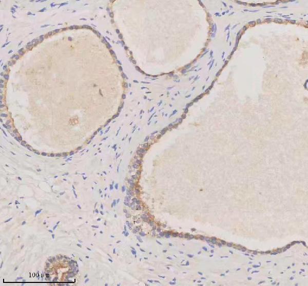
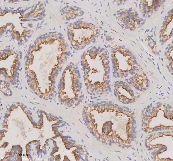
Real IHC data IHC analysis of Melanophilin/MLPH using anti-Melanophilin/MLPH antibody (A06081-2). Melanophilin/MLPH was detected in a paraffin-embedded section of human prostate cancer tissue. Heat mediated antigen retrieval was performed in EDTA buffer (pH 8.0, epitope retrieval solution). The tissue section was blocked with 10% goat serum. The tissue section was then incubated with 1:100 rabbit anti-Melanophilin/MLPH Antibody (A06081-2) overnight at 4°C. Peroxidase Conjugated Goat Anti-rabbit IgG was used as secondary antibody and incubated for 30 minutes at 37°C. The tissue section was developed using HRP Conjugated Rabbit IgG Super Vision Assay Kit (Catalog # SV0002) with DAB as the chromogen.
Anti-Melanophilin/MLPH Antibody
Cat # A06081-2
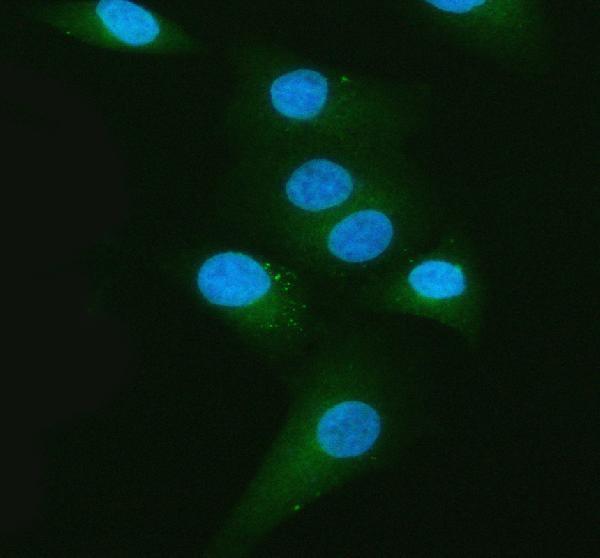
Real IF data IF analysis of Melanophilin/MLPH using anti-Melanophilin/MLPH antibody (A06081-3). Melanophilin/MLPH was detected in an immunocytochemical section of A549 cells. Enzyme antigen retrieval was performed using IHC enzyme antigen retrieval reagent (AR0022) for 15 mins. The cells were blocked with 10% goat serum. And then incubated with 5 μg/mL rabbit anti-Melanophilin/MLPH Antibody (A06081-3) overnight at 4°C. DyLight®488 Conjugated Goat Anti-Rabbit IgG (BA1127) was used as secondary antibody at 1:500 dilution and incubated for 30 minutes at 37°C. The section was counterstained with DAPI. Visualize using a fluorescence microscope and filter sets appropriate for the label used.
Anti-MLPH Antibody ®
Cat # A06081-3

A06081-2 is listed for IHC and IF/ICC, with an IHC image from paraffin-embedded human prostate cancer tissue (A06081-2 applications and IHC caption). A06081-3 is listed for IF/ICC, with an IF image from A549 cells (A06081-3 applications and IF caption).

Which to pick: For tissue IHC, choose A06081-2: this human-reactive polyclonal antibody lists IHC, and its own image documents EDTA retrieval at pH 8.0 in a paraffin-embedded section; the fixative is unreported (A06081-2 catalog and IHC caption). For IF/ICC, choose A06081-3: this human-reactive polyclonal antibody lists IF/ICC and has an A549-cell IF image, but no listed IHC application (A06081-3 catalog and IF caption). Neither has catalog support for a cross-species recommendation because both list only Human reactivity (catalog reactivity).

Each figure is that product's own IHC / IF validation image from its datasheet.

References

  1. UniProt Consortium. UniProt entry Q9BV36 (MELPH_HUMAN, Melanophilin).
  2. Human Protein Atlas. MLPH tissue IHC expression (reliability: Enhanced).
  3. Human Protein Atlas. MLPH subcellular location (ICC-IF): Highest expression in MCF-7: 209.5 nTPM.
  4. Human Protein Atlas. MLPH antibody validation summary (1 antibodies).
  5. Evolutionary selection of alleles in the melanophilin gene that impacts on prostate organ function and cancer risk. Evolution, medicine, and public health 2021 — PMC8573191.
  6. Putative Prostate Cancer Risk SNP in an Androgen Receptor-Binding Site of the Melanophilin Gene Illustrates Enrichment of Risk SNPs in Androgen Receptor Target Sites. Human mutation 2016 — PMC4715509.
  7. Overexpression of MLPH in Rectal Cancer Patients Correlates with a Poorer Response to Preoperative Chemoradiotherapy and Reduced Patient Survival. Diagnostics (Basel, Switzerland) 2021 — PMC8621396.
  8. Identification of gene expression signature in estrogen receptor positive breast carcinoma. Biomarkers in cancer 2010 — PMC3783308.
  9. PubMed PMID:14702039 — UniProt-cited evidence.
  10. PubMed PMID:15815621 — UniProt-cited evidence.
  11. PubMed PMID:15489334 — UniProt-cited evidence.