MMP11 / Stromelysin-3 · IHC design guide

Design Immunohistochemistry for MMP11

Plan MMP11 paraffin-section IHC using the reported cytoplasmic tissue pattern and high staining in placental trophoblasts (HPA tissue IHC). The catalog antibody has an IHC-P working range of 0.5–1 μg/ml (datasheet A04490-1); interpret cell-associated staining with its secreted location in mind (UniProt).

Evidence assembled Oct 2026 · For research use; verify linked source records and product datasheet before use
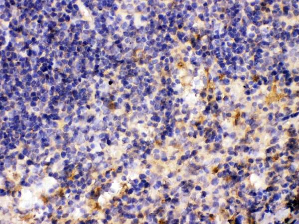
Immunohistochemistry protocol sheet for MMP11 (IHC for MMP11): expected localisation Cytoplasmic staining in several tissues (HPA tissue IHC), antibody A04490-1, validated IHC image, and IHC protocol steps
Printable MMP11 IHC protocol sheet — expected localisation Cytoplasmic staining in several tissues (HPA tissue IHC), antibody A04490-1, controls and protocol steps. Open the full MMP11 IHC guide →

MMP11 Immunohistochemistry Experimental Design Guide

Expected localisation, validated protocols, controls and antibodies — the at-a-glance facts below, then the full design guide.

Must know before staining
Expected localisation Cytoplasmic staining in several tissues (HPA tissue IHC)
Staining pattern Cytoplasmic staining; high in placental trophoblasts (HPA tissue IHC)
Antigen retrieval Citrate pH 6 HIER, heat-mediated (datasheet A04490-1)
Positive control ⓘ Placenta
Negative control ⓘ Appendix+4 more · see all
Important caveats
Reasons your staining may differ from the expected pattern.
Fixation Keep formalin fixation consistent across sections. (standard IHC practice; not target-specific)
Caveat Secreted MMP11 may stain away from its producing cells (HPA tissue IHC)
Regulation Expressed in breast carcinoma stroma (UniProt)
Isoform / epitope No isoforms; signal and propeptide are cleaved (UniProt)
Section 1

Recommended MMP11 IHC & IF Protocols

The catalog antibody protocol uses citrate pH 6 retrieval (datasheet: A04490-1). The published options below cover lung adenocarcinoma and inverted papilloma tissue (PMC9900007; PMC12770796).

Recommended immunohistochemistry (IHC-P) protocol parameters
SampleParaffin-embedded mouse spleen tissues; fixative not specified (datasheet A04490-1)
FixationImage fixative and duration unreported (datasheet A04490-1); verify before use.
Sectioning4–5 µm sections on charged slides (standard)
DeparaffinisationXylene, graded ethanol series to water (standard)
Antigen retrievalHeat retrieval: Citrate pH 6, 20 min (datasheet A04490-1)
Peroxidase block3% H2O2, 10 min, room temperature (standard)
Blocking10% goat serum (datasheet A04490-1)
Primary antibodyRabbit anti-MMP11, 0.5-1μg/ml (datasheet A04490-1)
Primary incubationOvernight at 4 °C (datasheet A04490-1)
DetectionStreptavidin-biotin complex (SABC), DAB chromogen (datasheet A04490-1)
CounterstainHematoxylin, blue, dehydrate and mount (standard)
Expected resultMMP11-positive staining in trophoblastic cells of placenta (HPA tissue IHC: High). HPA tissue profile: Cytoplasmic expression in several tissues. No signal in the no-primary control.
💡Decision noteStart with citrate pH 6 heat retrieval because the catalog datasheet specifies it (datasheet: A04490-1). ER2 retrieval for 20 min is a published alternative (PMC12770796).
Section 2

What Is the Expected MMP11 Staining Pattern?

MMP11 is a secreted protein associated with the extracellular space and matrix; it has no transmembrane segment (UniProt P24347). In tissue IHC, expect cytoplasmic staining, especially strong staining in placental trophoblastic cells (HPA tissue IHC: High). Stromal cells of breast carcinomas are another relevant cell population, although no staining intensity is specified for them here (UniProt P24347: tissue specificity). Interpret the pattern with HPA’s Approved reliability rating and its warning that presumed off-target binding was disregarded (HPA tissue IHC).

What am I looking at on my slide?
Strong cytoplasmic staining in placental trophoblastic cells.This matches the clearest supplied positive tissue pattern (HPA tissue IHC: High in placental trophoblastic cells). Score the cells and compartment together. A high signal elsewhere cannot inherit the same interpretation without supporting tissue evidence.
Predominantly nuclear or sharply membrane-restricted staining.These are poor matches for the reported cytoplasmic tissue pattern (HPA tissue IHC) and for a protein with no transmembrane segment (UniProt P24347 topology). Treat them as suspect; compare controls and staining distribution before assigning them to MMP11.
Strong staining in a cell population reported as undetected.For example, appendix glandular cells and breast adipocytes are listed as Not detected (HPA tissue IHC). Unexpected staining may reflect cross-reactivity or endogenous detection activity (general IHC practice). Check its compartment, distribution, and assay controls before interpreting it as target expression.
Diffuse color across tissue, stroma, and blank areas.A uniform haze that obscures cell boundaries is background, not a convincing cell-specific pattern (general IHC practice). Because MMP11 is secreted, extracellular signal is biologically plausible (UniProt P24347); that annotation alone cannot validate diffuse staining on a particular slide.
No signal in placental trophoblastic cells.This conflicts with the supplied positive reference (HPA tissue IHC: High). First assess tissue quality, retrieval, antibody dilution, detection, and counterstain using the chosen assay’s controls (general IHC practice). A negative result elsewhere is hard to interpret until the positive control works.
💡Expected MMP11 appearanceCall positive a clear, strong cytoplasmic signal in placental trophoblastic cells (HPA tissue IHC: High); isolated nuclear staining or uniform background is suspect against that reference pattern (HPA tissue IHC; general IHC practice).
How each factor affects the staining
Secretion and compartmentMMP11 is secreted to extracellular space and matrix and lacks a transmembrane segment (UniProt P24347). Tissue IHC nevertheless reports cytoplasmic expression in several tissues (HPA tissue IHC). Judge the observed cellular pattern without requiring every positive field to show a matrix deposit.
Protein processingThe precursor has a signal peptide at residues 1–31 and a propeptide at 32–97; the annotated chain spans 98–488 (UniProt P24347). Antibody recognition could depend on its epitope (general immunostaining principle), but no epitope location is supplied, so no processing-specific staining prediction is justified.
Tissue selection and intensityPlacental trophoblastic cells provide the documented High reference (HPA tissue IHC). Several other listed cell populations are Low or Not detected (HPA tissue IHC). These labels describe the named populations; they do not establish that every cell in the same tissue has that intensity.
Antibody validationThe tissue profile is Approved, with presumed off-target binding observed and disregarded (HPA tissue IHC). HPA068864 and CAB002593 have IHC Approved status, while HPA055204 has ICC Approved status (HPA antibodies). Keep IHC and ICC validation separate when weighing an unexpected compartment or cell type.
Why is my staining missing, weak or wrong?
SituationLikely causeNext action
Placental trophoblastic cells are blank.The expected positive reference is missing (HPA tissue IHC: High). The slide alone cannot identify which assay step failed.Check section integrity, antigen retrieval, primary-antibody conditions, detection reagents, and counterstain against the chosen IHC protocol and controls (general IHC practice). Reassess other negative fields after the positive control stains.
The whole section has a weak brown haze.Diffuse background can arise from nonspecific binding or detection activity (general IHC practice); a secreted annotation does not prove the haze is MMP11 (UniProt P24347).Inspect a no-primary control, verify blocking and washes, and compare the haze with cell boundaries and the placental positive pattern (general IHC practice; HPA tissue IHC: High in trophoblastic cells).
Undetected-reference cells stain strongly.Staining in appendix glandular cells or breast adipocytes conflicts with their Not detected entries (HPA tissue IHC). Cross-reactivity or endogenous detection activity is possible (general IHC practice).Compare a no-primary control and the validated positive tissue, then review detection blocking and whether the signal is truly cellular (general IHC practice; HPA tissue IHC: High in placental trophoblastic cells).
Signal appears only in nuclei or as a crisp cell rim.The compartment differs from the cytoplasmic tissue profile (HPA tissue IHC); a membrane rim also lacks support from the no-transmembrane topology (UniProt P24347).Check counterstain overlap, chromogen deposits, and control sections before scoring it as MMP11 (general IHC practice). Record the unexpected compartment separately from any convincing cytoplasmic staining.
Breast carcinoma stroma appears negative.UniProt identifies stromal cells of breast carcinomas as an expression site but supplies no IHC intensity for the examined section (UniProt P24347: tissue specificity).Confirm that the section contains assessable stromal cells and that a placental positive control stains (general IHC practice; HPA tissue IHC: High). Report the result for the sampled section without assigning an expected stromal intensity.
What should an IF/ICC result look like?ICC-IF and tissue IHC describe different observations: HPA reports mainly Golgi localization, with additional cytosol localization marked uncertain (HPA subcellular ICC-IF).For IF/ICC interpretation, consult its separate guide; do not transfer a tissue IHC protocol to ICC-IF. HPA055204 is ICC Approved, while HPA068864 and CAB002593 are IHC Approved (HPA antibodies).

Sample controls for MMP11 IHC & IF

🧪Run placenta first: trophoblastic cells should stain (HPA: High in trophoblastic cells). Use appendix glandular cells as the negative tissue (HPA: Not detected in glandular cells); on the placental slide, use morphologically distinct cells that remain unstained as internal background references, without assuming all other cell types are MMP11-negative (HPA: High in trophoblastic cells).
Positive control tissue: Placenta (Trophoblastic cells, HPA High)
Negative control tissue: Appendix (HPA Not detected)
ICC-IF cell lines (HPA subcellular resource): HPA ICC-IF images show MMP11 in SH-SY5Y, SiHa, U2OS, with annotated localisation: Golgi apparatus (approved) (HPA subcellular).
Technical controls: Include a no-primary, secondary-only control and a concentration-matched, clonality-matched nonimmune rabbit IgG control (caption: rabbit primary antibody); use an MMP11 knockout specimen or peptide-blocked primary as a biological specificity control if available. For placental chromogenic IHC, block endogenous peroxidase and check endogenous biotin background with the stated streptavidin–biotin detection system (caption: SABC with DAB).
⚠️Feasibility: A target-specific fixation window or fixation effect is unreported, and the selected A04490-1 tissue-IHC caption does not state the fixative (caption: fixative not stated). Citrate retrieval at pH 6 for 20 minutes was used in the paraffin-section example, but whether retrieval is required has not been established (caption: heat-mediated antigen retrieval). The supplied evidence does not establish that frozen sections or IF/ICC are easier; placental endogenous biotin warrants a background check with the caption’s SABC detection method (caption: SABC with DAB).

HPA tissue IHC evidence for MMP11

Comprehensive Human Protein Atlas IHC scoring per tissue (reliability: Approved — Secreted protein, tissue location of RNA and protein is expected to differ. Presumed off target binding observed and disregarded.). Rows are taken directly from the HPA tissue chart — click any row's HPA link to view the source.

Positive expression · recommended positive controls

TissueCell typeLevelEvidenceSource
Placenta Trophoblastic cells High Protein (IHC) HPA →

Undetected expression · recommended negative controls

TissueCell typeLevelEvidenceSource
Appendix Glandular cells Not detected Protein (IHC) HPA →
Bone marrow Hematopoietic cells Not detected Protein (IHC) HPA →
Breast Adipocytes Not detected Protein (IHC) HPA →
Caudate Glial cells Not detected Protein (IHC) HPA →
Cerebellum Cells in granular layer Not detected Protein (IHC) HPA →
Section 3

Advanced MMP11 IHC Tips

Use the catalog antibody’s tissue IHC example as a starting point, then judge MMP11 staining by compartment, cell type and control performance.

Which retrieval conditions should I start with for MMP11 paraffin-section IHC?
Start with heat-mediated antigen retrieval in citrate buffer at pH 6 (datasheet A04490-1). The catalog antibody’s mouse spleen paraffin-section example heated sections for 20 minutes before a 10% goat-serum block and primary incubation at 1 µg/mL overnight at 4°C (caption A04490-1). Keep retrieval time, section thickness and detection conditions matched across comparison sections (standard IHC practice). If staining is weak, adjust heating time on replicate sections while checking tissue integrity and control staining (standard IHC practice). The example reports paraffin embedding but does not identify the fixative, so retrieval success cannot establish performance across fixation methods (caption A04490-1).
How should I troubleshoot fixation-related variation in MMP11 staining?
Target-specific MMP11 sensitivity to any fixative or fixation duration is unknown from the supplied IHC evidence (caption A04490-1). The catalog example identifies paraffin-embedded mouse spleen but does not state its fixative; its citrate pH 6 retrieval for 20 minutes therefore cannot validate a particular fixation condition (caption A04490-1). Record the fixative and processing history for each specimen, and compare matched sections under the same retrieval and detection conditions (standard IHC practice). If staining differs, assess morphology and a concurrently processed positive-control section before changing primary-antibody concentration (standard IHC practice). Do not attribute a difference to MMP11 biology until processing variation has been assessed (standard IHC practice).
Should MMP11 staining appear inside cells or in extracellular matrix?
Interpret MMP11 as a secreted protein associated with extracellular space and matrix, with no annotated transmembrane segment (UniProt P24347). Cellular staining is also plausible: HPA reports cytoplasmic tissue expression and an approved Golgi location in its subcellular analysis (HPA tissue IHC; HPA subcellular). Score cellular and extracellular signal separately on the 1 µg/mL catalog-antibody IHC condition if that condition is used (caption A04490-1; standard IHC practice). Examine adjacent sections for staining that tracks recognizable cells or matrix rather than folds and section edges (standard IHC practice). Secreted protein can accumulate away from its producing cell, so staining location alone does not identify the source cell (UniProt P24347; HPA tissue IHC).
Could MMP11 processing change which structures the antibody detects?
MMP11 is annotated as a 488-residue precursor with signal peptide 1–31, propeptide 32–97 and mature chain 98–488 (UniProt P24347). The supplied record annotates 0 isoforms and 0 glycosylation sites; those annotations do not identify the catalog antibody’s epitope (UniProt P24347; caption A04490-1). Check the antibody’s epitope documentation before assigning staining specifically to precursor, proprotein or mature extracellular MMP11 (standard IHC practice). In paraffin sections, compare staining after the documented citrate pH 6, 20-minute retrieval with an appropriate control section (caption A04490-1; standard IHC practice). Treat a change in compartmental staining as a hypothesis requiring epitope evidence, not proof of a particular processing state (UniProt P24347; standard IHC practice).
How can IF help resolve ambiguous chromogenic MMP11 localisation?
Use IF as a separate localisation check alongside the paraffin-section IHC result, with a marker for the expected cell population in a distinct channel (standard IF practice). HPA reports high MMP11 staining in placental trophoblastic cells, while its subcellular IF analysis places signal mainly in the Golgi apparatus (HPA tissue IHC; HPA subcellular). Select fluorophores after checking tissue autofluorescence in unstained and single-label controls, and keep each detector channel free of detectable bleed-through (standard IF practice). If examining an intracellular Golgi epitope, permeabilise after fixation; for accessible extracellular epitopes, compare a condition without permeabilisation (HPA subcellular; UniProt P24347; standard IF practice). The catalog IHC caption supplies no IF fixation or permeabilisation condition to transfer (caption A04490-1).
What should I check when MMP11 DAB staining is widespread?
First compare the stained section with a no-primary control and inspect folds, edges, necrotic areas and vascular spaces (standard IHC practice). The catalog example uses 10% goat serum, a biotinylated goat anti-rabbit secondary, a streptavidin–biotin complex and DAB; background can arise at several stages of that detection sequence (caption A04490-1; standard IHC practice). Include a peroxidase block and, where relevant, evaluate endogenous biotin as general chromogenic IHC controls (standard IHC practice). HPA’s tissue assessment notes presumed off-target binding that was disregarded, so a positive-looking region is not automatically specific (HPA tissue IHC). Recheck background against the 1 µg/mL primary condition and a matched negative control (caption A04490-1; standard IHC practice).
How should I quantify MMP11 across paraffin sections? ⚠ ANSWER MARKED FOR VERIFICATION
Define the analysis region and cell population before scoring, and analyse sections using the same staining and imaging settings (standard IHC practice). For cellular DAB signal, report the percentage of positive cells and an H-score using intensity grades 0–3; normalise counts to the eligible cells in each region (standard IHC practice). For extracellular signal, report stained area or optical-density measurements per mm² of evaluable tissue, with folds and necrosis excluded (standard IHC practice). Keep cellular and matrix measurements separate because MMP11 is secreted and HPA also reports cytoplasmic tissue staining (UniProt P24347; HPA tissue IHC). Record control performance and processing conditions for each batch before comparing scores (standard IHC practice).
How can I distinguish true MMP11 signal from an IHC artefact?
Expect a pattern compatible with secreted or matrix-associated MMP11, while allowing cellular signal consistent with HPA’s cytoplasmic tissue staining (UniProt P24347; HPA tissue IHC). HPA reports high staining in placental trophoblastic cells and no detection in appendix glandular cells; these observations can guide control selection without establishing performance for every specimen (HPA tissue IHC). Question isolated staining in an unexpected cell type or compartment, especially when it follows a section edge, fold or necrotic area (standard IHC practice). Compare those regions with a no-primary section and a peroxidase-blocked control before interpreting DAB deposits as target signal (standard IHC practice). HPA explicitly notes presumed off-target binding, so require a coherent pattern across controls and intact tissue (HPA tissue IHC; standard IHC practice).
Boster reagents

Best MMP11 / Stromelysin-3 IHC Antibodies

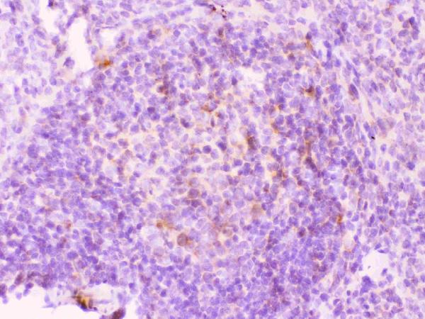
A04490-1 has IHC images from paraffin sections of mouse and rat spleen and human appendicitis tissue (catalog image captions); M04490 lists IHC and IF/ICC for human, mouse, and rat (catalog applications/reactivity).

Real IHC data IHC analysis of MMP11 using anti-MMP11 antibody (A04490-1). MMP11 was detected in paraffin-embedded section of mouse spleen tissues. Heat mediated antigen retrieval was performed in citrate buffer (pH6, epitope retrieval solution) for 20 mins. The tissue section was blocked with 10% goat serum. The tissue section was then incubated with 1μg/ml rabbit anti-MMP11 Antibody (A04490-1) overnight at 4°C. Biotinylated goat anti-rabbit IgG was used as secondary antibody and incubated for 30 minutes at 37°C. The tissue section was developed using Strepavidin-Biotin-Complex (SABC)(Catalog # SA1022) with DAB as the chromogen.
Anti-MMP11 Antibody ®
Cat # A04490-1

A04490-1 will render with its own IHC figure; its captions document staining in mouse and rat spleen and human appendicitis paraffin sections (catalog image captions). M04490 will render with IHC and IF/ICC listed for human, mouse, and rat, but no IHC or IF image is supplied (catalog applications/reactivity; image alts).

Which to pick: Choose A04490-1 for paraffin-section IHC because its own captions document staining in three species; the fixative is unreported (A04490-1 image captions). Choose M04490 for IF/ICC because that application is listed for this rabbit monoclonal antibody, although no IF image is supplied (M04490 catalog applications, host/clone, image alts). Both list human, mouse, and rat reactivity; A04490-1 has the cross-species IHC images (catalog reactivity; A04490-1 image captions).

Each figure is that product's own IHC / IF validation image from its datasheet.

References

  1. UniProt Consortium. UniProt entry P24347 (MMP11_HUMAN, Stromelysin-3).
  2. Human Protein Atlas. MMP11 tissue IHC expression (reliability: Approved).
  3. Human Protein Atlas. MMP11 subcellular location (ICC-IF): Mainly localized to the Golgi apparatus. In addition localized to the cytosol..
  4. Human Protein Atlas. MMP11 antibody validation summary (3 antibodies).
  5. MMP11 is associated with the immune response and immune microenvironment in EGFR-mutant lung adenocarcinoma. Frontiers in oncology 2023 — PMC9900007.
  6. MMP11 as a Prognostic Indicator for Inverted Papilloma Histologic Grade and Recurrence. The Laryngoscope 2026 — PMC12770796.
  7. Dual tumor-suppressors miR-139-5p and miR-139-3p targeting matrix metalloprotease 11 in bladder cancer. Cancer science 2016 — PMC5021030.
  8. Tumor-associated macrophages and survival in classic Hodgkin's lymphoma. The New England journal of medicine 2010 — PMC2897174.
  9. PubMed PMID:1701851 — UniProt-cited evidence.
  10. PubMed PMID:10591208 — UniProt-cited evidence.
  11. PubMed PMID:15489334 — UniProt-cited evidence.