This website uses cookies to ensure you get the best experience on our website.


- Table of Contents
MORC3 shows nuclear and cytoplasmic tissue staining (HPA tissue IHC), while its nuclear-body localization depends on ATP (UniProt). Use this guide to plan paraffin-section IHC with the catalog antibody at 0.5–1 μg/mL (datasheet A06075-4) and interpret the staining pattern.

Expected localisation, validated protocols, controls and antibodies — the at-a-glance facts below, then the full design guide.
| Expected localisation | Nuclear and cytoplasmic staining (HPA tissue IHC) | |
| Staining pattern | Nuclear and cytoplasmic staining across tissues (HPA tissue IHC) | |
| Antigen retrieval | EDTA pH 8.0 HIER, heat-mediated (datasheet A06075-4) | |
| Positive control | Adipose tissue+4 more · see all | |
| Negative control | None in HPA (detected in all 45 tissues); use no-primary + isotype controls |
| Fixation | Keep fixation consistent across paraffin sections. (standard IHC practice; not target-specific) | |
| Caveat | Antibody staining has low consistency with RNA expression (HPA tissue IHC) | |
| Regulation | ATP-dependent nuclear-body formation (UniProt) | |
| Isoform / epitope | No annotated isoforms or processing (UniProt) |
The catalog antibody uses EDTA pH 8.0 heat retrieval (datasheet A06075-4). The published protocol stains an OSCC tissue microarray (PMC11425343).
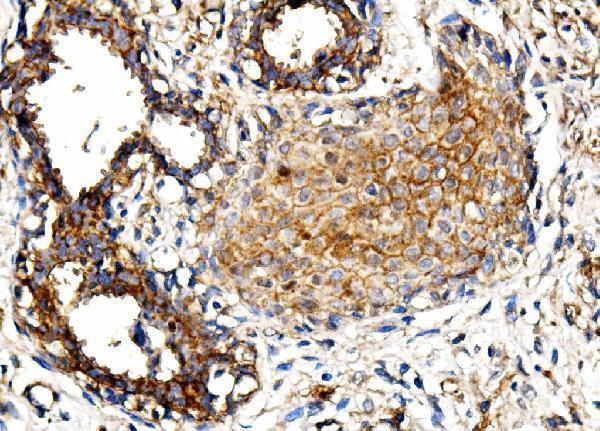
| Sample | Paraffin-embedded human mammary cancer tissue; fixative not specified (datasheet A06075-4) |
| Fixation | Image fixative and duration unreported (datasheet A06075-4); verify before use. |
| Sectioning | 4–5 µm sections on charged slides (standard) |
| Deparaffinisation | Xylene, graded ethanol series to water (standard) |
| Antigen retrieval | Heat retrieval: EDTA pH 8.0 (datasheet A06075-4); 20 min, 95–100 °C (standard) |
| Peroxidase block | 3% H2O2, 10 min, room temperature (standard) |
| Blocking | 10% goat serum (datasheet A06075-4) |
| Primary antibody | Rabbit anti-MORC3, 0.5-1μg/ml (datasheet A06075-4) |
| Primary incubation | Overnight at 4 °C (datasheet A06075-4) |
| Detection | Streptavidin-biotin complex (SABC), DAB chromogen (datasheet A06075-4) |
| Counterstain | Hematoxylin, blue, dehydrate and mount (standard) |
| Expected result | MORC3-positive staining in adipocytes of adipose tissue (HPA tissue IHC: High). HPA tissue profile: Nuclear and cytoplasmic expression in all tissues. No signal in the no-primary control. |
MORC3 is a nuclear protein found in the nucleoplasm, nuclear matrix and nuclear bodies (UniProt Q14149 localization); it has no transmembrane segment (UniProt Q14149 topology). In paraffin-section IHC, assess staining in the expected cells, including adipocytes, hematopoietic cells, glandular cells and neurons (HPA tissue IHC: High). HPA reports nuclear and cytoplasmic staining across tissues, but rates its IHC evidence Approved with low consistency between staining and RNA expression (HPA tissue IHC).
| Nuclei stain in expected cells, with occasional discrete nuclear foci. | Nuclear staining fits MORC3's nucleoplasmic, nuclear-matrix and nuclear-body locations (UniProt Q14149 localization). Foci are plausible, but their visibility need not be uniform: localization to nuclear bodies is ATP-dependent (UniProt Q14149 localization). Judge the result against cell identity and background across the section, not foci alone. |
| Cytoplasm stains strongly while nuclei remain unstained. | Treat a cytoplasmic-only result as suspect and check detection background and section quality. UniProt places MORC3 in nuclear compartments (UniProt Q14149 localization), although HPA reports nuclear and cytoplasmic tissue IHC (HPA tissue IHC). Cytoplasmic staining alone cannot establish specific MORC3 detection. |
| Signal predominates in cells outside the expected population within a selected tissue. | Compare neighboring cell types and controls before assigning MORC3 expression. The supplied high-staining examples name adipocytes, hematopoietic cells, glandular cells and neurons (HPA tissue IHC: High); staining elsewhere could reflect cross-reactivity or endogenous detection activity (general IHC practice). HPA's broad tissue profile is not a cell-by-cell exclusion list (HPA tissue IHC). |
| Color is widespread over tissue and blank areas, obscuring nuclei. | This is diffuse background rather than an interpretable compartment pattern (general IHC practice). Check a no-primary control for endogenous detection activity or nonspecific reagent signal, then reassess the nuclear pattern expected from UniProt (UniProt Q14149 localization). |
| No signal appears in a supplied high-staining tissue. | A blank result in adipose tissue, bone marrow or cerebral cortex conflicts with HPA's High staining examples (HPA tissue IHC: High). First check tissue preservation, detection and controls as general IHC workflow steps; one negative section does not overturn the reference pattern, especially given low staining–RNA consistency (HPA tissue IHC). |
| Reference compartment and topology | MORC3 is nucleoplasmic, nuclear-matrix and nuclear-body associated, with no transmembrane segment (UniProt Q14149 localization and topology). Prioritize nuclear morphology when scoring. HPA's tissue IHC profile also reports cytoplasmic staining (HPA tissue IHC), so record that component separately rather than assuming every cytoplasmic signal is specific. |
| Tissue evidence and reliability | HPA lists High staining in adipocytes, adrenal and breast glandular cells, bone-marrow hematopoietic cells, bronchial epithelial cells and neurons in sampled brain regions (HPA tissue IHC: High). Its IHC reliability is Approved, with low consistency between antibody staining and RNA expression (HPA tissue IHC); use these examples as comparisons, not absolute positive or negative controls. |
| Nuclear-body behavior | MORC3 forms nuclear bodies through an ATP-dependent mechanism and also occurs in PML-independent bodies (UniProt Q14149 localization and function). Fine nuclear puncta may fit the reference biology. Their presence or absence on a chromogenic section cannot, by itself, verify identity or exclude expression. |
| Antibody validation | The listed IHC antibody HPA018406 is Approved for IHC, while the separate antibody HPA034848 is Enhanced for ICC (HPA antibodies). These designations apply to their stated applications; the ICC status does not validate an IHC result obtained with another antibody. |
| IF/ICC Q&A: should mitochondrial signal count? | HPA's ICC-IF main location is enhanced nucleoplasm; its additional mitochondrial location is uncertain (HPA subcellular). Score a clear nuclear result first and treat mitochondrial-only fluorescence as unresolved. This IF/ICC observation does not provide a paraffin-IHC protocol or establish mitochondrial staining in tissue. |
| Situation | Likely cause | Next action |
|---|---|---|
| Cytoplasmic color dominates with little nuclear signal. | The compartment balance conflicts with UniProt's nuclear localization, despite HPA's reported cytoplasmic tissue staining (UniProt Q14149 localization; HPA tissue IHC). | Review nuclear counterstain and a no-primary control, then report nuclear and cytoplasmic components separately (general IHC practice). Avoid calling cytoplasmic-only color a confirmed MORC3 pattern. |
| Unexpected cells are the strongest stained population. | Cross-reactivity or endogenous detection activity is possible (general IHC practice); HPA's High entries identify specific cell populations within sampled tissues (HPA tissue IHC: High). | Identify the stained cells morphologically and compare them with adjacent expected cells and a no-primary control (general IHC practice). Record the discrepancy rather than assigning a new MORC3-positive population from this section alone. |
| Diffuse color covers tissue or blank regions. | Nonspecific reagent signal or endogenous detection activity can obscure cellular localization (general IHC practice). | Use a no-primary control, check blocking and detection steps, and adjust the general IHC workflow as needed; interpret MORC3 only after nuclei can be distinguished (general IHC practice; UniProt Q14149 localization). |
| A supplied high-staining tissue has no interpretable signal. | The section or detection workflow may have failed; HPA reports High staining in examples including adipocytes and bone-marrow hematopoietic cells (HPA tissue IHC: High). | Check tissue integrity, counterstain and a working positive control, then repeat the IHC workflow if needed (general IHC practice). Do not infer a MORC3-specific fixation effect; none is established by the supplied sources. |
| Nuclei stain, but no distinct nuclear foci are visible. | MORC3 also occupies the nucleoplasm and nuclear matrix, while localization to nuclear bodies is ATP-dependent (UniProt Q14149 localization). | Assess the overall nuclear pattern, cell identity and background. Do not reject an otherwise credible result solely because chromogenic staining does not resolve puncta (UniProt Q14149 localization; general IHC practice). |
| ICC-IF shows mitochondrial-only fluorescence. | HPA calls mitochondrial localization uncertain and nucleoplasm the enhanced main location (HPA subcellular). | Treat the mitochondrial-only result as unresolved, verify nuclear morphology and controls, and consult the separate IF/ICC guide for that application (HPA subcellular; general IF practice). |


Comprehensive Human Protein Atlas IHC scoring per tissue (reliability: Approved — Low consistency between antibody staining and RNA expression data.). Rows are taken directly from the HPA tissue chart — click any row's HPA link to view the source.
| Tissue | Cell type | Level | Evidence | Source |
|---|---|---|---|---|
| Adipose tissue | Adipocytes | High | Protein (IHC) | HPA → |
| Adrenal gland | Glandular cells | High | Protein (IHC) | HPA → |
| Bone marrow | Hematopoietic cells | High | Protein (IHC) | HPA → |
| Breast | Glandular cells | High | Protein (IHC) | HPA → |
| Bronchus | Respiratory epithelial cells | High | Protein (IHC) | HPA → |
| Tissue | Cell type | Level | Evidence | Source |
|---|---|---|---|---|
| None in HPA: MORC3 is detected in all 45 scored tissues. Use a no-primary (secondary-only) and an isotype control instead. | ||||
Troubleshoot MORC3 staining in paraffin sections by checking retrieval, nuclear localisation, controls and scoring before interpreting the chromogenic signal.
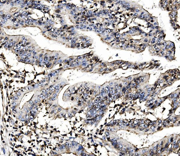
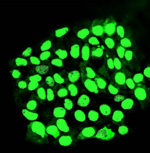
The IHC-validated antibody has paraffin-section IHC images from human mammary and rectal cancer tissue and an IF image from A431 cells (catalog image captions). Human, mouse and rat reactivity is listed (catalog reactivity).
A06075-4 will render with its own IHC image from a paraffin section of human mammary cancer tissue (A06075-4 IHC image caption). The catalog also shows IHC in human rectal cancer tissue and IF in A431 cells; IHC, IF and ICC are listed applications (A06075-4 image captions and applications).
Which to pick: Choose A06075-4 for paraffin-section tissue IHC: its IHC caption documents EDTA retrieval at pH 8.0 and 1 μg/ml primary antibody, but does not report the fixative (A06075-4 IHC image caption). The same SKU is listed for IF/ICC and has an IF image from A431 cells at 2 μg/ml (A06075-4 applications and IF image caption). It is also listed as reactive with human, mouse and rat, although the supplied IHC images show human tissue only; its clonality is unreported (A06075-4 reactivity, IHC image captions and catalog clone field).